
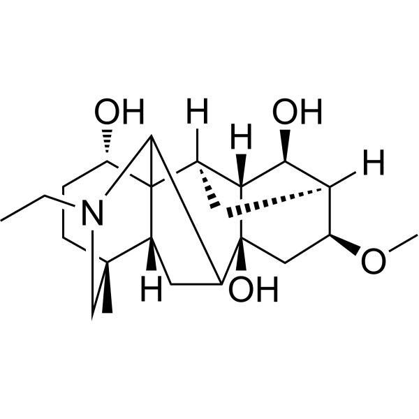
Karacoline
Based on 1 Customer Validation
Karacoline is an orally active PPARγ activator and ERK/JNK MAPK inhibitor. Karacoline restricts ROS production, maintains mitochondrial membrane potential, and inhibits pulmonary cell apoptosis. Karacoline inhibits NF-κB pathway activation, reduces acetylation levels of p65 and the expression of MMP14 and MMP9, enhances the expression of type II collagen (collagen II) and aggrecan (aggrecan), and suppresses extracellular matrix degradation. In a mouse model of sepsis-induced acute lung injury, Karacoline alleviates lung injury, inhibits the release of IL-1β, IL-6 and TNF-α, increases the Bcl-2/BAX ratio, and reduces caspase 3 expression. Karacoline can be used in research related to acute lung injury and intervertebral disc degeneration.
For research use only. We do not sell to patients.
- Purity : 99.58%
- CAS No.: 39089-30-0
- Formula: C22H35NO4
- Molecular Weight:377.52
-
Storage:
4°C, protect from light
* In solvent : -80°C, 6 months; -20°C, 1 month (protect from light)
All Caspase Isoforms
More
Biological Activity
Description
|
PPARγ |
MMP9 |
MMP14 |
IL-1β |
IL-6 |
Caspase 3 |
TNF-α |
In Vitro
Karacoline (1.6 μM; 24 h) inhibits LPS (HY-D1056)-induced apoptosis of MH-S cells by reducing ROS production and maintaining the stability of mitochondrial membrane potential[1].
Karacoline (1.6 μM; 24 h) upregulates the expression of PPARγ in LPS-stimulated MH-S cells, inhibits the JNK/ERK MAPK signaling pathway (excluding p38), and simultaneously suppresses the expression of MMP9[1].
Karacoline (0.001-1000.0 μM; 24 h) has an IC50 of 6.444 μM in primary rat nucleus pulposus cells, and Karacoline (1.25 μM; 0-3 days) partially reverses TNF-α-induced cytotoxicity in these cells[2].
Karacoline (1.25-12.88 μM; 24 h) reverses TNF-α-induced changes in gene expression in primary rat nucleus pulposus cells, downregulates MMP-14 expression and upregulates type II collagen expression, while the 1.25 μM concentration also upregulates aggrecan expression[2].
Karacoline (1.25-12.88 μM; 48 h) reverses TNF-α-induced extracellular matrix degradation in primary rat nucleus pulposus cells and increases aggrecan secretion; at the concentration of 1.25 μM, it also increases type II collagen secretion and reduces MMP-14 secretion[2].
Karacoline (1.25-12.88 μM; 4 days) increases the levels of type II collagen and proteoglycan, and decreases the level of MMP-14, in primary rat nucleus pulposus cells treated with TNF-α[2].
Karacoline (1.25 μM-12.88 μM; 24-48 h) reduces TNF-α-induced MMP-14 protein expression and inhibits the activation of the NF-κB pathway in primary rat nucleus pulposus cells by decreasing the acetylation level of p65[2].
Karacoline (1.25-12.88 μM; 48 h) reduces TNF-α-induced apoptosis in primary rat nucleus pulposus cells[2].
MedChemExpress (MCE) has not independently confirmed the accuracy of these methods. They are for reference only. Further protocols information, click here.
-
Cell Line:LPS-stimulated MH-S murine alveolar macrophages
-
Concentration:1.6 μM
-
Incubation Time:24 h
-
Result:Attenuated LPS-induced apoptosis, as shown by reduced cleaved caspase-3 expression, increased Bcl-2/BAX ratio, and decreased proportion of apoptotic cells.
Reduced LPS-induced intracellular ROS fluorescence intensity.
Reversed LPS-mediated MMP loss (restored red/green fluorescence ratio).
-
Cell Line:primary rat nucleus pulposus cells
-
Concentration:0, 1.25, 12.88 μM+100 ng/mL TNF-α
-
Incubation Time:24 h
-
Result:Reduced MMP-14 expression and increased collagen II expression, with 1.25 μM also increasing aggrecan expression.
-
Cell Line:primary rat nucleus pulposus cells
-
Concentration:0, 1.25, 12.88 μM+100 ng/mL TNF-α
-
Incubation Time:48 h
-
Result:Reduced TNF-α-induced MMP-14 protein expression.
-
Cell Line:primary rat nucleus pulposus cells
-
Concentration:0, 1.25, 12.88 μM+100 ng/mL TNF-α
-
Incubation Time:4 days
-
Result:Increased collagen II and aggrecan levels and reduced MMP-14 levels.
In Vivo
MedChemExpress (MCE) has not independently confirmed the accuracy of these methods. They are for reference only.
-
Animal Model:C57BL/6 (male, 6-8 weeks old, 20-25 g, sepsis-induced acute lung injury via CLP surgery)[1]
-
Dosage:10 mg/kg (survival monitoring); 20 mg/kg; 40 mg/kg (survival monitoring)
-
Administration:i.g. (single dose for survival monitoring); i.g. (1 h before surgery and 12 h after surgery)
-
Result:Improved 7-day survival of CLP-induced septic mice at 20 mg/kg and 10 mg/kg (40 mg/kg did not improve survival beyond 20 mg/kg).
Reduced lung wet-to-dry weight ratio at 20 mg/kg.
Decreased total protein and albumin levels in bronchoalveolar lavage fluid (BALF) at 20 mg/kg.
Lowered BALF concentrations of IL-1β, IL-6, and TNF-α at 20 mg/kg.
Reduced lung injury scores at 20 mg/kg.
Mitigated histopathological changes including alveolar disruption, interstitial edema, and inflammatory cell infiltration at 20 mg/kg.
Increased the Bcl-2/BAX ratio in lung tissue at 20 mg/kg.
Decreased cleaved-caspase-3 levels in lung tissue at 20 mg/kg.
Reduced BAX expression in lung tissue at 20 mg/kg.
Increased Bcl-2 expression in lung tissue at 20 mg/kg.
Lowered TUNEL-positive cell counts in lung tissue at 20 mg/kg.
Restored PPARγ mRNA and protein expression in lung tissue at 20 mg/kg.
Suppressed phosphorylation of JNK and ERK in lung tissue at 20 mg/kg.
Reduced MMP9 expression in lung tissue at 20 mg/kg.
Chemical Information
-
CAS No. 39089-30-0
-
Appearance Solid
-
Molecular Weight 377.52
-
Formula C22H35NO4
-
Color White to off-white
-
SMILES
O[C@@H]1C23C4C([C@]5(O)[C@]6([H])[C@@]3([H])C[C@@]([C@@H](OC)C5)([H])[C@@H]6O)C[C@]2([H])[C@@](CN4CC)(C)CC1
-
Structure Classification
-
Initial Source
-
Shipping
Room temperature in continental US; may vary elsewhere.
-
Storage
4°C, protect from light
* In solvent : -80°C, 6 months; -20°C, 1 month (protect from light)
Solvent & Solubility
In Vitro:
DMSO : 25 mg/mL (66.22 mM; Need ultrasonic; Hygroscopic DMSO has a significant impact on the solubility of product, please use newly opened DMSO)
Please refer to the solubility information to select the appropriate solvent. Once prepared, please aliquot and store the solution to prevent product inactivation from repeated freeze-thaw cycles.
Storage method and period of stock solution: -80°C, 6 months; -20°C, 1 month (protect from light). When stored at -80°C, please use it within 6 months. When stored at -20°C, please use it within 1 month.
Please refer to the solubility information to select the appropriate solvent. Once prepared, please aliquot and store the solution to prevent product inactivation from repeated freeze-thaw cycles.
Storage method and period of stock solution: -80°C, 6 months; -20°C, 1 month (protect from light). When stored at -80°C, please use it within 6 months. When stored at -20°C, please use it within 1 month.
Concentration (start) × Volume (start) = Concentration (final) × Volume (final)
In Vivo:
Select the appropriate dissolution method based on your experimental animal and administration route.
- For the following dissolution methods, please ensure to first prepare a clear stock solution using an In Vitro approach and then sequentially add co-solvents:
- To ensure reliable experimental results, the clarified stock solution can be appropriately stored based on storage conditions. As for the working solution for In Vivo experiments, it is recommended to prepare freshly and use it on the same day.
- The percentages shown for the solvents indicate their volumetric ratio in the final prepared solution. If precipitation or phase separation occurs during preparation, heat and/or sonication can be used to aid dissolution.
Add each solvent one by one: 10% DMSO 40% PEG300 5% Tween-80 45% Saline
Solubility: ≥ 2.5 mg/mL (6.62 mM); Clear solution
This protocol yields a clear solution of ≥ 2.5 mg/mL (saturation unknown).
Taking 1 mL working solution as an example, add 100 μL DMSO stock solution (25.0 mg/mL) to 400 μL PEG300, and mix evenly; then add 50 μL Tween-80 and mix evenly; then add 450 μL Saline to adjust the volume to 1 mL.
Preparation of Saline: Dissolve 0.9 g sodium chloride in ddH₂O and dilute to 100 mL to obtain a clear Saline solution.
Add each solvent one by one: 10% DMSO 90% (20% SBE-β-CD in Saline)
Solubility: ≥ 2.5 mg/mL (6.62 mM); Clear solution
This protocol yields a clear solution of ≥ 2.5 mg/mL (saturation unknown).
Taking 1 mL working solution as an example, add 100 μL DMSO stock solution (25.0 mg/mL) to 900 μL 20% SBE-β-CD in Saline, and mix evenly.
Preparation of 20% SBE-β-CD in Saline (4°C, storage for one week): 2 g SBE-β-CD powder is dissolved in 10 mL Saline, completely dissolve until clear.
In Vivo Dissolution Calculator
Please enter the basic information of animal experiments:
-
-
-
-
Recommended: Prepare an additional quantity of animals to account for potential losses during experiments.
Please enter your animal formula composition:
-
%DMSO +
Recommended: Keep the proportion of DMSO in working solution below 2% if your animal is weak.
-
%+
-
+%Tween-80 + +
-
%Saline +
The co-solvents required include: DMSO, . All of co-solvents are available by MedChemExpress (MCE). , Tween 80. All of co-solvents are available by MedChemExpress (MCE).
Working solution concentration: 0.22 mg/mL
Method for preparing stock solution: mg drug dissolved in μL DMSO. Stock solution concentration: mg/mL. * In solvent : -80°C, 6 months; -20°C, 1 month (protect from light)
1. Take μL DMSO stock solution;
2. Add μL .
μL , mix evenly;
3. Then add μL Tween 80, mix evenly;
4. Then add μL
Please ensure that the stock solution in the first step is dissolved to a clear state, and add co-solvents in sequence. You can use ultrasonic heating (ultrasonic cleaner, recommended frequency 20-40 kHz), vortexing, etc. to assist dissolution.
Protocols
-
RNA extraction experimental
By lysing cells, releasing RNA, and removing impurities such as proteins and DNA, high-purity RNA products are finally obtained. The commonly used traditional method is the guanidine isothiocyanate/phenol/chloroform method (Trizol), which is suitable for a variety of animal materials including animal tissues, microorganisms, cultured cells, etc., and most plant materials.
-
Collagen: Sirius Red Staining
Sirius Red or picrosirius red staining is a histochemical method for visualizing collagen-rich extracellular matrix in tissue sections, and collagen fibers are detected as red-stained structures under bright-field microscopy with enhanced birefringence under polarized light. Picrosirius red is useful for assessing total collagen organization, distribution, and fibrosis burden, but polarized color should not be interpreted as a definitive collagen type I versus type III readout because color is affected by fiber orientation, thickness, and packing.
-
Mitochondrial membrane-potential fluorescent assay
Mitochondrial membrane potential fluorescent assays estimate ΔΨm in living cells using lipophilic cationic dyes such as TMRM, TMRE, rhodamine 123, and JC-1, which accumulate in mitochondria according to membrane polarization; loss of signal after FCCP or CCCP treatment is interpreted as mitochondrial depolarization. TMRM/TMRE and rhodamine 123 are commonly used for semi-quantitative live-cell microscopy or flow cytometry, while JC-1 can report a shift from red aggregate fluorescence to green monomer fluorescence during depolarization; interpretation requires controls because dye concentration, quenching mode, cell type, dye efflux, and mitochondrial mass can affect fluorescence independently of ΔΨm.
-
Annexin V plus membrane-impermeant dye apoptosis staining
Annexin V-based apoptosis assays rely on the detection of phosphatidylserine (PS) externalization from the inner leaflet of the plasma membrane to the outer leaflet, an early biochemical hallmark of apoptosis. Fluorescently labeled Annexin V binds PS in a calcium-dependent manner, enabling identification of early apoptotic cells by flow cytometry or fluorescence microscopy. When combined with a membrane-impermeant DNA-binding dye (e. g. , propidium iodide), this approach allows discrimination between viable (Annexin V−/dye−), early apoptotic (Annexin V+/dye−), and late apoptotic or necrotic (Annexin V+/dye+) cell populations by assessing membrane integrity and PS exposure.
-
ROS/oxidative-stress fluorescent staining
ROS/oxidative-stress fluorescent staining uses cell-permeant fluorogenic probes that become fluorescent after oxidation inside cells or tissues; commonly used examples include DCFH-DA/DCFDA for broad cellular oxidant detection, DHE for superoxide-related signal detection, MitoSOX for mitochondrial superoxide-related signal detection, and CellROX probes for oxidative-stress-associated fluorescence readouts. The assay detects probe oxidation rather than a single ROS species unless the probe and analysis method have been chemically validated for that species. DCFH-DA enters cells, is deacetylated by intracellular esterases to DCFH, and produces fluorescent DCF after oxidation, so the readout is used as an operational measure of total cellular oxidative stress rather than a species-specific ROS measurement. DHE and MitoSOX can report superoxide-related oxidation, but red fluorescence alone can include non-specific ethidium-like oxidation products; HPLC or optimized spectral approaches are
-
ECM-Embedded Organoid (Matrigel/Dome) Culture
ECM-embedded organoid dome culture embeds epithelial stem cells, crypts, organoid fragments, or tumor-derived epithelial cells in a basement-membrane-like hydrogel such as Matrigel, allowing 3D growth, self-organization, lumen formation, budding or cystic morphogenesis, and lineage maintenance under defined niche-factor-containing medium. The primary readouts are organoid establishment efficiency, growth, morphology, passaging capacity, lineage-marker expression, and, when fluorescently labeled lines are used, microscopy- or flow-cytometry-based quantification of population behavior in 3D culture.
-
Research Protocol for Inflammation-related Diseases
The NLRP3 inflammasome is a cytosolic innate immune signaling platform that integrates priming signals and danger-signal activation to promote caspase-1 activation, maturation of IL-1β and IL-18, and gasdermin D-mediated pyroptotic cell death. The core experimental logic is to determine whether inflammatory disease phenotypes are driven by increased NLRP3 expression, ASC-containing inflammasome assembly, caspase-1 cleavage, GSDMD cleavage, and extracellular release of IL-1β/IL-18 rather than by nonspecific cell injury alone. The pathway is strongly linked to inflammation-related disease phenotypes because monosodium urate crystals activate NALP3/NLRP3 inflammasome signaling in gout-like crystal inflammation, cholesterol crystals activate NLRP3 inflammasomes in atherogenesis models, and DSS-induced intestinal inflammation has been reported to involve NLRP3 inflammasome activity. However, experimental colitis studies also show context-dependent protective effects of NLRP3 inflammasome co
-
Matrigel Transwell/Boyden Chamber Invasion Assay
Matrigel Transwell/Boyden chamber invasion assay measures the ability of cells to degrade or traverse an extracellular matrix-coated porous membrane and move from an upper chamber toward a chemoattractant in a lower chamber. Invasion is distinguished from migration by coating the membrane with Matrigel or basement membrane matrix; uncoated inserts measure migration, while coated inserts require cells to cross an ECM barrier before reaching the underside of the membrane.
-
How to Choose the Right Model Animal
Choosing the right model animal is a validity-driven decision in which the species, strain, sex, age, genetic background, disease-induction method, outcome measures, and welfare burden must match the scientific question rather than laboratory tradition or convenience. A model should be selected by judging face validity, construct validity, and predictive validity: whether it resembles the human phenotype, whether it reproduces relevant mechanisms, and whether results are likely to predict human biology or treatment response. Animal studies often fail to translate because of species differences, weak disease resemblance, poor experimental design, inadequate reporting, publication bias, and underuse of randomization, blinding, and sample-size justification. Unresolved questions include how to rank competing models objectively, how much human-disease complexity must be reproduced for a given objective, and when non-animal systems such as organoids, ex vivo tissue, or computational models
-
Fibrosis/Collagen Morphometry
Fibrosis and collagen morphometry is based on the quantitative visualization of fibrillar collagen deposition in tissue sections using histochemical stains such as Sirius Red (Picrosirius Red) or Masson's trichrome, followed by image-based or polarization-enhanced analysis to estimate collagen proportional area as a surrogate of extracellular matrix accumulation during fibrotic remodeling. Sirius Red combined with polarized light microscopy enhances detection of collagen fibers due to birefringence properties, enabling more specific visualization of collagen type I and III fibrils compared to conventional bright-field histology, while whole-section or region-restricted digital morphometry reduces field-selection bias in fibrosis assessment. Alternative quantitative approaches include second harmonic generation (SHG) and two-photon excited fluorescence microscopy, which enable label-free detection of fibrillar collagen and have been validated against histological staining and biochemica
-
Invadopodia/Fluorescent Gelatin Degradation Assay
Invadopodia/fluorescent gelatin degradation assay detects proteolytic extracellular matrix degradation by cancer-cell invadopodia, which are actin-rich protrusive structures associated with matrix remodeling, invasion, and metastasis. The readout is generated by culturing cells on fluorescent gelatin and measuring dark degraded areas where fluorescent substrate has been locally removed, often together with immunofluorescent detection of invadopodia markers such as F-actin, cortactin, and TKS5.
-
Apoptosis
Apoptosis, also called programmed cell death, is generally characterized by distinct morphological characteristics.
-
TUNEL staining for apoptotic DNA fragmentation
TUNEL staining detects DNA strand breaks by using terminal deoxynucleotidyl transferase to add labeled nucleotides to exposed 3′-OH DNA termini, generating either microscopic staining in fixed cells or tissue sections, or fluorescence/cytometric signal in cell suspensions. TUNEL positivity reflects DNA fragmentation but should not be interpreted alone as definitive apoptosis, because TUNEL can also label necrotic, autolytic, mechanically damaged, or DNA-repair-associated DNA breaks.
-
LPS-Induced Endotoxemia/Systemic Inflammation
Lipopolysaccharide (LPS)-induced endotoxemia is a widely used in vivo model of acute systemic inflammation in which LPS, a Gram-negative bacterial endotoxin, activates innate immune signaling primarily through TLR4, leading to rapid and transient induction of pro-inflammatory cytokines such as TNF-α, IL-6, and IL-1β in circulation and tissues. This cytokine surge is commonly used as a measurable readout of systemic inflammatory activation and immune dysregulation, and is typically assessed within hours after intraperitoneal LPS administration in mouse models of endotoxemia. The model captures key features of systemic inflammatory response syndrome, including cytokine release, immune cell activation, and downstream tissue responses, and has been used to evaluate anti-inflammatory interventions such as cytokine modulation, lipid mediators, and immune cell-targeting therapies.
-
Fluorescent plasma-membrane potential dye assay
Fluorescent plasma-membrane potential dye assays measure changes in cell membrane potential using voltage-sensitive dyes whose fluorescence changes when cells depolarize or hyperpolarize. Anionic bis-oxonol dyes such as DiBAC4(3) enter depolarized cells more readily and show increased fluorescence after intracellular binding, while hyperpolarization reduces dye accumulation and fluorescence. FMP/FLIPR membrane-potential dyes are used for faster, homogeneous microplate assays of ion-channel or receptor-mediated membrane-potential changes.
-
Mitochondrial membrane-potential and mitochondrial mass staining
Mitochondrial membrane potential staining measures the electrochemical polarization across the mitochondrial inner membrane in live cells using lipophilic cationic fluorescent probes; early rhodamine-based work showed that selective mitochondrial dye accumulation is lost when the mitochondrial transmembrane potential is dissipated. JC-1 reports mitochondrial polarization by shifting from green monomer fluorescence to red J-aggregate fluorescence as dye concentration increases within energized mitochondria; therefore, the red/green fluorescence ratio is used as a relative readout of mitochondrial membrane potential. TMRE or TMRM staining provides a single-channel relative readout because these cationic rhodamine esters accumulate in polarized mitochondria, and lower fluorescence indicates reduced mitochondrial polarization when acquisition and dye-loading conditions are controlled. Mitochondrial mass staining is commonly performed with MitoTracker Green FM or related MitoTracker dyes as
-
Apoptosis Solutions
Apoptosis is a regulated, generally non-lytic cell-death pathway that removes unwanted, damaged, infected, or abnormal cells through coordinated morphological changes, caspase activation, DNA fragmentation, and membrane remodeling. The intrinsic apoptosis pathway is controlled mainly by mitochondrial outer membrane permeabilization, BCL-2 family proteins, cytochrome c release, apoptosome formation, caspase-9 activation, and downstream executioner caspase-3/7 activation. The extrinsic apoptosis pathway is initiated by death receptors such as Fas, TNFR, and TRAIL receptors, which recruit adaptor proteins and activate caspase-8 before engaging executioner caspases or mitochondrial amplification through BID cleavage. Apoptosis is linked to many phenotypes, including cancer cell killing, tissue homeostasis, immune regulation, neurodegeneration, infection response, and treatment-induced cytotoxicity; unresolved questions include how apoptosis interacts with necroptosis, pyroptosis, ferroptos
-
Matrigel/ECM Transwell Invasion Assay
The Matrigel/ECM Transwell invasion assay measures the ability of cells to move toward a chemoattractant while crossing an extracellular-matrix barrier placed on a porous membrane; therefore, the readout reflects both chemotactic motility and matrix invasion rather than migration alone. Matrigel is a basement-membrane-rich matrix derived from Engelbreth-Holm-Swarm mouse sarcoma and has been used as a reconstituted basement membrane barrier in chemoinvasion assays. The assay readout is generated by quantifying cells that reach the underside of the insert membrane or lower compartment after incubation, commonly by staining and counting invaded cells or by fluorescence-based quantification.
-
Inhalation Toxicity Study
Inhalation toxicity studies expose rodents to a controlled aerosol, vapor, gas, or smoke atmosphere and assess respiratory and systemic toxicity using exposure-atmosphere characterization, clinical observations, body and organ weights, bronchoalveolar lavage fluid, histopathology, blood chemistry, hematology, and, when included, molecular endpoints such as transcriptomics, proteomics, lipidomics, or tissue burden analysis. The primary biological readouts are airway irritation, pulmonary inflammation, cytotoxicity, altered surfactant or lipid homeostasis, impaired particle clearance, and tissue remodeling, reflected by BALF cell differentials, BALF protein, LDH, phosphatase activities, cytokines, lung weight, microscopic respiratory-tract lesions, and retained lung burden.
-
Pyroptosis Solutions
Pyroptosis is a lytic inflammatory cell-death pathway executed by gasdermin pores, most classically through inflammasome-mediated activation of caspase-1, cleavage of gasdermin D, membrane pore formation, LDH release, and secretion of IL-1β and IL-18. The canonical pathway is commonly modeled by priming cells with an inflammatory signal such as LPS to induce pro-IL-1β and inflammasome components, followed by an activation signal such as ATP or nigericin to activate NLRP3, ASC speck formation, caspase-1 cleavage, GSDMD cleavage, cytokine release, and pyroptotic membrane rupture. The non-canonical pathway is triggered when cytosolic LPS activates mouse caspase-11 or human caspase-4/5, leading to GSDMD cleavage and pyroptosis, and this can secondarily activate NLRP3-dependent IL-1β release. Pyroptosis is linked to inflammatory injury, infection, cancer, liver disease, ocular disease, placental inflammation, and other disease phenotypes, but unresolved questions include which gasdermin fam
Purity & Documentation
-
Data Sheet (288 KB)
-
SDS (252 KB)
- English - EN (252 KB)
- Français - FR (252 KB)
- Deutsch - DE (252 KB)
- Norwegian - NO (252 KB)
- Español - ES (252 KB)
- Swedish - SV (252 KB)
- Italian - IT (252 KB)
- Korean - KR (252 KB)
- Portuguese - PT (252 KB)
-
Handling Instructions (2659 KB)
References
[1]. Lei Z, et al. Karacoline attenuates sepsis-induced acute lung injury by suppressing apoptosis via PPARγ-associated inhibition of JNK/ERK MAPK signaling. Respiratory research. 2026 Jun 15. [Content Brief]
[2]. Zhou X, et al. Karacoline, identified by network pharmacology, reduces degradation of the extracellular matrix in intervertebral disc degeneration via the NF-κB signaling pathway. Journal of pharmaceutical analysis. 2020 Feb;10(1):13-22. [Content Brief]
Complete Stock Solution Preparation Table
Please refer to the solubility information to select the appropriate solvent. Once prepared, please aliquot and store the solution to prevent product inactivation from repeated freeze-thaw cycles.
Storage method and period of stock solution: -80°C, 6 months; -20°C, 1 month (protect from light). When stored at -80°C, please use it within 6 months. When stored at -20°C, please use it within 1 month.
| Optional Solvent | Concentration Solvent Mass | 1 mg | 5 mg | 10 mg | 25 mg |
|---|---|---|---|---|---|
| DMSO | 1 mM | 2.6489 mL | 13.2443 mL | 26.4887 mL | 66.2217 mL |
| 5 mM | 0.5298 mL | 2.6489 mL | 5.2977 mL | 13.2443 mL | |
| 10 mM | 0.2649 mL | 1.3244 mL | 2.6489 mL | 6.6222 mL | |
| 15 mM | 0.1766 mL | 0.8830 mL | 1.7659 mL | 4.4148 mL | |
| 20 mM | 0.1324 mL | 0.6622 mL | 1.3244 mL | 3.3111 mL | |
| 25 mM | 0.1060 mL | 0.5298 mL | 1.0595 mL | 2.6489 mL | |
| 30 mM | 0.0883 mL | 0.4415 mL | 0.8830 mL | 2.2074 mL | |
| 40 mM | 0.0662 mL | 0.3311 mL | 0.6622 mL | 1.6555 mL | |
| 50 mM | 0.0530 mL | 0.2649 mL | 0.5298 mL | 1.3244 mL | |
| 60 mM | 0.0441 mL | 0.2207 mL | 0.4415 mL | 1.1037 mL |
Keywords
- Karacoline
- 39089-30-0
- NF-κB
- PPAR
- ERK
- JNK
- p38 MAPK
- Reactive Oxygen Species (ROS)
- Apoptosis
- MMP
- Collagen
- Interleukin Related
- Bcl-2 Family
- Caspase
- TNF Receptor
- male C57BL/6 mice
- ERK/JNK MAPK
- intervertebral disc degeneration
- MH-S cells
- PPARγ
- NF-κB pathway
- mitochondrial membrane potential
- sepsis-induced acute lung injury
- ROS
- primary rat nucleus pulposus cells
- Inhibitor
- inhibitor
- inhibit

