



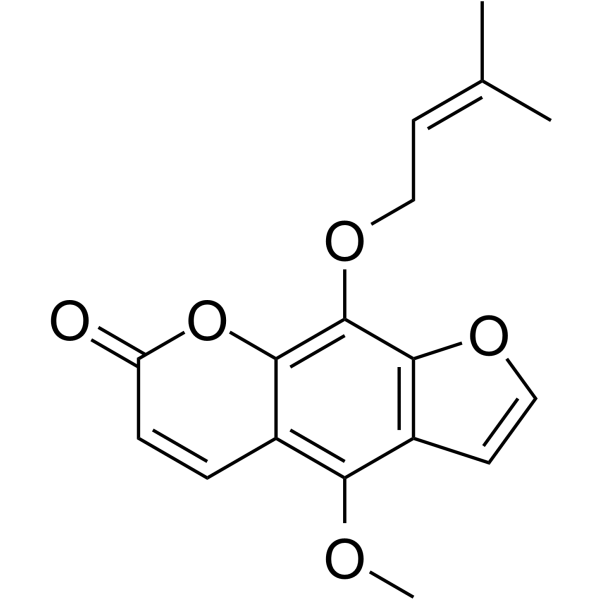
Phellopterin

Based on 1 Customer Validation
Phellopterin, an orally active furocoumarin with multiple biological activities. Phellopterin is a partial agonist of the central benzodiazepine receptors. Phellopterin exerts anti-inflammatory effects by upregulating SIRT1, downregulating ICAM-1 (reducing chronic inflammation, aiding diabetic ulcer healing), inhibiting STAT3 phosphorylation (easing atopic dermatitis inflammation), regulating Akt/PKC pathways (lowering TNF-α-induced VCAM-1 to block monocyte adhesion), and inhibiting TLR4/NF-κB pathway and macrophage M2 polarization (alleviating colitis-related cancers). Phellopterin suppresses ovarian cancer progression via inhibiting the PU.1/CLEC5A/PI3K-AKT loop (inducing cell cycle arrest, apoptosis, DNA damage). Phellopterin alleviates murine diabetes by promoting adipocyte differentiation and increasing PPARγ. Phellopterin also has anti-HSV-1 activity. Phellopterin can be used for studying anti-inflammation, anti-cancer (e.g., ovarian cancer, colitis cancer), blood glucose lowering, anti-diabetes, and anti-virus.
For research use only. We do not sell to patients.
- Purity : 99.94%
- CAS No.: 2543-94-4
- Formula: C17H16O5
- Molecular Weight:300.31
-
Storage:Powder -20°C, 3 years , 4°C, 2 years ; In solvent -80°C, 6 months , -20°C, 1 month

To place orders, for customer services and technical support, please contact: MedChemExpress USA
Tel: 609-228-6898 E-mail: [email protected] [email protected]


-
 Biological Activity
-
Chemical Information
-
Solvent & Solubility
- Protocols
- Purity & Documentation
- References
-
 Help & FAQs





Biological Activity
Description
|
PI3K |
SIRT3 |
PKC |
PPARγ |
Akt |
ERK |
TLR4 |
In Vitro
Phellopterin (1-50 μM, 24 h) significantly inhibits VCAM-1 expression and Akt and PKC phosphorylation in a dose-dependent manner in TNF-α-stimulated HUVECs, and effectively prevents monocyte adhesion to TNF-α-stimulated ECs by regulating VCAM-1 expression[1].
Phellopterin (1-25 μM, 24 h) upregulates the expression of SIRT1 and downregulates the expression of ICAM-1 in HaCaT cells, thereby reversing the proliferation inhibition caused by IFN-γ[2].
Phellopterin (1-16 μM, 24 h) inhibits IL-4-induced activation of STAT3, which leaded to suppress the STAT3-mediated transcription of TSLP and IL-33 in HaCaT cells[3].
Phellopterin (1-100 μg/mL, 48 h) attenuates the proliferation of ovarian cancer cells with IC50s for OV90 and SKOV3 cells of 18.67 and 27.75 μg/mL[4].
Phellopterin (25-50 μg/mL) attenuates ovarian cancer progression by inhibiting cell proliferation through modulating DNA replication, cell cycle (G0/G1), and apoptosis in OV90 cells and SKOV3 cells[4].
Phellopterin (25 μg/mL, 48 h) inactivates CLEC5A/PI3K/AKT signaling in OV90 cells and SKOV3 cells [4].
Phellopterin (12.5-100 μg/mL) induces adipocyte differentiation and increases the mRNA expression of peroxisome proliferator-activated receptors γ (PPARγ)[5].
Phellopterin (1-500 μg/mL, 72 h) reduces the HSV-1 replication by 3.01 log at the concentration of 7.81 mg/mL, and has a significant cytotoxicity towards Vero cells with CC50 of 14.61 μg/mL[6].
Phellopterin (40 min) inhibits [3H]diazepam and [3H]Ro 15-1788 binding to the benzodiazepine site of the rat brain 3,-aminobutyric acidA (GABAA) receptor in vitro with IC50 values of 400 and 680 nM, respectively[7].
MedChemExpress (MCE) has not independently confirmed the accuracy of these methods. They are for reference only. Further protocols information, click here.
-
Cell Line:TNF-α-stimulated HUVECs
-
Concentration:1, 5, 10 and 50 μM
-
Incubation Time:24 h
-
Result:Did not inhibit TNF-α-induced ICAM-1 expression, whereas it inhibited VCAM-1 expression from 5 μM, and completely suppressed it at 50 μM concentration.
Had no effect on ERK1/2 phosphorylation, whereas they significantly inhibited the phosphorylation of Akt and PKC in dose-dependent manner.
-
Cell Line:TNF-α-stimulated HUVECs
-
Concentration:1, 5, 10 and 50 μM
-
Incubation Time:24 h
-
Result:Showed significant reduction of adherent cells to ECs from 4-fold to 2.5-fold.
-
Cell Line:TNF-γ-stimulated HaCaT cells
-
Concentration:1, 5 and 25 μM
-
Incubation Time:24 h
-
Result:Stimulated HaCaT cell proliferation.
Was unable to restore cell proliferation to normal levels.
-
Cell Line:TNF-γ-stimulated HaCaT cells
-
Concentration:2, 4, 8 and 16 μM
-
Incubation Time:24 h
-
Result:Suppressed the STAT3-mediated transcription of TSLP and IL-33.
-
Cell Line:IL-4-stimulated HaCaT cells
-
Concentration:2, 4, 8 and 16 μM
-
Incubation Time:24 h
-
Result:Suppressed the STAT3-mediated expression of TSLP and IL-33.
-
Cell Line:OV90 and SKOV3 cells
-
Concentration:25 μg/mL
-
Incubation Time:48 h
-
Result:Significantly reduced the protein levels of CLEC5A, p-PI3K, and p-AKT.
Reduced the expression of PU.1 protein.
In Vivo
Phellopterin (0.5-4.5 mg/kg, ear topically spread, twice a day for 10 days) improves the atopic dermatitis (AD)-like lesions in mice[3].
Phellopterin (50 mg/kg, i,g., five times a week for 4 weeks) suppresses cancer growth in mice ovarian cancer xenograft model[4].
Phellopterin (0.5-2 mg/kg, i,g., once daily for 4 weeks) significantly lowers blood sugar levels thus prevents High-fat diet/Streptozotocin (HFD/STZ) (HY-13753)-induced type Ⅱ diabetes in mice[5].
Phellopterin (0.5-2 mg/kg, i,g.) improves the symptoms and inflammatory response of colitis-associated cancer (CAC) and inhibits the occurrence of colon cancer by inhibiting M2 polarization of macrophages and activation of the TLR4/NF-κB pathway in mice[8].
MedChemExpress (MCE) has not independently confirmed the accuracy of these methods. They are for reference only.
-
Animal Model:Streptozotocin (STZ) -induced diabetic model established in five-week-old C57BL/6J male mice[2]
-
Dosage:0.6, 1.2 and 2.4 mg/kg
-
Administration:Intravenous injection (i.v.), for 14 days
-
Result:Seemed to have the greatest effect on wound healing at medium dose.
Promoted diabetic wound healing by accelerating epidermic re-epithelialization.
Downregulated ICAM-1 expression via SIRT1 in mice with diabetic ulcers.
-
Animal Model:Calcipotriol (MC-903) (HY-10001) induced AD like skin model established in male C57BL/6 mice around 8 to 10 weeks of age[3]
-
Dosage:0.5, 1.5 and 4.5 mg/kg
-
Administration:Ear topically spread, twice a day for 10 days
-
Result:The concentration at 1.5 mg/kg showed the optimal therapeutic effect.
Exhibited significant reduction in epidermal thickness and scales as well as serum IgE levels.
Decreased the infiltrated eosinophils and mast cells.
The protein levels of TSLP and IL-33 in epidermal keratinocytes were decreased.
The expression of IL-4 was remarkably elevated in the AD-like skin lesions, but not significantly changed its expression.
-
Animal Model:SKOV3 cells induced ovarian cancer xenograft model established in nude mice[4]
-
Dosage:50 mg/kg
-
Administration:Intragastric administration (i.g.), five times a week for 4 weeks
-
Result:Attenuated cancer growth.
No significant difference in mice body weight and the histopathological changes of the liver and kidney.
Downregulated Ki67 levels in tumor.
-
Animal Model:High-fat diet/Streptozotocin (HFD/STZ)-induced diabetic established in male ICR strain mice[5]
-
Dosage:0.5, 1 and 2 mg/kg
-
Administration:Intragastric administration (i.g.), once daily for 4 weeks
-
Result:Decreased total cholesterol from 1154.8 to 630.4 mg/dL.
Dropped triglycerides dropped from 541.6 to 346.7 mg/dL.
Observed no obvious toxic reactions and the organ coefficients were within the normal range.
-
Animal Model:Azoxymethane (AOM) (HY-111375)/DSS (Dapsone) (HY-B0688) induced colitis-associated cancer (CAC) model established in C57BL/6 mice[8]
-
Dosage:0.5, 1 and 2 mg/kg
-
Administration:Intragastric administration (i.g.)
-
Result:Dose-dependently improved the weight loss in mice and increased the length of their colons.
Reduced the rate of tumor formation.
Increased CD4+ and CD8+, and decreased pro-inflammatory factors (IL-6, IL-1β, TNF-α).
Reduced the levels of M2-type markers (such as CD163, CD206, etc.)
Decreased the expression of TLR4 and NF-κB p65.


Chemical Information
-
CAS No. 2543-94-4
-
Appearance Solid
-
Molecular Weight 300.31
-
Formula C17H16O5
-
Color Off-white to light yellow
-
SMILES
O=C1C=CC2=C(OC)C3=C(OC=C3)C(OC/C=C(C)/C)=C2O1
-
Structure Classification
-
Shipping
Room temperature in continental US; may vary elsewhere.
-
Storage
Powder -20°C 3 years 4°C 2 years In solvent -80°C 6 months -20°C 1 month


Solvent & Solubility
In Vitro:
DMSO : 100 mg/mL (332.99 mM; Need ultrasonic; Hygroscopic DMSO has a significant impact on the solubility of product, please use newly opened DMSO)
Please refer to the solubility information to select the appropriate solvent. Once prepared, please aliquot and store the solution to prevent product inactivation from repeated freeze-thaw cycles.
Storage method and period of stock solution: -80°C, 6 months; -20°C, 1 month. When stored at -80°C, please use it within 6 months. When stored at -20°C, please use it within 1 month.
Please refer to the solubility information to select the appropriate solvent. Once prepared, please aliquot and store the solution to prevent product inactivation from repeated freeze-thaw cycles.
Storage method and period of stock solution: -80°C, 6 months; -20°C, 1 month. When stored at -80°C, please use it within 6 months. When stored at -20°C, please use it within 1 month.
Concentration (start) × Volume (start) = Concentration (final) × Volume (final)
In Vivo:
Select the appropriate dissolution method based on your experimental animal and administration route.
- For the following dissolution methods, please ensure to first prepare a clear stock solution using an In Vitro approach and then sequentially add co-solvents:
- To ensure reliable experimental results, the clarified stock solution can be appropriately stored based on storage conditions. As for the working solution for In Vivo experiments, it is recommended to prepare freshly and use it on the same day.
- The percentages shown for the solvents indicate their volumetric ratio in the final prepared solution. If precipitation or phase separation occurs during preparation, heat and/or sonication can be used to aid dissolution.
In Vivo Dissolution Calculator
Please enter the basic information of animal experiments:
-
-
-
-
Recommended: Prepare an additional quantity of animals to account for potential losses during experiments.
Please enter your animal formula composition:
-
%DMSO +
Recommended: Keep the proportion of DMSO in working solution below 2% if your animal is weak.
-
%+
-
+%Tween-80 + +
-
%Saline +
The co-solvents required include: DMSO, . All of co-solvents are available by MedChemExpress (MCE). , Tween 80. All of co-solvents are available by MedChemExpress (MCE).
Working solution concentration: 0.22 mg/mL
Method for preparing stock solution: mg drug dissolved in μL DMSO. Stock solution concentration: mg/mL.
1. Take μL DMSO stock solution;
2. Add μL .
μL , mix evenly;
3. Then add μL Tween 80, mix evenly;
4. Then add μL
Please ensure that the stock solution in the first step is dissolved to a clear state, and add co-solvents in sequence. You can use ultrasonic heating (ultrasonic cleaner, recommended frequency 20-40 kHz), vortexing, etc. to assist dissolution.

Protocols
-
Western Blot
Western blotting (WB) is a commonly used experimental method in molecular biology, biochemistry, and immunogenetics for identifying and quantifying target proteins. It combines gel electrophoresis with immunoassay, enabling researchers to analyze protein expression, post-translational modifications, and molecular weight.
-
Annexin V plus membrane-impermeant dye apoptosis staining
Annexin V-based apoptosis assays rely on the detection of phosphatidylserine (PS) externalization from the inner leaflet of the plasma membrane to the outer leaflet, an early biochemical hallmark of apoptosis. Fluorescently labeled Annexin V binds PS in a calcium-dependent manner, enabling identification of early apoptotic cells by flow cytometry or fluorescence microscopy. When combined with a membrane-impermeant DNA-binding dye (e. g. , propidium iodide), this approach allows discrimination between viable (Annexin V−/dye−), early apoptotic (Annexin V+/dye−), and late apoptotic or necrotic (Annexin V+/dye+) cell populations by assessing membrane integrity and PS exposure.
-
BrdU Incorporation Assay
Bromodeoxyuridine (BrdU) incorporation assay is based on the principle that BrdU, a thymidine analog, is incorporated into newly synthesized DNA during the S phase of the cell cycle, thereby serving as a marker of DNA replication and cellular proliferation. Incorporated BrdU can be detected using anti-BrdU antibodies following DNA denaturation, enabling visualization or quantification of proliferating cells through immunochemical detection methods such as immunofluorescence or immunohistochemistry.
-
Protocol for Cell Cycle
Cell-cycle analysis by flow cytometry measures DNA content in single cells to estimate the fraction of cells in G0/G1, S, and G2/M phases. Propidium iodide intercalates into DNA, and after RNA removal with RNase, fluorescence intensity reflects cellular DNA content: 2N cells are assigned to G0/G1, cells between 2N and 4N to S phase, and 4N cells to G2/M. DNA-content analysis alone cannot reliably separate G0 from G1 or G2 from M. Ki-67 can distinguish quiescent G0 cells from cycling cells, EdU or BrdU incorporation marks active DNA synthesis in S phase, and phospho-histone H3 staining identifies mitotic cells within the 4N population.
-
Research Protocol for Inflammation-related Diseases
The NLRP3 inflammasome is a cytosolic innate immune signaling platform that integrates priming signals and danger-signal activation to promote caspase-1 activation, maturation of IL-1β and IL-18, and gasdermin D-mediated pyroptotic cell death. The core experimental logic is to determine whether inflammatory disease phenotypes are driven by increased NLRP3 expression, ASC-containing inflammasome assembly, caspase-1 cleavage, GSDMD cleavage, and extracellular release of IL-1β/IL-18 rather than by nonspecific cell injury alone. The pathway is strongly linked to inflammation-related disease phenotypes because monosodium urate crystals activate NALP3/NLRP3 inflammasome signaling in gout-like crystal inflammation, cholesterol crystals activate NLRP3 inflammasomes in atherogenesis models, and DSS-induced intestinal inflammation has been reported to involve NLRP3 inflammasome activity. However, experimental colitis studies also show context-dependent protective effects of NLRP3 inflammasome co
-
Research Protocol for Metabolic Diseases
AMP-activated protein kinase, AMPK, is a conserved cellular energy sensor that responds to reduced cellular energy status and coordinates metabolism by increasing ATP-generating catabolic pathways while suppressing ATP-consuming anabolic processes. In metabolic disease research, the AMPK pathway is experimentally relevant because it regulates hepatic lipid synthesis, fatty acid oxidation, glucose production, skeletal-muscle glucose disposal, mTORC1-linked biosynthesis, autophagy, mitochondrial homeostasis, and whole-body energy balance. The central pathway logic is that energy stress, metformin, exercise-like stimulation, or direct AMPK activators increase AMPKα Thr172 phosphorylation and downstream substrate phosphorylation, including ACC and RAPTOR. Phosphorylation of ACC suppresses lipogenesis and supports fatty acid oxidation, whereas phosphorylation of RAPTOR suppresses mTORC1 signaling and links cellular energy status to growth and protein synthesis control. The pathway is linked
-
Kinase activity and phosphorylation assays
Kinase activity assays measure the ability of kinases to transfer phosphate groups from ATP to specific substrates, while phosphorylation assays detect the presence and levels of phosphorylated proteins. Common methods include radiolabeled ATP incorporation (e. g. ,), ADP release detection via bioluminescence (e. g. ,[3]), enzyme-linked immunosorbent assays (ELISA) for phospho-specific epitopes (e. g. ,[6]), and microtiter-based formats for high-throughput screening (e. g. ,[8]). The ADP-Glo assay quantifies kinase activity by measuring ADP produced during phosphorylation using a luciferase-based system. Radiometric assays involve autoradiography or scintillation counting after incorporation of 32P-labeled ATP into substrate proteins. ELISA-based approaches rely on phospho-specific antibodies to detect activated kinases in cell lysates or purified samples.
-
3T3-L1 preadipocyte-to-adipocyte differentiation
3T3-L1 preadipocytes are induced to differentiate after growth arrest using adipogenic media containing insulin, dexamethasone, and IBMX; differentiation is assessed by lipid-droplet accumulation, triglyceride increase, Oil Red O staining, and adipocyte-marker induction such as PPARγ and C/EBPα.
-
Apoptosis
Apoptosis, also called programmed cell death, is generally characterized by distinct morphological characteristics.
-
TUNEL staining for apoptotic DNA fragmentation
TUNEL staining detects DNA strand breaks by using terminal deoxynucleotidyl transferase to add labeled nucleotides to exposed 3′-OH DNA termini, generating either microscopic staining in fixed cells or tissue sections, or fluorescence/cytometric signal in cell suspensions. TUNEL positivity reflects DNA fragmentation but should not be interpreted alone as definitive apoptosis, because TUNEL can also label necrotic, autolytic, mechanically damaged, or DNA-repair-associated DNA breaks.
-
Flow cytometric DNA-content cell-cycle staining
Flow cytometric DNA-content cell-cycle staining measures the fluorescence intensity of DNA-bound fluorochromes in single cells or nuclei to estimate DNA content distributions, allowing assignment of populations to G0/G1, S, and G2/M phases by DNA histogram deconvolution. Propidium iodide (PI) intercalates into DNA, and PI fluorescence is proportional to cellular DNA content when staining is performed under conditions that make DNA accessible and minimize non-DNA signal. Cells with G2/M DNA content are expected to show approximately twice the fluorescence intensity of G0/G1 cells, while S-phase cells occupy intermediate fluorescence values. PI-based DNA-content analysis can also detect cells with fractional DNA content, often reported as sub-G1, when DNA fragmentation and extraction during staining reduce retained DNA signal in apoptotic cells. DAPI is an alternative DNA fluorochrome for univariate DNA-content analysis, while bivariate approaches combining DNA content with proliferation
-
Cotton Pellet Granuloma
Cotton pellet granuloma is a classical in vivo chronic inflammation model used to evaluate the anti-inflammatory potential of test substances by measuring their ability to inhibit granuloma tissue formation around an implanted foreign body (cotton pellet) in rodents. The method is based on the biological response to a sterile implanted material, which induces proliferative phase inflammation characterized by fibroblast proliferation and collagen-rich granuloma formation, and the final readout reflects the extent of chronic inflammatory tissue growth surrounding the pellet. In multiple preclinical pharmacological evaluations, inhibition of cotton pellet-induced granuloma formation has been used as an indicator of anti-inflammatory activity in both synthetic and natural product screening contexts.
-
DSS-Induced Colitis
Dextran sulfate sodium (DSS)-induced colitis is generated by administering DSS in mouse drinking water, producing epithelial injury, barrier disruption, weight loss, diarrhea, fecal blood, colon shortening, histologic mucosal damage, and inflammatory mediator changes; the model is mainly used to study acute or chronic intestinal inflammation resembling selected features of ulcerative colitis. DSS injury is interpreted through clinical and tissue readouts rather than a single molecular endpoint: daily body weight, stool consistency, and bleeding are combined into a disease activity index, while colon length, histology, cytokines, myeloperoxidase activity, intestinal permeability, and tight-junction markers provide complementary measures of inflammation and barrier damage.
-
Large-size fat particle sorting
Large-size fat particle sorting is widely used to isolate cells up to 200 μm in diameter. Single-cell flow sorting will allow greater insight into adipocyte heterogeneity by identifying gene expression, protein composition, and metabolic signatures at the single-cell level.
-
TPA/Croton Oil Ear Edema and Dermatitis
The TPA (12-O-tetradecanoylphorbol-13-acetate) and croton oil-induced mouse ear edema model is a well-established acute cutaneous inflammation system used to evaluate topical anti-inflammatory activity by measuring edema formation, neutrophil infiltration, vascular permeability, and cytokine-mediated skin responses in vivo. The inflammatory response is triggered by topical application of phorbol esters (TPA) or croton oil constituents, leading to rapid activation of protein kinase C signaling, leukocyte recruitment, and increased vascular permeability, which can be quantified by ear thickness, weight, dye extravasation, and biochemical markers such as myeloperoxidase (MPO) activity and pro-inflammatory mediators in ear tissue homogenates. This model is widely used for screening anti-inflammatory agents, where reductions in edema and inflammatory biomarkers reflect suppression of acute dermal inflammation and immune cell infiltration. Histological evaluation typically confirms epidermal
-
TNBS-Induced Colitis
TNBS-induced colitis is produced by intrarectal delivery of 2,4,6-trinitrobenzene sulfonic acid in ethanol, where ethanol disrupts the mucosal barrier and TNBS haptenates colonic proteins, generating immune-mediated colonic inflammation with weight loss, diarrhea, ulceration, transmural injury, inflammatory-cell infiltration, and cytokine responses. The model is used as an experimental intestinal inflammation model with Crohn’s disease–like features, especially when Th1-type responses, IL-12–dependent inflammation, chronic relapsing inflammation, or fibrosis-related endpoints are studied.
-
Apoptosis Solutions
Apoptosis is a regulated, generally non-lytic cell-death pathway that removes unwanted, damaged, infected, or abnormal cells through coordinated morphological changes, caspase activation, DNA fragmentation, and membrane remodeling. The intrinsic apoptosis pathway is controlled mainly by mitochondrial outer membrane permeabilization, BCL-2 family proteins, cytochrome c release, apoptosome formation, caspase-9 activation, and downstream executioner caspase-3/7 activation. The extrinsic apoptosis pathway is initiated by death receptors such as Fas, TNFR, and TRAIL receptors, which recruit adaptor proteins and activate caspase-8 before engaging executioner caspases or mitochondrial amplification through BID cleavage. Apoptosis is linked to many phenotypes, including cancer cell killing, tissue homeostasis, immune regulation, neurodegeneration, infection response, and treatment-induced cytotoxicity; unresolved questions include how apoptosis interacts with necroptosis, pyroptosis, ferroptos
-
Protocol for Kinase activity and phosphorylation assays
Kinase activity assays measure transfer of phosphate from ATP to a protein or peptide substrate, generating phosphorylated substrate, ADP, or incorporated radiolabeled phosphate as the readout; phosphorylation assays measure site-specific phosphorylation in cells or tissues as a proxy for kinase-pathway activation, inhibition, or substrate regulation. Phosphorylation can be detected by phospho-specific Western blot, immunoprecipitation kinase assay, phospho-immunofluorescence, phospho-flow cytometry, luminescent ADP detection, radiolabeled ATP incorporation, or reporter-based pathway assays, and these readouts can be applied to cancer cells, primary neurons, mouse tumors, organoids, inflammatory macrophages, ferroptosis studies, and mitophagy studies when the kinase target is biologically relevant.
-
Genotoxicity/Mutagenicity Study
The bacterial reverse mutation assay detects point mutations that restore amino-acid prototrophy in auxotrophic Salmonella typhimurium or Escherichia coli tester strains; after exposure to a test article, mutagenic activity is read out as an increased number of revertant colonies on minimal agar compared with the vehicle control. The assay uses tester strains with different mutation targets so that base-substitution and frameshift mutagens can be detected, and testing is performed with and without exogenous mammalian metabolic activation because some chemicals require biotransformation to become mutagenic.


Purity & Documentation
-
Data Sheet (295 KB)
-
SDS (392 KB)
- English - EN (392 KB)
- Français - FR (392 KB)
- Deutsch - DE (392 KB)
- Norwegian - NO (392 KB)
- Español - ES (392 KB)
- Swedish - SV (392 KB)
- Italian - IT (392 KB)
- Korean - KR (392 KB)
- Portuguese - PT (392 KB)
-
Handling Instructions (2659 KB)
References
[1]. Nizamutdinova IT, et al. Hesperidin, hesperidin methyl chalone and phellopterin from Poncirus trifoliata (Rutaceae) differentially regulate the expression of adhesion molecules in tumor necrosis factor-alpha-stimulated human umbilical vein endothelial cells. Int Immunopharmacol. 2008 May;8(5):670-8. [Content Brief]
[2]. Zou J, et al. Phellopterin cream exerts an anti-inflammatory effect that facilitates diabetes-associated cutaneous wound healing via SIRT1. Phytomedicine. 2022 Dec;107:154447. [Content Brief]
[3]. Guo A, Lin J, Zhong P, Chen J, Wang L, Lin X, Feng M. Phellopterin attenuates ovarian cancer proliferation and chemoresistance by inhibiting the PU.1/CLEC5A/PI3K-AKT feedback loop. Toxicol Appl Pharmacol. 2023 Oct 15;477:116691. [Content Brief]
[4]. Han HS, Jeon H, Kang SC. Phellopterin isolated from Angelica dahurica reduces blood glucose level in diabetic mice. Heliyon. 2018 Mar 19;4(3):e00577. [Content Brief]
[5]. Nakamura M, et al. Stimulation of phosphorylation of ERK and CREB by phellopterin and auraptene isolated from Citrusjunos. Nat Prod Commun. 2014 Oct;9(10):1491-4. [Content Brief]
[6]. Rajtar B, et al. Antiviral effect of compounds derived from Angelica archangelica L. on Herpes simplex virus-1 and Coxsackievirus B3 infections. Food Chem Toxicol. 2017 Nov;109(Pt 2):1026-1031. [Content Brief]
[7]. Dekermendjian K, et al. Characterisation of the furanocoumarin phellopterin as a rat brain benzodiazepine receptor partial agonist in vitro. Neurosci Lett. 1996 Nov 29;219(3):151-4. [Content Brief]
[8]. Xu X, et al. The therapeutic effect of phellopterin on colitis-associated cancer and its effects on TLR4/NF-κB pathway and macrophage M2 polarization. Cell Mol Biol (Noisy-le-grand). 2023 Dec 31;69(15):51-57. [Content Brief]


Complete Stock Solution Preparation Table
Please refer to the solubility information to select the appropriate solvent. Once prepared, please aliquot and store the solution to prevent product inactivation from repeated freeze-thaw cycles.
Storage method and period of stock solution: -80°C, 6 months; -20°C, 1 month. When stored at -80°C, please use it within 6 months. When stored at -20°C, please use it within 1 month.
| Optional Solvent | Concentration Solvent Mass | 1 mg | 5 mg | 10 mg | 25 mg |
|---|---|---|---|---|---|
| DMSO | 1 mM | 3.3299 mL | 16.6495 mL | 33.2989 mL | 83.2473 mL |
| 5 mM | 0.6660 mL | 3.3299 mL | 6.6598 mL | 16.6495 mL | |
| 10 mM | 0.3330 mL | 1.6649 mL | 3.3299 mL | 8.3247 mL | |
| 15 mM | 0.2220 mL | 1.1100 mL | 2.2199 mL | 5.5498 mL | |
| 20 mM | 0.1665 mL | 0.8325 mL | 1.6649 mL | 4.1624 mL | |
| 25 mM | 0.1332 mL | 0.6660 mL | 1.3320 mL | 3.3299 mL | |
| 30 mM | 0.1110 mL | 0.5550 mL | 1.1100 mL | 2.7749 mL | |
| 40 mM | 0.0832 mL | 0.4162 mL | 0.8325 mL | 2.0812 mL | |
| 50 mM | 0.0666 mL | 0.3330 mL | 0.6660 mL | 1.6649 mL | |
| 60 mM | 0.0555 mL | 0.2775 mL | 0.5550 mL | 1.3875 mL | |
| 80 mM | 0.0416 mL | 0.2081 mL | 0.4162 mL | 1.0406 mL | |
| 100 mM | 0.0333 mL | 0.1665 mL | 0.3330 mL | 0.8325 mL |


Phellopterin Related Classifications
HY-N2110 Related Classifications
 Powered by Bioz