



SYHA1813

Based on 1 Customer Validation
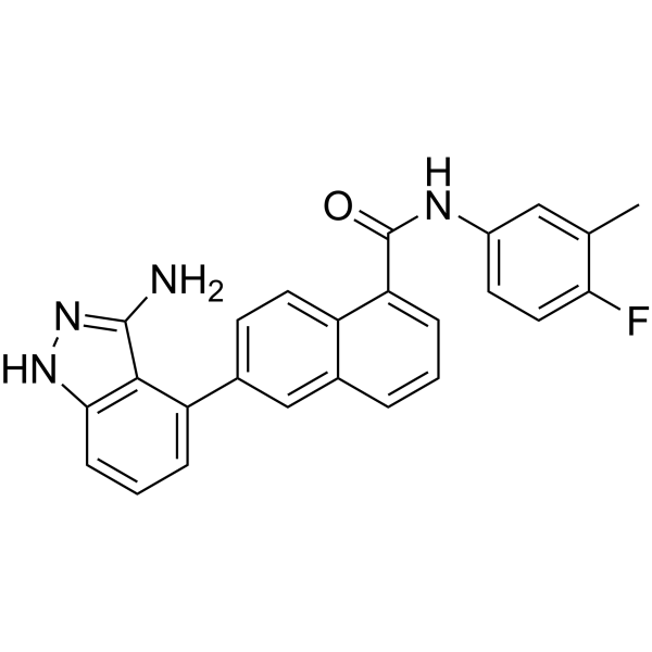
SYHA1813 is a blood-brain barrier-penetrant, orally effective dual VEGFR and CSF1R receptor tyrosine kinase inhibitor. SYHA1813 exhibits IC50 values of 2.8, 0.3, 4.3, and 19.3 nM against VEGFR-1, VEGFR-2, VEGFR-3, and CSF1R, respectively. SYHA1813 blocks VEGFR and CSF1R kinase activity, inhibits pro-tumor macrophage polarization and angiogenesis, and modulates the tumor immune microenvironment. SYHA1813 activates the ATM/CHK/P53 signaling pathway, impairs DNA damage repair, and induces G2/M phase arrest, apoptosis, and senescence. SYHA1813 also inhibits c-Kit and PDGFRα kinases. SYHA1813 can be used for research on glioblastoma, malignant meningioma, melanoma, and diffuse large B-cell lymphoma.
Nur für Forschungszwecke. Wir verkaufen nicht an Patienten.
- Reinheit : 99.46%
- CAS. Nr.: 1807466-30-3
- Formel: C25H19FN4O
- Molecular Weight:410.44
-
Speicherung:
4°C, protect from light
* In solvent : -80°C, 6 months; -20°C, 1 month (protect from light)

To place orders, for customer services and technical support, please contact: MedChemExpress USA
Tel: 609-228-6898 E-mail: [email protected] [email protected]


-
 Biologische Aktivität
-
Chemical Information
-
Lösungsmittel & Löslichkeit
- Protokoll
- Reinheit & Dokumentation
- Verweise
-
 Help & FAQs




Biologische Aktivität
Beschreibung
|
VEGFR-1 2.8 nM (IC50) |
VEGFR-2 0.3 nM (IC50) |
VEGFR-3 4.3 nM (IC50) |
PDGFRα 232.2 nM (IC50) |
PDGFRαD842V 70.3 nM (IC50) |
PDGFRαV561D 2.4 nM (IC50) |
CSF1R 19.3 nM (IC50) |
c-Kit 162.5 nM (IC50) |
c-Kit (D816H) 134.0 nM (IC50) |
Chk1 |
Chk2 |
Bax |
Caspase-3 |
Bcl-2 |
Cellular Effect
|
Cell Line

|
Type | Value | Description | References |
|---|---|---|---|---|
| BMDM | IC50 |
93 nM
|
Inhibition of CSF1-stimulated mouse BMDM viability incubated 72 hrs by CCK-8 assay.
Inhibition of CSF1-stimulated mouse BMDM viability incubated 72 hrs by CCK-8 assay.
|
38045044 |
| HUVEC | IC50 |
13 nM
|
Inhibition of VEGF-stimulated HUVEC viability incubated 72 hrs by CCK-8 assay.
Inhibition of VEGF-stimulated HUVEC viability incubated 72 hrs by CCK-8 assay.
|
38045044 |
| GL261 | IC50 |
> 20 μM
|
Negligible inhibition of GL261 cell viability with IC50 greater than 20 mM incubated 72 hrs by CCK-8 assay.
Negligible inhibition of GL261 cell viability with IC50 greater than 20 mM incubated 72 hrs by CCK-8 assay.
|
38045044 |
| D283 Med | IC50 |
> 20 μM
|
Negligible inhibition of D283 cell viability with IC50 greater than 20 mM incubated 72 hrs by CCK-8 assay.
Negligible inhibition of D283 cell viability with IC50 greater than 20 mM incubated 72 hrs by CCK-8 assay.
|
38045044 |
| U-87MG ATCC | IC50 |
> 20 μM
|
Negligible inhibition of U87MG cell viability with IC50 greater than 20 mM incubated 72 hrs by CCK-8 assay.
Negligible inhibition of U87MG cell viability with IC50 greater than 20 mM incubated 72 hrs by CCK-8 assay.
|
38045044 |
| U-251 | IC50 |
> 20 μM
|
Negligible inhibition of U251 cell viability with IC50 greater than 20 mM incubated 72 hrs by CCK-8 assay.
Negligible inhibition of U251 cell viability with IC50 greater than 20 mM incubated 72 hrs by CCK-8 assay.
|
38045044 |
In Vitro
SYHA1813 (2.5-10 μM; 48 h) increases γ-H2AX and TUNEL-positive cells in IOMM-Lee and CH157 cells, activates ATM/CHK2/CHK1, and upregulates P53 and P21 while downregulating DNA damage response pathway proteins such as CDK2 and PCNA[2].
SYHA1813 (100 μM) inhibits most of the 328 kinases in the panel, with inhibition rates below 80%, and subsequent IC50 determination reveals c-Kit (IC50 = 162.5 nM), c-Kit D816H (IC50 = 134.0 nM), and PDGFRα V561D (IC50 = 2.4 nM) as its additional active targets beyond VEGFR-2 (IC50 = 0.3 nM)[1].
SYHA1813 (up to 10 μM; 72 h) dose-dependently inhibits the viability of CSF1-stimulated BMDMs, with an IC50 of 93 nM[1].
SYHA1813 (up to 10 μM; 72 h) dose-dependently inhibits VEGF-stimulated HUVEC viability with an IC50 of 13 nM[1].
SYHA1813 (1 nM-10 μM; after 2 h pretreatment followed by VEGF stimulation) inhibits VEGF-induced phosphorylation of VEGFR-2 and ERK in HUVECs[1].
SYHA1813 (72 h) does not directly inhibit the viability of GL261, DBT, D283, U87MG, or U251 cells, with IC50 values greater than 20 μM[1].
MedChemExpress (MCE) has not independently confirmed the accuracy of these methods. They are for reference only.
-
Cell Line:HUVECs
-
Concentration:1, 10, 100, 1000, and 10000 nM
-
Incubation Time:2 h
-
Result:Markedly inhibited VEGF-induced VEGFR-2 activation and ERK phosphorylation in HUVECs.
-
Cell Line:IOMM-Lee and CH157 human meningioma cell lines
-
Concentration:0.25, 0.5, 1, 2.5, 5, 10, 25, 50, 100 µM
-
Incubation Time:48 h
-
Result:Inhibited cell viability in a dose-dependent manner in both IOMM-Lee and CH157 cells.
At 2.5 µM, reduced cell viability in both cell lines after 48 h.
-
Cell Line:IOMM-Lee and CH157 human meningioma cell lines
-
Concentration:2.5, 5, 10 µM
-
Incubation Time:48 h
-
Result:Markedly decreased the fraction of EdU-positive IOMM-Lee and CH157 cells compared with the vehicle control at 48 h.
-
Cell Line:IOMM-Lee and CH157 human meningioma cell lines
-
Concentration:2.5, 5, 10 µM
-
Incubation Time:24 h
-
Result:Inhibited migration of IOMM-Lee and CH157 cells in a dose-dependent manner, with reduced area recovery at 24 h compared with the vehicle control.
-
Cell Line:IOMM-Lee and CH157 human meningioma cell lines
-
Concentration:2.5, 5, 10 µM
-
Incubation Time:12 h
-
Result:Suppressed invasion of both IOMM-Lee and CH157 cells, with lower migration rates than the vehicle control.
-
Cell Line:IOMM-Lee and CH157 human meningioma cell lines
-
Concentration:2.5, 5, 10 µM
-
Incubation Time:48 h
-
Result:Increased the percentage of cells in G2/M phase, accompanied by decreased percentages of cells in G0/G1 and S phase in a concentration-dependent manner in both IOMM-Lee and CH157 cells.
-
Cell Line:IOMM-Lee and CH157 human meningioma cell lines
-
Concentration:2.5, 5, 10 µM
-
Incubation Time:48 h
-
Result:Downregulated Cyclin B1 and Cyclin A2 and upregulated Cyclin E1 in IOMM-Lee and CH157 cells.\nIncreased Caspase 3 and Bax levels and decreased Bcl-2 and pro-caspase 3 levels in IOMM-Lee and CH157 cells.\nIncreased γH2AX protein levels in IOMM-Lee and CH157 cells.\nIncreased p-ATM and p-CHK2 without affecting total ATM or CHK2, and also increased p-CHK1.
Upregulated P53 and P21 and reduced CDK2, E2F1, PCNA, and p-H3 protein levels in IOMM-Lee and CH157 cells.
-
Cell Line:IOMM-Lee and CH157 human meningioma cell lines
-
Concentration:2.5, 5, 10 µM
-
Incubation Time:48 h
-
Result:Induced apoptosis in both IOMM-Lee and CH157 cells in a concentration-dependent manner, with a pronounced increase in late apoptotic cells.\nTUNEL staining revealed DNA fragmentation and nuclear condensation in IOMM-Lee and CH157 cells, with an increased rate of TUNEL-positive cells compared with controls.
-
Cell Line:IOMM-Lee human meningioma cells
-
Concentration:5 µM
-
Incubation Time:48 h
-
Result:Decreased mRNA expression of DNA repair-associated genes MCM4, MCM5, MCM6, and MCM7.
Decreased mRNA expression of cell cycle-related genes CDC6, CDC7, CDC20, CDC25a, and CDC25c.
-
Cell Line:IOMM-Lee and CH157 human meningioma cell lines
-
Concentration:2.5, 5, 10 µM
-
Incubation Time:48 h
-
Result:Increased the number of γH2AX-positive IOMM-Lee and CH157 cells compared with the vehicle control.
In Vivo
SYHA1813 (20-40 mg/kg; p.o.; twice daily) enters the brain compartment and prolongs the survival of mice bearing intracranial U87MG-luc tumors[1].
SYHA1813 (10 mg/kg; p.o.; once daily; 21 days) inhibits tumor growth in the malignant meningioma IOMM-Lee subcutaneous xenograft tumor model[2].
SYHA1813 (2.5 mg/kg; p.o.; twice daily; 31 days) reduces systemic melanoma metastasis in the intracardiac injection A375 model, with metastatic burden decreased by 76.6% on day 31[3].
SYHA1813 (2.5-5 mg/kg; p.o.; twice daily; 30 days) inhibits the growth of BRAF V600E-mutant A375 melanoma xenografts, with 5 mg/kg achieving a 79.8% tumor growth inhibition rate and reducing angiogenesis, M2 macrophages, and proliferation markers[3].
SYHA1813 (2.5-5 mg/kg; p.o.; twice daily; 12 days) produces dose-dependent antitumor efficacy in the BTK A428D TMD8-ibruR1 DLBCL xenograft tumor model, with TGI of 38.8% and 54.6%, respectively[4].
SYHA1813 (10 mg/kg; p.o.; twice daily; 17 days) inhibits U87MG xenograft tumor growth and reduces angiogenesis markers and M2 macrophage markers[1].
SYHA1813 (2.5-5 mg/kg; p.o.; twice daily; 24 days) dose-dependently inhibits tumor growth in the BTK wild-type OCI-LY10 DLBCL xenograft tumor model and reduces VEGFR2/CSF1R phosphorylation and vascular/macrophage markers[4].
MedChemExpress (MCE) has not independently confirmed the accuracy of these methods. They are for reference only.
-
Animal Model:Nude mice[1]
-
Dosage:5 mg/kg and 10 mg/kg
-
Administration:p.o.; twice daily; 17 days
-
Result:Tumor volume was reduced by 67.7% at 5 mg/kg and 81.0% at 10 mg/kg.
Phosphorylation of CSF1R and VEGFR-2 in U251 tumor tissue was markedly inhibited.
CD31, CD105, and CD206 expression in U251 tumors was decreased by immunohistochemistry.
-
Animal Model:Nude mice[1]
-
Dosage:10 mg/kg
-
Administration:p.o.; twice daily; 17 days
-
Result:U87MG subcutaneous tumor growth was inhibited.
CD31, CD105, CD206, CD163, and Ki67 expression was decreased in U87MG tumors.
-
Animal Model:NOD-SCID mice[1]
-
Dosage:10 mg/kg
-
Administration:p.o.; twice daily; 14 days
-
Result:Tumor growth in the TMZ-insensitive D283 xenograft was inhibited at 10 mg/kg.
-
Animal Model:BALB/c nude mice (female, 4 weeks old)[2]
-
Dosage:10 mg/kg
-
Administration:p.o.; once daily; 21 days
-
Result:Tumor volume was lower than in the vehicle group over the 21-day administration period, with reduced tumor volume at day 21.
Excised tumor weight was also reduced.
Immunohistochemistry showed fewer Ki67-positive tumor cells and increased γ-H2AX and P53 staining.
-
Animal Model:NOD-SCID mice (male, 4-5 weeks old)[3]
-
Dosage:2.5 mg/kg; 5 mg/kg
-
Administration:p.o.; twice daily; 28 days
-
Result:At 5 mg/kg, 72.5% tumor growth inhibition was achieved.
At 2.5 and 5 mg/kg, MeWo tumor growth was inhibited dose-dependently and tumor weight/volume was reduced.
Immunohistochemistry showed reduced CD105, CD31, F4/80, and Ki67 expression at both doses, with greater reduction at 5 mg/kg.
-
Animal Model:NOD-SCID mice (male, 4-5 weeks old)[3]
-
Dosage:2.5 mg/kg; 5 mg/kg
-
Administration:p.o.; twice daily; 30 days
-
Result:At 5 mg/kg, 79.8% tumor growth inhibition was achieved.
At 2.5 and 5 mg/kg, A375 tumor growth was inhibited dose-dependently.
Immunohistochemistry showed reduced CD105, CD31, F4/80, CD206, and Ki67 expression.
-
Animal Model:NOD-SCID mice (male, 4-5 weeks old)[3]
-
Dosage:2.5 mg/kg (SYHA1813); 20 mg/kg (vemurafenib)
-
Administration:p.o.; twice daily (SYHA1813); once daily (vemurafenib); 32 days
-
Result:The combination of 2.5 mg/kg plus vemurafenib achieved 72.9% tumor growth inhibition by day 32.
CD105, CD31, F4/80, and CD206 expression was reduced; the combination further suppressed these markers and Ki67 expression.
-
Animal Model:BALB/c nude mice[3]
-
Dosage:2.5 mg/kg
-
Administration:p.o.; twice daily; 31 days
-
Result:On day 31, systemic metastasis was reduced by 76.6% at 2.5 mg/kg, with lower bioluminescence than control.
GFP signal was reduced in femur, skull, parietal bone, and brain, indicating curtailed bone and brain metastasis.
-
Animal Model:NOD-SCID mice[4]
-
Dosage:2.5 mg/kg and 5 mg/kg
-
Administration:p.o.; twice daily; 18 days
-
Result:Inhibited LY10-016R xenograft growth with TGI of 54.7% at 2.5 mg/kg and 61.1% at 5 mg/kg.
Tumor weight was reduced at endpoint in both dose groups.
p-VEGFR2 and p-CSF1R were decreased in tumor tissues.
CD31, CD105, and CD206 were reduced in tumor tissues.
-
Animal Model:NOD-SCID mice[4]
-
Dosage:2.5 mg/kg and 5 mg/kg
-
Administration:p.o.; twice daily; 24 days
-
Result:Dose-dependently suppressed tumor growth in OCI-LY10 xenografts.
p-VEGFR2 and p-CSF1R were decreased in tumor tissues.
CD31, CD105, and CD206 were reduced in tumor tissues.
-
Animal Model:NOD-SCID mice[4]
-
Dosage:2.5 mg/kg and 5 mg/kg
-
Administration:p.o.; twice daily; 12 days
-
Result:Inhibited TMD8-ibruR1 xenograft growth with TGI of 38.8% at 2.5 mg/kg and 54.6% at 5 mg/kg.
Tumor weight was reduced at endpoint in both dose groups.
CD31, CD105, and CD206 were reduced in tumor tissues.
- Human pluripotent stem cell endothelial-cell differentiation
- Vascular/Branching Fractal Analysis
- Annexin V plus membrane-impermeant dye apoptosis staining
- Protocol for Cell Cycle
- Senescence-associated β-galactosidase staining
- Kinase activity and phosphorylation assays
- Phagocytosis Functional Assay
- Detection of Apoptosis
- TUNEL staining for apoptotic DNA fragmentation
- Flow cytometric DNA-content cell-cycle staining
- Endothelial Tube Formation Assay
- Primary monocyte-to-macrophage differentiation
- Apoptosis Solutions
- Protocol for Kinase activity and phosphorylation assays
- Endothelial Cell Migration/Angiogenic Sprouting Assay
- Genotoxicity/Mutagenicity Study
Parmacokinetics
| Species | Dose | Route | T1/2 (Plasma) | Tmax (Plasma) | T1/2 (Brain) | Tmax (Brain) | Cmax (Brain) |
|---|---|---|---|---|---|---|---|
| Mice[1] | 10 mg/kg | p.o. | 2.15 h | 1 h | 2.06 h | 2 h | 1214 ng/mL |
Clinical Trial
| NCT Number | Sponsor | Condition | Start Date |
Phase
|
|---|---|---|---|---|
| NCT01329991 | 2011-05 | PHASE1 |


Chemical Information
-
CAS. Nr. 1807466-30-3
-
Appearance Solid
-
Molecular Weight 410.44
-
Formel C25H19FN4O
-
Color White to off-white
-
SMILES
NC1=NNC2=C1C(C3=CC=C4C(C=CC=C4C(NC5=CC=C(F)C(C)=C5)=O)=C3)=CC=C2
-
Versand
Room temperature in continental US; may vary elsewhere.
-
Speicherung
4°C, protect from light
* In solvent : -80°C, 6 months; -20°C, 1 month (protect from light)


Lösungsmittel & Löslichkeit
In Vitro:
DMSO : 100 mg/mL (243.64 mM; Need ultrasonic; Hygroscopic DMSO has a significant impact on the solubility of product, please use newly opened DMSO)
Please refer to the solubility information to select the appropriate solvent. Once prepared, please aliquot and store the solution to prevent product inactivation from repeated freeze-thaw cycles.
Storage method and period of stock solution: -80°C, 6 months; -20°C, 1 month (protect from light). When stored at -80°C, please use it within 6 months. When stored at -20°C, please use it within 1 month.
Please refer to the solubility information to select the appropriate solvent. Once prepared, please aliquot and store the solution to prevent product inactivation from repeated freeze-thaw cycles.
Storage method and period of stock solution: -80°C, 6 months; -20°C, 1 month (protect from light). When stored at -80°C, please use it within 6 months. When stored at -20°C, please use it within 1 month.
Konzentration (Stammlösung) × Volumen (Stammlösung) = Konzentration (Ziellösung) × Volumen (Ziellösung)
In Vivo:
Select the appropriate dissolution method based on your experimental animal and administration route.
- For the following dissolution methods, please ensure to first prepare a clear stock solution using an In Vitro approach and then sequentially add co-solvents:
- To ensure reliable experimental results, the clarified stock solution can be appropriately stored based on storage conditions. As for the working solution for In Vivo experiments, it is recommended to prepare freshly and use it on the same day.
- The percentages shown for the solvents indicate their volumetric ratio in the final prepared solution. If precipitation or phase separation occurs during preparation, heat and/or sonication can be used to aid dissolution.
In Vivo Dissolution Calculator
Please enter the basic information of animal experiments:
-
-
-
-
Recommended: Prepare an additional quantity of animals to account for potential losses during experiments.
Please enter your animal formula composition:
-
%DMSO +
Recommended: Keep the proportion of DMSO in working solution below 2% if your animal is weak.
-
%+
-
+%Tween-80 + +
-
%Saline +
The co-solvents required include: DMSO, . All of co-solvents are available by MedChemExpress (MCE). , Tween 80. All of co-solvents are available by MedChemExpress (MCE).
Working solution concentration: 0.22 mg/mL
Method for preparing stock solution: mg drug dissolved in μL DMSO. Stock solution concentration: mg/mL. * In solvent : -80°C, 6 months; -20°C, 1 month (protect from light)
1. Take μL DMSO stock solution;
2. Add μL .
μL , mix evenly;
3. Then add μL Tween 80, mix evenly;
4. Then add μL
Please ensure that the stock solution in the first step is dissolved to a clear state, and add co-solvents in sequence. You can use ultrasonic heating (ultrasonic cleaner, recommended frequency 20-40 kHz), vortexing, etc. to assist dissolution.


Reinheit & Dokumentation


Verweise
[1]. Liu Y, et al. Preclinical and early clinical studies of a novel compound SYHA1813 that efficiently crosses the blood-brain barrier and exhibits potent activity against glioblastoma. Acta Pharm Sin B. 2023 Dec;13(12):4748-4764. [Content Brief]
[4]. Song PR, et al. Discovery of a novel BTK inhibitor S-016 and identification of a new strategy for the treatment of lymphomas including BTK inhibitor-resistant lymphomas. Acta pharmacologica Sinica. 2024 Oct;45(10):2163-2173. [Content Brief]

Complete Stock Solution Preparation Table
Please refer to the solubility information to select the appropriate solvent. Once prepared, please aliquot and store the solution to prevent product inactivation from repeated freeze-thaw cycles.
Storage method and period of stock solution: -80°C, 6 months; -20°C, 1 month (protect from light). When stored at -80°C, please use it within 6 months. When stored at -20°C, please use it within 1 month.
| Optional Solvent | Concentration Solvent Mass | 1 mg | 5 mg | 10 mg | 25 mg |
|---|---|---|---|---|---|
| DMSO | 1 mM | 2.4364 mL | 12.1820 mL | 24.3641 mL | 60.9102 mL |
| 5 mM | 0.4873 mL | 2.4364 mL | 4.8728 mL | 12.1820 mL | |
| 10 mM | 0.2436 mL | 1.2182 mL | 2.4364 mL | 6.0910 mL | |
| 15 mM | 0.1624 mL | 0.8121 mL | 1.6243 mL | 4.0607 mL | |
| 20 mM | 0.1218 mL | 0.6091 mL | 1.2182 mL | 3.0455 mL | |
| 25 mM | 0.0975 mL | 0.4873 mL | 0.9746 mL | 2.4364 mL | |
| 30 mM | 0.0812 mL | 0.4061 mL | 0.8121 mL | 2.0303 mL | |
| 40 mM | 0.0609 mL | 0.3046 mL | 0.6091 mL | 1.5228 mL | |
| 50 mM | 0.0487 mL | 0.2436 mL | 0.4873 mL | 1.2182 mL | |
| 60 mM | 0.0406 mL | 0.2030 mL | 0.4061 mL | 1.0152 mL | |
| 80 mM | 0.0305 mL | 0.1523 mL | 0.3046 mL | 0.7614 mL | |
| 100 mM | 0.0244 mL | 0.1218 mL | 0.2436 mL | 0.6091 mL |


SYHA1813 Related Classifications
HY-161025 Related Classifications
 Powered by Bioz
Keywords
- SYHA1813
- 1807466-30-3
- SYHA 1813
- SYHA-1813
- c-Fms
- VEGFR
- Apoptosis
- CDK
- Akt
- ERK
- Caspase
- Bcl-2 Family
- Checkpoint Kinase (Chk)
- MDM-2/p53
- ATM/ATR
- c-Kit
- PDGFR
- CSF-1 receptor
- colony stimulating factor 1 receptor
- CSF-1R
- CSF1R
- Vascular endothelial growth factor receptor
- Cyclin dependent kinase
- PKB
- Protein kinase B
- Extracellular signal regulated kinases
- Ataxia telangiectasia mutated
- ATM and RAD3 related
- SCFR
- CD117
- Platelet-derived growth factor receptor
- ATM/CHK/P53 signaling
- DNA damage repair
- G2/M arrest
- PDGFRα
- angiogenesis
- apoptosis
- blood-brain barrier
- Inhibitor
- inhibitor
- inhibit