



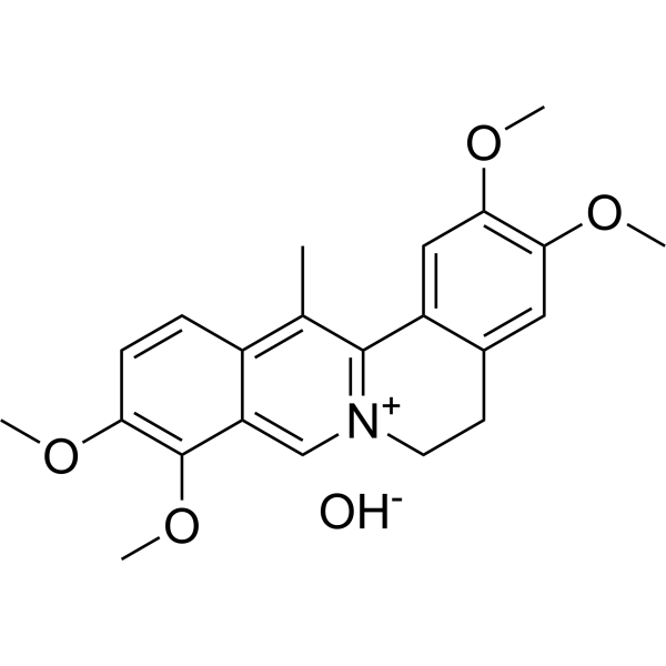
Dehydrocorydaline (hydroxyl)
Based on 46 publication(s) in Google Scholar
Dehydrocorydaline (13-Methylpalmatine) (hydroxyl) is an orally active alkaloid that inhibits acetylcholinesterase (AChE) (IC50 = 0.62 μM). Dehydrocorydaline (hydroxyl) inhibits synaptosomal reuptake of serotonin/dopamine/norepinephrine, the NLRP3 inflammasome, microglial activation, biofilm formation, and Plasmodium/Trypanosoma growth, and has pain-relieving effects. Dehydrocorydaline (hydroxyl) activates p38 MAPK/MyoD-driven myoblast differentiation, binds SPTA1 (Kd = 2.18 µM), inhibits aldose reductase, hOCT2/3, hPMAT, MEK/ERK, NF-κB/TRAF6, IRE1/PERK, SMAD2/3, and NR2B phosphorylation, and modulates Bcl-2/Bax, caspase-8/7, and PARP signaling. Dehydrocorydaline (hydroxyl) can be used in research on pain, breast cancer, melanoma, non-small cell lung cancer, bacterial infections, parasitic infections, pulmonary fibrosis, and atherosclerosis.
For research use only. We do not sell to patients.
- Purity : 99.52%
- Formula: C22H25NO5
- Molecular Weight:383.44
-
Storage:Powder -20°C, 3 years , 4°C, 2 years ; In solvent -80°C, 6 months , -20°C, 1 month

To place orders, for customer services and technical support, please contact: MedChemExpress USA
Tel: 609-228-6898 E-mail: [email protected] [email protected]


-
 Biological Activity
-
Chemical Information
-
Solvent & Solubility
- Protocols
- Purity & Documentation
- References
-
 Help & FAQs


Publications Citing Use of MedChemExpress (MCE) Dehydrocorydaline (hydroxyl)
More- J Nanobiotechnology. 2025 Jul 22;23(1):535. [Abstract]
- J Clin Invest. 2024 Jun 18;134(16):e178303. [Abstract]
- Phytomedicine. 2021 Nov:92:153740. [Abstract]
- Mater Today Bio. 2026 Mar 24:38:103059. [Abstract]
- New Phytol. 2024 Jul;243(1):381-397. [Abstract]
- Int J Biol Macromol. 2024 May;266(Pt 1):130939. [Abstract]
- Phytother Res. 2026 May;40(5):2473-2490. [Abstract]
- J Ethnopharmacol. 2025 Jan 30;337(Pt 1):118738. [Abstract]
- J Agric Food Chem. 2023 Oct 25;71(42):15538-15552. [Abstract]
- Ecotoxicol Environ Saf. 2025 Jul 15:300:118460. [Abstract]
- Biochem Pharmacol. 2025 May:235:116820. [Abstract]
- Biochem Pharmacol. 2024 Dec;230(Pt 1):116574. [Abstract]
- Mol Metab. 2026 Apr:106:102341. [Abstract]
- Eur J Pharmacol. 2026 Jan 20:1012:178500. [Abstract]
- Respir Res. 2025 Apr 12;26(1):136. [Abstract]
- Eur J Pharmacol. 2021 Jun 5:900:174069. [Abstract]
- Int Immunopharmacol. 2026 Jul 15:181:116715. [Abstract]
- Int Immunopharmacol. 2026 Jun 1:178:116594. [Abstract]
- Int Immunopharmacol. 2024 Sep 30:139:112720. [Abstract]
- Bone Joint Res. 2023 Nov 1;12(11):677-690. [Abstract]
- Arthritis Res Ther. 2025 May 31;27(1):118. [Abstract]
- Front Pharmacol. 2021 May 31:12:653272. [Abstract]
- J Inflamm Res. 2021 Sep 16;14:4669-4686. [Abstract]
- FASEB J. 2025 Aug 31;39(16):e70945. [Abstract]
- J Nutr. 2020 Jul 1;150(7):1731-1737. [Abstract]
- Front Biosci (Landmark Ed). 2025 Apr 25;30(4):25221. [Abstract]
- J Food Sci. 2025 Apr;90(4):e70217. [Abstract]
- J Cell Physiol. 2019 Dec;234(12):22463-22476. [Abstract]
- Genomics. 2025 Jul;117(4):111067. [Abstract]
- Mol Carcinog. 2024 Nov;63(11):2190-2204. [Abstract]
- Andrology. 2025 Jun 16. [Abstract]
- Tissue Cell. 2023 Dec:85:102218. [Abstract]
- Exp Eye Res. 2023 Feb:227:109365. [Abstract]
- BMC Musculoskelet Disord. 2022 May 30;23(1):514. [Abstract]
- Infect Genet Evol. 2022 Jan:97:105158. [Abstract]
- J Mol Histol. 2021 Jun;52(3):521-537. [Abstract]
- Biochem Biophys Res Commun. 2018 Sep 5;503(2):467-473. [Abstract]
- Biochem Biophys Res Commun. 2018 May 23;499(4):743-750. [Abstract]
- Eur J Histochem. 2025 Jan 21;69(1). [Abstract]
- J Clin Neurosci. 2020 Aug:78:365-370. [Abstract]
- J Cardiothorac Surg. 2024 Dec 20;19(1):665. [Abstract]
- bioRxiv. 2026 May 7.
- Bioengineered. 2022 May;13(5):12847-12862. [Abstract]
- Aging (Albany NY). 2021 Oct 7;13(19):23133-23148. [Abstract]
- Comput Math Methods Med. 2021 Aug 2:2021:3337514. [Abstract]
- Aging (Albany NY). 2021 May 11;13(9):12780-12799. [Abstract]
 Customer Validation & Images

-
Histological Imaging/Staining
-
RT-PCR
-
WB
-
IF
-
Cell Proliferation/Viability Assay
All Parasite Isoforms
MoreAll Caspase Isoforms
MoreAll 5-HT Receptor Isoforms
MoreAll Dopamine Receptor Isoforms
More

Biological Activity
Description
IC50 & Target
IC50: 38 nM (P. falciparum 3D7 strain)[3]
In Vitro
Dehydrocorydaline (40 μM; 48 h) (hydroxyl) reduces BrdU-positive A375 and MV3 melanoma cells[14].
Dehydrocorydaline (40 μM; 0-72 h) (hydroxyl) inhibits wound closure in A375 and MV3 melanoma cells[14].
Dehydrocorydaline (40 μM) (hydroxyl) reduces A375 and MV3 migration and Matrigel invasion[14].
Dehydrocorydaline (40 μM; 2-3 weeks) (hydroxyl) reduces colony formation in A375 and MV3 soft agar cultures[14].
Dehydrocorydaline (40-80 µM; 24 h) (hydroxyl) reduces H1299 motility in the wound healing assay[15].
Dehydrocorydaline (40-80 µM; 16 h) (hydroxyl) inhibits H1299 transwell migration[15].
Dehydrocorydaline (40-80 µM) (hydroxyl) suppresses MMP7 and MMP9 mRNA in H1299 cells[15].
Dehydrocorydaline (40-120 µM) (hydroxyl) reduces MMP9 and MMP7 protein expression in H1299 cells[15].
Dehydrocorydaline ((40-80 µM; 24 h) (hydroxyl) shows an anti-migratory effect that is blocked by Bcl-2 overexpression in H1299 cells[15].
Dehydrocorydaline (200 μM; 24-72 h) (hydroxyl) induces time-dependent apoptotic nuclear morphology in MCF-7 cells[4].
Dehydrocorydaline (0-200 μM; 72 h) (hydroxyl) increases DNA fragmentation in MCF-7 cells in a time-dependent manner, especially at 72 h[4].
Dehydrocorydaline (0-200 μM) (hydroxyl) regulates Bax/Bcl-2 and activates caspase-7, caspase-8, and PARP without activating caspase-9 in MCF-7 cells[4].
Dehydrocorydaline (40-120 µM) (hydroxyl) decreases Bcl-2 and increases Bax protein in H1299, H596, H460, and A549 NSCLC cells[15].
Dehydrocorydaline (20-80 μM; 48 h; 24-72 h) (hydroxyl) decreases CDK6 and cyclin D1 protein levels in A375 and MV3 melanoma cells in dose- and time-dependent manners[14].
Dehydrocorydaline (20-80 μM; 48 h; 24-72 h) (hydroxyl) increases E-cadherin and decreases vimentin and β-catenin in A375 and MV3 melanoma cells[14].
Dehydrocorydaline (20-80 μM; 48 h; 24-72 h) (hydroxyl) decreases phospho-MEK1/2 and phospho-ERK1/2 levels in A375 and MV3 melanoma cells[14].
Dehydrocorydaline (5-20 μg/mL) (hydroxyl) reverses LPS-induced iNOS upregulation and Nrf2/HO-1 suppression in H9C2 cardiomyocytes[10].
Dehydrocorydaline (5-20 μg/mL) (hydroxyl) dose-dependently attenuates LPS-induced TRAF6/NF-κB activation in H9C2 cardiomyocytes[10].
Dehydrocorydaline ((hydroxyl)) (0-4 mg/mL; 24 h) (hydroxyl) inhibits L. monocytogenes ATCC 7644 planktonic growth in a concentration-dependent manner, with MIC 1 mg/mL and MBC 2 mg/mL suppressing growth over 24 h[8].
Dehydrocorydaline (0-2 mg/mL; 0-4 h) (hydroxyl) disrupts L. monocytogenes ATCC 7644 cytoplasmic membrane integrity, causing nucleic acid and protein leakage at MIC 1 mg/mL and MBC 2 mg/mL[8].
Dehydrocorydaline (0-2 mg/mL; 4 h) (hydroxyl) at MIC 1 mg/mL and MBC 2 mg/mL causes membrane shrinkage and cell-wall/membrane destruction in L. monocytogenes ATCC 7644[8].
Dehydrocorydaline (0-1 mg/mL; 24 h) (hydroxyl) reduces L. monocytogenes ATCC 7644 biofilm formation at 0.25, 0.5, and 1 mg/mL over 24 h[8].
Dehydrocorydaline (0-1 mg/mL) (hydroxyl) reduces metabolic activity in L. monocytogenes ATCC 7644 biofilms at 0.25, 0.5, and 1 mg/mL[8].
Dehydrocorydaline (0-1 mg/mL; 24 h) (hydroxyl) inhibits L. monocytogenes ATCC 7644 swimming motility at 0.25, 0.5, and 1 mg/mL and swarming motility at 0.5 and 1 mg/mL[8].
Dehydrocorydaline (10-30 μM) (hydroxyl) reduces TGF-β1-induced Fibronectin, Collagen 1, and α-SMA protein in HPFs[12].
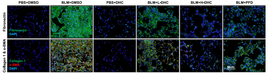
Dehydrocorydaline (10-30 μM) (hydroxyl) attenuates TGF-β1-induced fibrotic marker immunofluorescence in HPFs[12].
Dehydrocorydaline (2 h) (hydroxyl) suppresses TGF-β1-induced SMAD2/3 phosphorylation in HPFs without affecting PI3K-AKT-mTOR or MAPK/ERK[12].
Dehydrocorydaline (100 μM) (hydroxyl) inhibits LPS-induced p65 and ERK1/2 phosphorylation without affecting p38 or JNK phosphorylation in BMDMs[9].
Dehydrocorydaline (hydroxyl) inhibits LPS-induced p65 nuclear translocation in BMDMs[9].
Dehydrocorydaline (5-20 μg/mL) (hydroxyl) selectively reduces TRAF6 expression in H9C2 cardiomyocytes[10].
Dehydrocorydaline (50-100 μM; 6-12 h) (hydroxyl) decreases IL-1β and IL-18 mRNA in resting BMDMs in a time- and concentration-dependent manner[9].
Dehydrocorydaline (50-100 μM; 12-24 h) (hydroxyl) decreases CD80 and IL-18 protein levels in unstimulated BMDMs[9].
Dehydrocorydaline (hydroxyl) reduces inflammatory factor mRNA and VCAM1 protein in rat VSMCs[11].
Dehydrocorydaline (50 μM; 48 h) (hydroxyl) attenuates LPS-induced p65 and TNF-α protein expression in SV40-transfected human chondrocytes, consistent with inhibition of NF-κB pro-inflammatory activation, while IL-6 could not be reliably detected[13].
Dehydrocorydaline (50-100 μM; 12-24 h) (hydroxyl) increases Cnn1, Myh11, Sm22α, and Acta2 mRNA in a time- and concentration-dependent manner in rat VSMCs[11].
Dehydrocorydaline (50-100 μM; 12-24 h) (hydroxyl) upregulates Cnn1 and SM-MHC protein in rat VSMCs[11].
Dehydrocorydaline (100 μM; 24 h) (hydroxyl) increases α-SMA immunofluorescence in rat VSMCs[11].
Dehydrocorydaline (100 μM) (hydroxyl) upregulates contractile markers in rat VSMCs; Spta1 knockdown prevents this upregulation, indicating that Spta1 mediates the effect[11].
Dehydrocorydaline (96 h) (hydroxyl) inhibits the multidrug-resistant P. falciparum Dd2 strain with an IC50 of 161 nM and 94.5% growth inhibition[1].
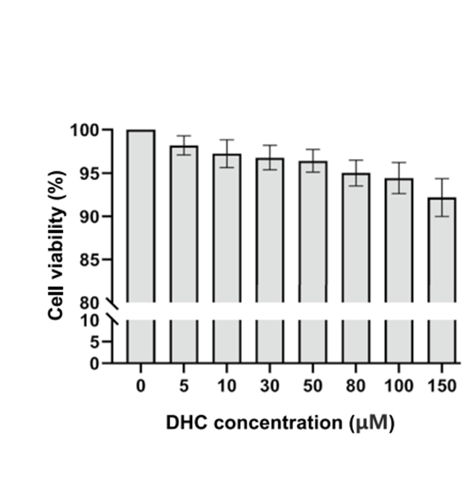
Dehydrocorydaline (0.001-10 µM; 96 h) (hydroxyl) shows low cytotoxicity to HEK 293T cells (>90% viability up to 10 µM) with a parasite selectivity of >14,900[1].
Dehydrocorydaline (0-48 h) (hydroxyl) inhibits P. falciparum 3D7 development at the trophozoite and schizont stages[1].
Dehydrocorydaline (72 h) (hydroxyl) inhibits luciferase-expressing T. cruzi trypomastigotes/amastigotes in NMH coculture with an IC50 of 3.47 μM[2].
Dehydrocorydaline (72 h) (hydroxyl) inhibits intracellular T. cruzi amastigotes in NMH cells with an IC50 of 7.89 μM[2].
Dehydrocorydaline (72 h) (hydroxyl) inhibits T. cruzi epimastigotes with an IC50 of 17.3 μM[2].
Dehydrocorydaline (10 nM; 24-48 h) (hydroxyl) is the active phytochemical from Corydalis tuber in the C2C12 MyoD reporter and MHC induction screen[3].
Dehydrocorydaline (125-500 nM; 2-3 days) (hydroxyl) increases MHC, MyoD, and myogenin expression in C2C12 myoblasts without markedly changing Cdo[3].
Dehydrocorydaline (125-500 nM; 3 days) (hydroxyl) promotes multinucleated myotube formation in C2C12 myoblasts[3].
Dehydrocorydaline (125-500 nM; 2-3 days) (hydroxyl) activates p38 MAPK in differentiating C2C12 myoblasts, with elevated p-p38 at day 2 and day 3[3].
Dehydrocorydaline (500 nM; 1-3 days) (hydroxyl) increases p-p38 immunofluorescence 2.34-fold in differentiating C2C12 myoblasts[3].
Dehydrocorydaline (500 nM; differentiation day 2) (hydroxyl) enhances MyoD-E2A interaction in differentiating C2C12 myoblasts[3].
Dehydrocorydaline (500 nM; 24 h) (hydroxyl) increases MyoD-E2A binding in MyoD-transfected 10T1/2 fibroblasts[3].
Dehydrocorydaline (500 nM; 24 h) (hydroxyl) increases MyoD-E2A binding in MyoD-transfected 293T cells[3].
Dehydrocorydaline (500 nM; 2 days) (hydroxyl) rescues Cdo depletion-induced reductions in muscle-specific protein expression and p38 MAPK activation in C2C12 myoblasts[3].
Dehydrocorydaline (500 nM; 3 days) (hydroxyl) improves defective myotube formation in Cdo-depleted C2C12 myoblasts[3].
Dehydrocorydaline (0.0001-100 μM; 3-30 min) (hydroxyl) selectively inhibits uptake-2 transporters hOCT2, hOCT3, and hPMAT in transfected MDCK cells with IC50 values of 0.137-3.70 μM, without inhibiting hSERT or hDAT at 100 μM[5].
Dehydrocorydaline (12.5-50 μM; 10 min) (hydroxyl) inhibits synaptosomal 5-HT, DA, and NE reuptake in mouse brain synaptosomes, producing approximately 52.2%, 39.9%, and 38.3% inhibition at 50 μM[5].
Dehydrocorydaline (5-20 μg/mL) (hydroxyl) dose-dependently restores LPS-reduced viability and proliferation in H9C2 cardiomyocytes[10].
Dehydrocorydaline (hydroxyl) binds SPTA1 with a Kd of 2.18 μM in an SPR assay[11].
Dehydrocorydaline (100 μM; 24 h) (hydroxyl) upregulates Spta1/SPTA1 expression, with Spta1 identified among the top induced differentially expressed genes by RNA-seq in rat VSMCs[11].
MedChemExpress (MCE) has not independently confirmed the accuracy of these methods. They are for reference only.
-
Cell Line:C2C12 mouse myoblasts
-
Concentration:125, 250, and 500 nM (2-day differentiation); 500 nM (differentiation day 2 and day 3)
-
Incubation Time:2 days (differentiation); day 2 and day 3 (500 nM)
-
Result:Increased MHC, MyoD, and myogenin expression in a dose-dependent manner compared with DMSO control cells; Cdo levels were not markedly altered.
At 500 nM, increased MHC, MyoD, and myogenin at day 2 and day 3.
-
Cell Line:C2C12 mouse myoblasts
-
Concentration:125, 250, and 500 nM
-
Incubation Time:3 days (differentiation)
-
Result:Formed larger myotubes with more nuclei per myotube than control cells, with an increase in myotubes containing ≥10 nuclei relative to vehicle control cells.
-
Cell Line:C2C12 mouse myoblasts
-
Concentration:125, 250, and 500 nM (differentiation); 500 nM (differentiation day 2 and day 3)
-
Incubation Time:during differentiation; day 2 and day 3 (500 nM)
-
Result:Upregulated p-p38 in a dose-dependent manner.
At 500 nM, elevated p-p38 was observed at differentiation day 2 and day 3 compared with vehicle control cells.
-
Cell Line:C2C12 mouse myoblasts
-
Concentration:500 nM
-
Incubation Time:1 day (Results); 2 or 3 days (Methods)
-
Result:Increased p-p38-positive cells compared with control cells.
p-p38 signal intensity was 2.34-fold higher relative to DMSO control cells.
-
Cell Line:C2C12 mouse myoblasts (Cdo-depleted and control)
-
Concentration:500 nM
-
Incubation Time:2 days (differentiation)
-
Result:Cdo-depleted cells had decreased Cdo, MHC, MyoD, and p-p38 compared with C2C12/pSuper control cells.
Restored MHC, MyoD, and myogenin expression in C2C12/Cdo shRNA cells to levels similar to those in C2C12/pSuper cells.
C2C12/Cdo shRNA cells exhibited slightly higher p-p38 levels than C2C12/pSuper cells, whereas total p38 remained constant.
-
Cell Line:C2C12 mouse myoblasts (Cdo-depleted and control)
-
Concentration:500 nM
-
Incubation Time:3 days (differentiation)
-
Result:C2C12/pSuper cells exhibited enhanced myotube formation, including myotubes containing ≥10 nuclei, compared with control C2C12/pSuper cells.
Vehicle control C2C12/Cdo shRNA cells exhibited reduced myotube formation, which was markedly improved almost to control cell levels.
-
Cell Line:H9C2 rat embryonic ventricular cardiomyocytes
-
Concentration:20 μg/mL (alone); 5, 10, 20 μg/mL (with LPS 10 μg/mL); 20 µM (DCFDA)
-
Incubation Time:45 min (DCFDA)
-
Result:At 5, 10, and 20 μg/mL with LPS, reduced IL-1β and TNFα and increased SOD and GSH-PX dose-dependently compared with LPS.
At 5, 10, and 20 μg/mL with LPS, reduced ROS dose-dependently compared with LPS.
-
Cell Line:SV40-transfected human immortalized chondrocytes (CTCC-001-0662)
-
Concentration:50 μM
-
Incubation Time:48 h (after 24 h LPS)
-
Result:Relative p65 protein expression was increased in the LPS group versus Control and significantly reduced relative to LPS at 50 μM.
Relative TNF-α protein expression was increased in the LPS group versus Control and significantly reduced relative to LPS at 50 μM.
IL-6 data required further validation because no clear detectable band was observed.
-
Cell Line:human melanoma A375 and MV3 cells; normal melanocyte PIG1 cells
-
Concentration:20, 40, 80 μM (A375/MV3); 20, 40, 80, 160 μM (PIG1); six doses (IC50); 40 and 80 μM (growth curves)
-
Incubation Time:48 h (A375/MV3, PIG1, IC50); 7 days (growth curves)
-
Result:IC50 values were 39.73 μM in A375, 42.34 μM in MV3, and 262.6 μM in PIG1.
Proliferation was inhibited in A375 and MV3 in a dose-dependent manner at 20, 40, and 80 μM after 48 h.
Growth was markedly suppressed in A375 and MV3 at 40 and 80 μM over 7 days.
No significant proliferation inhibition was observed in PIG1 after 20 or 40 μM for 48 h.
-
Cell Line:human melanoma A375 and MV3 cells
-
Concentration:40 μM; BrdU 10 μg/mL
-
Incubation Time:48 h; 0.5 h (BrdU)
-
Result:The ratio of BrdU-positive A375 and MV3 cells was significantly decreased compared with control groups at 40 μM after 48 h.
-
Cell Line:human melanoma A375 and MV3 cells; normal melanocyte PIG1 cells
-
Concentration:20, 40, 80 μM (A375/MV3); 40 μM (PIG1)
-
Incubation Time:48 h
-
Result:Significant G0/G1 phase arrest was induced in A375 and MV3 cells in a dose-dependent manner at 20, 40, and 80 μM after 48 h.
No significant cell cycle arrest was induced in PIG1 cells at 40 μM after 48 h.
-
Cell Line:human melanoma A375 and MV3 cells
-
Concentration:20, 40, 80 μM (dose response)
-
Incubation Time:48 h (dose response); 24, 48, 72 h (time course)
-
Result:CDK6 and cyclin D1 protein levels were decreased in both dose- and time-dependent manners in A375 and MV3 cells.\nE-cadherin expression was upregulated and vimentin and β-catenin expression was downregulated in A375 and MV3 cells in both dose- and time-dependent manners.\nPhosphorylation of MEK1/2 at Ser221 and ERK1/2 at Thr202/Tyr204 was downregulated in A375 and MV3 cells in both dose- and time-dependent manners.
-
Cell Line:human melanoma A375 and MV3 cells
-
Concentration:40 μM
-
Incubation Time:0, 24, 48, 72 h
-
Result:The rate of wound closure was significantly blocked in A375 and MV3 cells compared with control groups at 40 μM.
-
Cell Line:human melanoma A375 and MV3 cells
-
Concentration:40 μM
-
Incubation Time:2 weeks (MV3); 3 weeks (A375)
-
Result:A375 and MV3 cells formed smaller and less numerous colonies than control groups at 40 μM.
-
Cell Line:human melanoma A375 and MV3 cells
-
Concentration:50 μM (tBHQ)
-
Incubation Time:0, 2, 4, 6 days
-
Result:tBHQ at 50 μM recovered inhibition of A375 and MV3 cell proliferation.
-
Cell Line:H1299, H596, A549, and H460 NSCLC cells
-
Concentration:0, 10, 20, 40, 80, 120, 160 µM
-
Incubation Time:24 h
-
Result:Weak cytotoxicity occurred in H1299, H596, A549, and H460 NSCLC cells at 0, 10, 20, 40, 80, 120, and 160 µM after 24 h.
-
Cell Line:H1299 NSCLC cells
-
Concentration:40, 80 µM
-
Incubation Time:24 h
-
Result:At 80 µM, H1299 cell motility was significantly reduced compared with untreated control.
-
Cell Line:H1299 NSCLC cells
-
Concentration:40, 80 µM
-
Incubation Time:16 h
-
Result:At 40 and 80 µM, an anti-migratory effect reduced migrated cell numbers in H1299 cells compared with untreated control.
-
Cell Line:pcDNA- and pcDNA-Bcl-2-transfected H1299 NSCLC cells
-
Concentration:40, 80 µM
-
Incubation Time:24 h
-
Result:At 40 and 80 µM, Bcl-2 overexpression blocked the anti-migratory effect in pcDNA-Bcl-2-transfected H1299 cells compared with pcDNA control.
In Vivo
Dehydrocorydaline (3.6-10 mg/kg; i.p.; single dose; 15 min before acetic acid) (hydroxyl) dose-dependently reduces acetic acid-induced writhing in mice[6].
Dehydrocorydaline (10 mg/kg; i.p.; single dose; 15 min before formalin) (hydroxyl) antinociception in the formalin test is opioid receptor-dependent, being completely blocked by naloxone in Phase 1 and partially blocked in Phase 2 in mice[6].
Dehydrocorydaline (5-10 mg/kg; i.p.; every other day; days 12, 14, 16, 18, and 20) (hydroxyl) attenuates bleomycin-induced pulmonary fibrosis in C57BL/6 mice in a dose-dependent manner, reducing fibrosis scores, hydroxyproline content, and fibrotic marker expression[12].
MedChemExpress (MCE) has not independently confirmed the accuracy of these methods. They are for reference only.
-
Animal Model:ICR (male, 6 weeks old, 18-20 g)[5]
-
Dosage:1.5 mg/kg; 3 mg/kg
-
Administration:intraperitoneal injection; once daily; 21 days
-
Result:In the sucrose preference test, 3 mg/kg increased sucrose preference relative to the CUMS control group.
In the open-field test, 1.5 and 3 mg/kg increased time in the central region, frequency of passing across the center, and frequency of upright behavior relative to the CUMS control group to different extents.
In the forced swimming test, 1.5 and 3 mg/kg decreased immobility duration relative to the CUMS control group.
In prefrontal cortex biochemistry, 3 mg/kg increased 5-HT levels and decreased the 5-HIAA/5-HT ratio relative to the CUMS control group.
1.5 and 3 mg/kg increased NE levels and decreased the MHPG/NE ratio.
3 mg/kg decreased DA levels and increased the DOPAC/DA ratio relative to the CUMS control group.
-
Animal Model:ICR (adult male, 25-35 g)[6]
-
Dosage:3.6 mg/kg; 6 mg/kg; 10 mg/kg
-
Administration:i.p.; single dose; 15 min before acetic acid
-
Result:Delayed the onset time of writhing dose-dependently.
Reduced acetic acid-induced writhing episodes by 16.5%, 34.0%, and 52.6% at 3.6, 6, and 10 mg/kg, respectively, compared with control.
-
Animal Model:ICR (adult male, 25-35 g)[6]
-
Dosage:3.6 mg/kg; 6 mg/kg; 10 mg/kg
-
Administration:i.p.; single dose; 15 min before formalin
-
Result:Produced a dose-dependent reduction in Phase 2 licking/biting time; only the 10 mg/kg dose showed an antinociceptive effect in Phase 1.
At 10 mg/kg, reduced formalin-induced paw edema by 66.8%, with paw thickness of 0.67 mm.
Dose-dependently down-regulated spinal p-CASP6, a-CASP6, TNF-α, IL-1β, and IL-6; at 10 mg/kg, p-CASP6 was decreased by 59%, a-CASP6 by 67%, TNF-α by 77%, IL-1β by 65%, and IL-6 by 81%.
-
Animal Model:ICR (adult male, 25-35 g)[6]
-
Dosage:10 mg/kg; 2 mg/kg (naloxone)
-
Administration:i.p.; single dose; 15 min before formalin; naloxone i.p. before Dehydrocorydaline (nitrate)
-
Result:10 mg/kg alone produced antinociceptive activity in both Phase 1 and Phase 2.
Naloxone completely prevented the Phase 1 antinociceptive effect and partially prevented the Phase 2 effect.
-
Animal Model:C3H/HeN (male, 4-6 weeks old, 20-25 g)[7]
-
Dosage:10 mg/kg
-
Administration:i.p.; single administration; day 14 after osteosarcoma cell implantation
-
Result:At 1 h after administration, PWMT was higher and NSF was lower than in vehicle-treated mice.
At 4 h, PWMT remained higher and NSF remained lower than in vehicle-treated mice.
By 8 h both returned to preinjection levels.
Spinal iNOS was reduced at 1 h and 4 h.
Spinal Arg-1 was increased at 1 h.
CD16/32 was reduced at 1 h and 4 h with recovery by 8 h.
CD206 was increased at 1 h with return to preinjection levels by 4 h.
Iba-1 did not substantially change.
Arg-1 immunofluorescence changed similarly to CD206.
Spinal IL-1β was reduced at 1 h and 4 h.
Spinal IL-10 was increased at 1 h and 4 h, with recovery by 8 h.
ELISA showed corresponding IL-1β and IL-10 changes in spinal cord and blood.
-
Animal Model:ApoE−/− (male, 8 weeks old)[9]
-
Dosage:5 mg/kg
-
Administration:i.p.; daily; 12 weeks; i.p.; daily; 1 week (short-term aortic inflammation assessment)
-
Result:Peak diastolic velocity was decreased in the ascending aorta, aortic arch, and brachiocephalic artery.
B-mode echocardiography showed reduced atherosclerotic plaque formation in the aorta.
Aortic root lesion area, oil red O+ area, and F4/80+ macrophage area were reduced.
Collagen content was increased.
α-SMA+ area was not altered.
The plaque vulnerability index was decreased.
In the 5 mg/kg group, plasma TG was 1.3 mmol/L, TC was 28.4 mmol/L, LDL-C was 11.3 mmol/L, HDL-C was 1.5 mmol/L, and body weight was 29.7 g (n = 7 mice per group).
TG decreased and LDL-C increased, while HDL-C, TC, and body weight were comparable.
Plasma TNFα and IL-1β levels were reduced.
Short-term i.p. administration for 1 week reduced aortic TNFα, NLRP3, and IL-18 mRNA levels.
-
Animal Model:C57B6/L (male, 8-10 weeks old, 20-22 g)[10]
-
Dosage:5 mg/kg, 10 mg/kg, 20 mg/kg
-
Administration:intraperitoneal; once every 24 h; for three consecutive days; starting 30 min after E. coli injection
-
Result:Survival rate increased, with significance when the dose was over 10 mg/kg.
Reduced bacterial counts in peritoneal lavage fluid and serum.
Reduced serum creatinine, urea nitrogen, AST, and ALT.
H&E and Masson staining showed reduced disordered myocardial structure, inflammatory cell infiltration, and collagen fibers.
TUNEL staining showed fewer TUNEL-positive cardiomyocytes.
In heart tissue, Bax and Caspase3 decreased and Bcl2 increased in a dose-dependent manner.
Reduced plasma IL-1β, TNFα, IL-6, and IFNγ and increased SOD and GSH-PX in a dose-dependent manner.
In heart tissue, iNOS decreased and cytoplasmic Nrf2, HO-1, and nuclear Nrf2 were promoted.
Reduced TRAF6 expression in heart tissue and decreased cytoplasmic and nuclear p-NF-κB.
-
Animal Model:Apoe−/− (male)[11]
-
Dosage:5 mg/kg
-
Administration:i.p.; daily
-
Result:In Apoe−/− mice with carotid injury, carotid plaque area and F4/80 macrophage infiltration in plaques were reduced, and plaque α-SMA and SM-MHC mean fluorescence intensity was increased compared with vehicle.
-
Animal Model:C57BL/6 (male; 6-8 weeks old; average weight 25 g)[12]
-
Dosage:5 mg/kg; 10 mg/kg
-
Administration:i.p.; every other day on days 12, 14, 16, 18, and 20
-
Result:In bleomycin-induced pulmonary fibrosis mice, lung damage and collagen deposition on H&E, Masson’s trichrome, and Sirius Red staining were reduced in a dose-dependent manner at 5 mg/kg and 10 mg/kg.
Reduced Ashcroft scores relative to BLM+DMSO.
Reduced lung hydroxyproline content relative to BLM+DMSO.
Reduced Collagen 1, Collagen 3, α-SMA, and Fibronectin expression by Western blot and RT-qPCR relative to BLM+DMSO.
Immunofluorescence showed reduced Collagen 1, Fibronectin, and α-SMA relative to BLM+DMSO.
-
Animal Model:BALB/c-nu (nude) (female, 4 weeks old; n=6)[14]
-
Dosage:100 mg/kg
-
Administration:p.o.; every 24 h; 12 days
-
Result:Tumor volume was suppressed in A375 and MV3 melanoma xenografts compared with DMSO controls.
Excised tumor weights were lower than in DMSO control mice.
Immunohistochemistry showed reduced Ki-67, Cyclin D1, and vimentin expression in A375 and MV3 xenograft tumors.
H&E staining showed inhibited cell proliferation in xenografts.
-
Animal Model:C57BL/6J (adult male, specific-pathogen-free, 20-25 g)[16]
-
Dosage:10 mg/kg
-
Administration:intrathecal injection into the L5-L6 intervertebral space; single dose on day 14 after CCI
-
Result:A single intrathecal injection increased PWMT in CCI mice at 3-5 h after injection compared with CCI controls; PWMT decreased by 6 h after injection.
Downregulated spinal protein expression of iNOS, TNF-α, and IL-6 compared with CCI.
Reduced Iba1-positive microglial and GFAP-positive astrocyte activation in the spinal cord compared with CCI.
Reduced the spinal p-NR2B/NR2B ratio compared with CCI.
- Western Blot
- Collagen: Sirius Red Staining
- Connective Tissue: Masson's Trichrome/Collagen Trichrome Staining
- Research Protocol for Cardiovascular Diseases
- Research Protocol for Inflammation-related Diseases
- Breast Cancer Modeling
- Kinase activity and phosphorylation assays
- Fibrosis/Collagen Morphometry
- C2C12 myoblast-to-myotube differentiation
- Apoptosis Solutions
- Protocol for Kinase activity and phosphorylation assays
- Fungal Biofilm Culture
- Pyroptosis Solutions


Chemical Information
-
Appearance Solid
-
Molecular Weight 383.44
-
Formula C22H25NO5
-
Color Light yellow to yellow
-
SMILES
CC1=C(C=CC(OC)=C2OC)C2=C[N+]3=C1C4=CC(OC)=C(OC)C=C4CC3.[OH-]
-
Synonyms
13-Methylpalmatine (hydroxyl)
-
Structure Classification
-
Initial Source
-
Shipping
Room temperature in continental US; may vary elsewhere.
-
Storage
Powder -20°C 3 years 4°C 2 years In solvent -80°C 6 months -20°C 1 month


Publications (46)
-
Journal Impact Factor
-
Most Recent
-
J Nanobiotechnology
A fibrin gel-loaded Gouqi-derived nanovesicle (GqDNV) repairs the heart after myocardial infarction by inhibiting p38 MAPK/NF-κB p65 pathway. [Abstract]2025 Jul 22;23(1):535. PMID: 40696398 -
J Clin Invest
Myostatin regulates energy homeostasis through autocrine- and paracrine-mediated microenvironment communications. [Abstract]2024 Jun 18;134(16):e178303. PMID: 38889010 -
Phytomedicine
The role of daurisoline treatment in hepatocellular carcinoma: Inhibiting vasculogenic mimicry formation and enhancing sensitivity to sorafenib. [Abstract]2021 Nov:92:153740. PMID: 34600176 -
Mater Today Bio
Dual targeted gene delivery strategy mediated by GalNAc-modified lipid nanoparticles enhances liver regeneration through specific knockdown of MKK4. [Abstract]2026 Mar 24:38:103059. PMID: 41970250 -
New Phytol
LbSakA-mediated phosphorylation of the scaffolding protein LbNoxR in the ectomycorrhizal basidiomycete Laccaria bicolor regulates NADPH oxidase activity, ROS accumulation and symbiosis development. [Abstract]2024 Jul;243(1):381-397. PMID: 38741469 -
Int J Biol Macromol
2024 May;266(Pt 1):130939. PMID: 38493816 -
Phytother Res
Dehydrocorydaline Promotes STAT3/Bcl-2 Axis-Mediated Macrophage Efferocytosis to Attenuate Apoptosis in Atherosclerotic Plaques. [Abstract]2026 May;40(5):2473-2490. PMID: 41699991 -
J Ethnopharmacol
Dehydrocorydaline attenuates myocardial ischemia-reperfusion injury via the FoXO signalling pathway: A multimodal study based on network pharmacology, molecular docking, and experimental study. [Abstract]2025 Jan 30;337(Pt 1):118738. PMID: 39222757 -
J Agric Food Chem
Sulforaphene Inhibits Periodontitis through Regulating Macrophage Polarization via Upregulating Dendritic Cell Immunoreceptor. [Abstract]2023 Oct 25;71(42):15538-15552. PMID: 37823224 -
Ecotoxicol Environ Saf
Dibutyl phthalate promotes central precocious puberty through primordial follicle activation by downregulating BMP15. [Abstract]2025 Jul 15:300:118460. PMID: 40472697 -
Biochem Pharmacol
STM2457 decreases m6A methylation to reduce cisplatin-induced ototoxicity via MAPK signaling. [Abstract]2025 May:235:116820. PMID: 39983847 -
Biochem Pharmacol
Ursodeoxycholic acid alleviates fat embolism syndrome-induced acute lung injury by inhibiting the p38 MAPK/NF-κB signalling pathway through FXR. [Abstract]2024 Dec;230(Pt 1):116574. PMID: 39396648 -
Mol Metab
2026 Apr:106:102341. PMID: 41747880 -
Eur J Pharmacol
Therapeutic potential of oral edaravone in diabetic endothelial dysfunction through NF-κB and p38 pathway inhibition. [Abstract]2026 Jan 20:1012:178500. PMID: 41421410 -
Respir Res
Dehydrocorydaline attenuates bleomycin-induced pulmonary fibrosis by inhibiting fibroblast activation. [Abstract]2025 Apr 12;26(1):136. PMID: 40221718
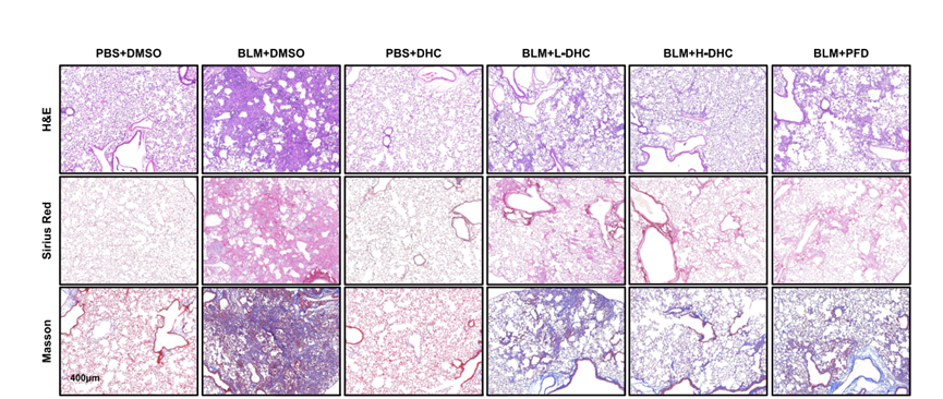
Dehydrocorydaline (hydroxyl) purchased from MedChemExpress. Usage Cited in: Respir Res. 2025 Apr 12;26(1):136. [Abstract]
Images of mice lung tissue slices stained with H&E, Masson, and Sirius Red treated with L-DHC (Dehydrocorydaline) 5 mg/kg, H-DHC: 10 mg/kg.
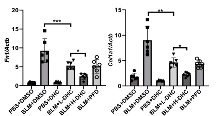
Dehydrocorydaline (hydroxyl) purchased from MedChemExpress. Usage Cited in: Respir Res. 2025 Apr 12;26(1):136. [Abstract]
RT-qPCR results for Col1a1 and Fn1 expression levels of mice lung tissue treated with L-DHC (Dehydrocorydaline) 5 mg/kg, H-DHC: 10 mg/kg.
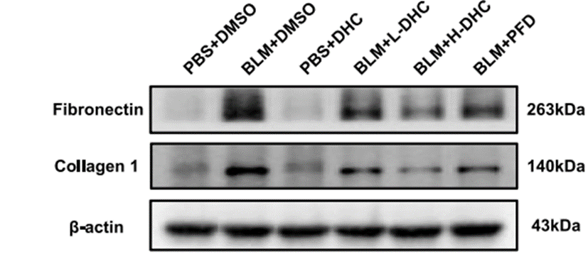
Dehydrocorydaline (hydroxyl) purchased from MedChemExpress. Usage Cited in: Respir Res. 2025 Apr 12;26(1):136. [Abstract]
Collagen 1 and Fibronectin of mice lung tissues are analyzed using Western blot treated with L-DHC (Dehydrocorydaline) 5 mg/kg, H-DHC: 10 mg/kg.
Dehydrocorydaline (hydroxyl) purchased from MedChemExpress. Usage Cited in: Respir Res. 2025 Apr 12;26(1):136. [Abstract]
Immunofluorescence images of α-SMA, Collagen 1, and Fibronectin in mice lung tissue slices treated with L-DHC (Dehydrocorydaline) 5 mg/kg, H-DHC: 10 mg/kg.
Dehydrocorydaline (hydroxyl) purchased from MedChemExpress. Usage Cited in: Respir Res. 2025 Apr 12;26(1):136. [Abstract]
CCK8 assay to determine the impact of Dehydrocorydaline (DHC) (5, 10, 30, 50, 80, 100, 150 μM) on HPFs viability.
-
Eur J Pharmacol
Eriodictyol inhibits glioblastoma migration and invasion by reversing EMT via downregulation of the P38 MAPK/GSK-3β/ZEB1 pathway. [Abstract]2021 Jun 5:900:174069. PMID: 33811837 -
Int Immunopharmacol
MCPIP1 modulates host antifungal immunity and exacerbates lethal Candida albicans infection. [Abstract]2026 Jul 15:181:116715. PMID: 42066549 -
Int Immunopharmacol
Salvianolic acid a inhibits neuroinflammation and ameliorates Alzheimer's disease pathology via the p38 MAPK/NF-κB pathway based on network pharmacology and experimental validation. [Abstract]2026 Jun 1:178:116594. PMID: 41930874 -
Int Immunopharmacol
Eupatilin suppresses osteoclastogenesis and periodontal bone loss by inhibiting the MAPKs/Siglec-15 pathway. [Abstract]2024 Sep 30:139:112720. PMID: 39047450 -
Bone Joint Res
Melatonin induces RAW264.7 cell apoptosis via the BMAL1/ROS/MAPK-p38 pathway to improve postmenopausal osteoporosis. [Abstract]2023 Nov 1;12(11):677-690. PMID: 37907083 -
Arthritis Res Ther
Roles of VWA1 and Rac2 in compensatory and decompensatory responses via FGF9/FGFR3 and p38 MAPK signaling pathways in condylar chondrocytes under distinct mechanical stress. [Abstract]2025 May 31;27(1):118. PMID: 40450276 -
Front Pharmacol
Inhibition of ASIC1a-Mediated ERS Improves the Activation of HSCs and Copper Transport Under Copper Load. [Abstract]2021 May 31:12:653272. PMID: 34135753 -
J Inflamm Res
The Effects of Adoptively Transferred IL-23/IL-18-Polarized Neutrophils on Tumor and Collagen-Induced Arthritis in Mice. [Abstract]2021 Sep 16;14:4669-4686. PMID: 34557012 -
FASEB J
USP44 Regulates Chemoresistance Induced by ROS and the MAPK/NF-κB Pathway Through the Stabilization of ITGB4 in Gastric Cancer. [Abstract]2025 Aug 31;39(16):e70945. PMID: 40824171 -
J Nutr
A-Type Cinnamon Procyanidin Oligomers Protect Against 1-Methyl-4-Phenyl-1,2,3,6-Tetrahydropyridine-Induced Neurotoxicity in Mice Through Inhibiting the P38 Mitogen-Activated Protein Kinase/P53/BCL-2 Associated X Protein Signaling Pathway. [Abstract]2020 Jul 1;150(7):1731-1737. PMID: 32386222 -
Front Biosci (Landmark Ed)
2025 Apr 25;30(4):25221. PMID: 40302325 -
J Food Sci
Curcumin Alleviates DON-Induced Intestinal Epithelial Barrier Disruption by Improving Ribotoxic Stress-Associated p38 Pathway-Mediated TJ Injury, Apoptosis, and Cell Cycle Arrest. [Abstract]2025 Apr;90(4):e70217. PMID: 40271829 -
J Cell Physiol
STRIP2 silencing inhibits vascular smooth muscle cell proliferation and migration via P38-AKT-MMP-2 signaling pathway. [Abstract]2019 Dec;234(12):22463-22476. PMID: 31093976 -
Genomics
MiR-100-5p as a biomarker in hand-arm vibration disease: Mediating pathogenesis through angiogenesis suppression. [Abstract]2025 Jul;117(4):111067. PMID: 40460899 -
Mol Carcinog
ITGB3-enriched extracellular vesicles mediate the formation of osteoclastic pre-metastatic niche to promote lung adenocarcinoma bone metastasis. [Abstract]2024 Nov;63(11):2190-2204. PMID: 39136603 -
Andrology
Expression of IL-33 in rodent testes and its role in Leydig cell steroidogenesis and aging. [Abstract]2025 Jun 16. PMID: 40521800 -
Tissue Cell
Placental mesenchymal stem cells-secreted proenkephalin suppresses the p38 MAPK signaling to block hyperproliferation of keloid fibroblasts. [Abstract]2023 Dec:85:102218. PMID: 37913601 -
Exp Eye Res
Embryonic stem cell extracellular vesicles reverse the senescence of retinal pigment epithelial cells by the p38MAPK pathway. [Abstract]2023 Feb:227:109365. PMID: 36577484 -
BMC Musculoskelet Disord
Knockdown of PAR2 alleviates cancer-induced bone pain by inhibiting the activation of astrocytes and the ERK pathway. [Abstract]2022 May 30;23(1):514. PMID: 35637468 -
Infect Genet Evol
Knockdown of GBP1 inhibits BCG-induced apoptosis in macrophage RAW 264.7 cells via p38/JNK pathway. [Abstract]2022 Jan:97:105158. PMID: 34826624 -
J Mol Histol
Nogo-A-Δ20/EphA4 interaction antagonizes apoptosis of neural stem cells by integrating p38 and JNK MAPK signaling. [Abstract]2021 Jun;52(3):521-537. PMID: 33555537 -
Biochem Biophys Res Commun
Deficiency of unc-51 like kinase 1 (Ulk1) protects against mice traumatic brain injury (TBI) by suppression of p38 and JNK pathway. [Abstract]2018 Sep 5;503(2):467-473. PMID: 29680658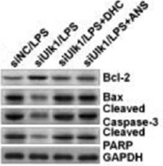
Dehydrocorydaline (hydroxyl) purchased from MedChemExpress. Usage Cited in: Biochem Biophys Res Commun. 2018 Sep 5;503(2):467-473. [Abstract]
The increase of Bcl-2 and decrease of Bax, cleaved Caspase-3 and PARP induced by Ulk1- inhibition in LPS-exposed AST are abrogated by the Dehydrocorydaline chloride (DHC) or Anisomycin (ANS) pre-treatment
-
Biochem Biophys Res Commun
NFATc3 deficiency protects against high fat diet (HFD)-induced hypothalamus inflammation and apoptosis via p38 and JNK suppression. [Abstract]2018 May 23;499(4):743-750. PMID: 29596828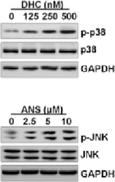
Dehydrocorydaline (hydroxyl) purchased from MedChemExpress. Usage Cited in: Biochem Biophys Res Commun. 2018 May 23;499(4):743-750. [Abstract]
Astrocytes are cultured with p38 and JNK activator of DHC and ANS, respectively, at the indicated concentrations for 24 h. Then, all cells are harvested for western blot analysis of p-p38 and p-JNK. Astrocytes are pretreated with DHC (500 nM) or ANS (10 mM) for 24 h, followed by Fru (5 mM) for an additional 24 h.
-
Eur J Histochem
Adipose-derived stem cells promote the recovery of intestinal barrier function by inhibiting the p38 MAPK signaling pathway. [Abstract]2025 Jan 21;69(1). PMID: 39836101 -
J Clin Neurosci
Effects of p38 MAPK signaling pathway on cognitive function and recovery of neuronal function after hypoxic-ischemic brain injury in newborn rats. [Abstract]2020 Aug:78:365-370. PMID: 32360159 -
J Cardiothorac Surg
Aconitine promotes ROS-activated P38/MAPK/Nrf2 pathway to inhibit autophagy and promote myocardial injury. [Abstract]2024 Dec 20;19(1):665. PMID: 39707526 -
-
Bioengineered
Deleted in lymphocytic leukemia 2 induces retinoic acid receptor beta promoter methylation and mitogen activated kinase-like protein activation to enhance viability and mobility of colorectal cancer cells. [Abstract]2022 May;13(5):12847-12862. PMID: 35611845 -
Aging (Albany NY)
Short-term high-fat diet favors the appearances of apoptosis and gliosis by activation of ERK1/2/p38MAPK pathways in brain. [Abstract]2021 Oct 7;13(19):23133-23148. PMID: 34620734 -
Comput Math Methods Med
Efficacy of Low Molecular Heparin on Preeclampsia by Inhibiting Apoptosis of Trophoblasts via the p38MAPK Signaling Pathway. [Abstract]2021 Aug 2:2021:3337514. PMID: 34394705 -
Aging (Albany NY)
2021 May 11;13(9):12780-12799. PMID: 33973871


Solvent & Solubility
In Vitro:
DMSO : 12.5 mg/mL (32.60 mM; ultrasonic and warming and heat to 60°C; Hygroscopic DMSO has a significant impact on the solubility of product, please use newly opened DMSO)
Please refer to the solubility information to select the appropriate solvent. Once prepared, please aliquot and store the solution to prevent product inactivation from repeated freeze-thaw cycles.
Storage method and period of stock solution: -80°C, 6 months; -20°C, 1 month. When stored at -80°C, please use it within 6 months. When stored at -20°C, please use it within 1 month.
Please refer to the solubility information to select the appropriate solvent. Once prepared, please aliquot and store the solution to prevent product inactivation from repeated freeze-thaw cycles.
Storage method and period of stock solution: -80°C, 6 months; -20°C, 1 month. When stored at -80°C, please use it within 6 months. When stored at -20°C, please use it within 1 month.
Concentration (start) × Volume (start) = Concentration (final) × Volume (final)
In Vivo Dissolution Calculator
Please enter the basic information of animal experiments:
-
-
-
-
Recommended: Prepare an additional quantity of animals to account for potential losses during experiments.
Working solution concentration: 0.22 mg/mL


Purity & Documentation
-
Data Sheet (335 KB)
-
SDS (251 KB)
- English - EN (251 KB)
- Français - FR (251 KB)
- Deutsch - DE (251 KB)
- Norwegian - NO (251 KB)
- Español - ES (251 KB)
- Swedish - SV (251 KB)
- Italian - IT (251 KB)
- Korean - KR (251 KB)
- Portuguese - PT (251 KB)
-
Handling Instructions (2659 KB)
References
[1]. Nonaka M, et al. Screening of a library of traditional Chinese medicines to identify anti-malarial compounds and extracts. Malaria journal. 2018 Jun 25;17(1):244. [Content Brief]
[3]. Yoo M, et al. Dehydrocorydaline promotes myogenic differentiation via p38 MAPK activation. Molecular medicine reports. 2016 Oct;14(4):3029-36. [Content Brief]
[4]. Xu Z, et al. Dehydrocorydaline inhibits breast cancer cells proliferation by inducing apoptosis in MCF-7 cells. The American journal of Chinese medicine. 2012;40(1):177-85. [Content Brief]
[6]. Yin ZY, et al. Antinociceptive effects of dehydrocorydaline in mouse models of inflammatory pain involve the opioid receptor and inflammatory cytokines. Scientific reports. 2016 Jun 07;6:27129. [Content Brief]
[9]. Wen B, et al. Antiatherosclerotic effect of dehydrocorydaline on ApoE mice: inhibition of macrophage inflammation. Acta pharmacologica Sinica. 2022 Jun;43(6):1408-1418. [Content Brief]
[10]. Li Y, et al. Dehydrocorydaline Protects Against Sepsis-Induced Myocardial Injury Through Modulating the TRAF6/NF-κB Pathway. Front Pharmacol. 2021 Aug 19;12:709604. [Content Brief]
[12]. He J, et al. Dehydrocorydaline attenuates bleomycin-induced pulmonary fibrosis by inhibiting fibroblast activation. Respir Res. 2025 Apr 12;26(1):136. [Content Brief]
[14]. Hu H, et al. Dehydrocorydaline inhibits cell proliferation, migration and invasion via suppressing MEK1/2-ERK1/2 cascade in melanoma. Onco Targets Ther. 2019 Jul 2;12:5163-5175. [Content Brief]


Complete Stock Solution Preparation Table
Please refer to the solubility information to select the appropriate solvent. Once prepared, please aliquot and store the solution to prevent product inactivation from repeated freeze-thaw cycles.
Storage method and period of stock solution: -80°C, 6 months; -20°C, 1 month. When stored at -80°C, please use it within 6 months. When stored at -20°C, please use it within 1 month.
| Optional Solvent | Concentration Solvent Mass | 1 mg | 5 mg | 10 mg | 25 mg |
|---|---|---|---|---|---|
| DMSO | 1 mM | 2.6080 mL | 13.0398 mL | 26.0797 mL | 65.1992 mL |
| 5 mM | 0.5216 mL | 2.6080 mL | 5.2159 mL | 13.0398 mL | |
| 10 mM | 0.2608 mL | 1.3040 mL | 2.6080 mL | 6.5199 mL | |
| 15 mM | 0.1739 mL | 0.8693 mL | 1.7386 mL | 4.3466 mL | |
| 20 mM | 0.1304 mL | 0.6520 mL | 1.3040 mL | 3.2600 mL | |
| 25 mM | 0.1043 mL | 0.5216 mL | 1.0432 mL | 2.6080 mL | |
| 30 mM | 0.0869 mL | 0.4347 mL | 0.8693 mL | 2.1733 mL |


Dehydrocorydaline (hydroxyl) Related Classifications
HY-N0674B Related Classifications
 Powered by Bioz
Keywords
- Dehydrocorydaline (hydroxyl)
- 13-Methylpalmatine (hydroxyl)
- Cholinesterase (ChE)
- Parasite
- Bcl-2 Family
- Caspase
- PARP
- p38 MAPK
- Apoptosis
- Bacterial
- NOD-like Receptor (NLR)
- 5-HT Receptor
- Dopamine Receptor
- poly ADP ribose polymerase
- Serotonin Receptor
- 5-hydroxytryptamine Receptor
- NLRP3 inflammasome
- Plasmodium falciparum
- SPTA1
- Trypanosoma cruzi
- acetylcholinesterase inhibitor
- aldose reductase
- atherosclerosis
- blood-brain barrier
- myoblast differentiation
- Inhibitor
- inhibitor
- inhibit