



Daphnetin
Based on 10 publication(s) in Google Scholar
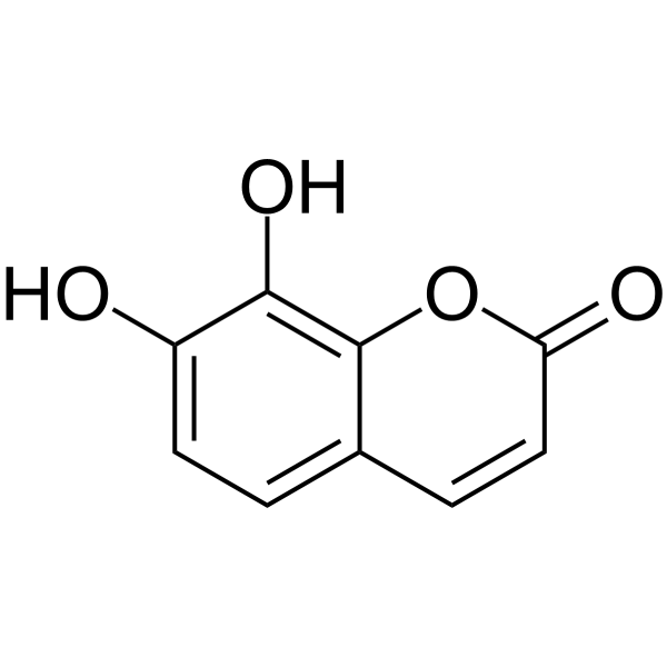
Daphnetin (7,8-dihydroxycoumarin), one coumarin derivative can be found in plants of the Genus Daphne, is a potent, oral active protein kinase inhibitor, with IC50s of 7.67 μM, 9.33 μM and 25.01 μM for EGFR, PKA and PKC in vitro, respectively. Daphnetin triggers ROS-induced cell apoptosis and induces cytoprotective autophagy by modulating the AMPK/Akt/mTOR pathway. Daphnetin has anti-inflammation activitity and inhibits TNF-α, IL-1β, ROS, and MDA production. Daphnetin has schizontocidal activity against malaria parasites. Daphnetin can be used for rheumatoid arthritis , cancer and anti-malarian research.
For research use only. We do not sell to patients.
- Purity : 99.77%
- CAS No.: 486-35-1
- Formula: C9H6O4
- Molecular Weight:178.14
-
Storage:Powder -20°C, 3 years , 4°C, 2 years ; In solvent -80°C, 6 months , -20°C, 1 month

To place orders, for customer services and technical support, please contact: MedChemExpress USA
Tel: 609-228-6898 E-mail: [email protected] [email protected]


-
 Biological Activity
-
Chemical Information
-
Solvent & Solubility
- Protocols
- Purity & Documentation
- References
-
 Help & FAQs


-

HY-10219
RapamycinmTOR Inhibitor -
HY-10529
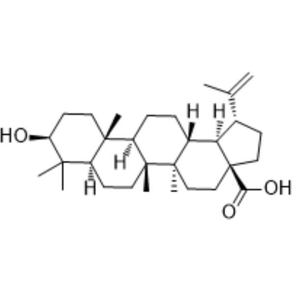
Betulinic acidApoptosis Inducer -
HY-15142
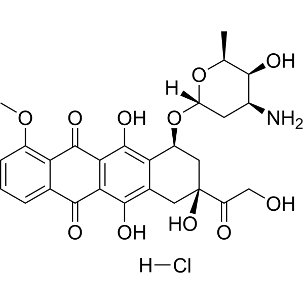
Doxorubicin hydrochlorideTopoisomerase Inhibitor -
HY-N0005
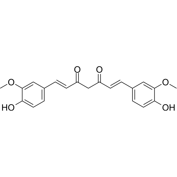
Curcuminp300 Histone Acetylatransferase Inhibitor -
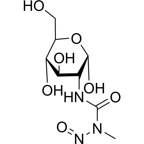
HY-13753
StreptozotocinDiabetes Modeling Agent -
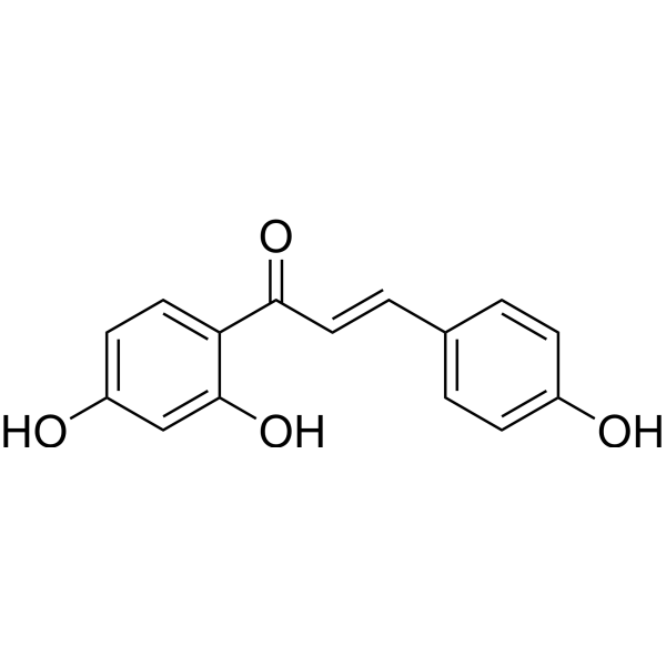
HY-N0102
IsoliquiritigeninAutophagy Inducer -
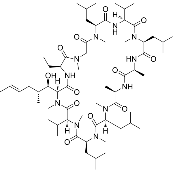
HY-B0579
Cyclosporin ACalcineurin Inhibitor -
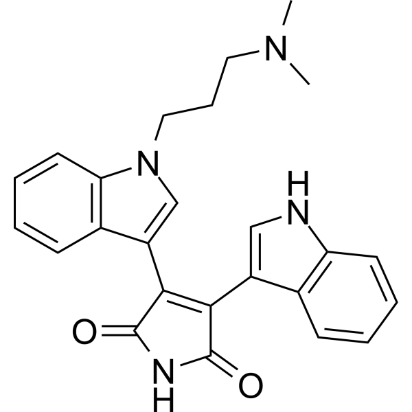
HY-13867
Bisindolylmaleimide IPKC Inhibitor -
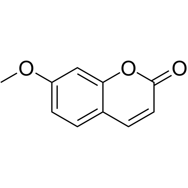
HY-N1366
HerniarinAnticancer Agent -
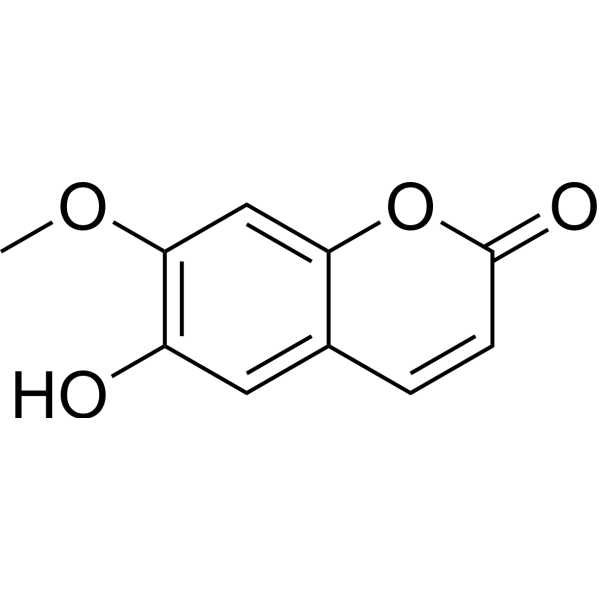
HY-N1365
IsoscopoletinHBV Inhibitor -
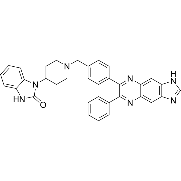
HY-10355
AKT inhibitor VIIIAKT Inhibitor -
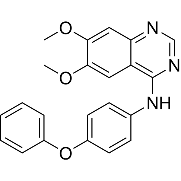
HY-101053
Src Inhibitor 1Src Inhibitor
Publications Citing Use of MedChemExpress (MCE) Daphnetin
More- Phytomedicine. 2026 Sep:159:158517.
- Int Immunopharmacol. 2026 Jun 17:185:116986. [Abstract]
- Int Immunopharmacol. 2024 May 30:133:112004. [Abstract]
- Biol Res. 2025 Dec 29;58(1):77. [Abstract]
- Am J Pathol. 2022 Dec;192(12):1725-1744. [Abstract]
- Rheumatology (Oxford). 2025 Aug 13:keaf437. [Abstract]
- J Cell Commun Signal. 2025 Apr 28;19(2):e70011. [Abstract]
- Pharmacology. 2021;106(7-8):369-383. [Abstract]
- Histol Histopathol. 2025 Sep 3:18978. [Abstract]
- In Vitro Cell Dev Biol Anim. 2026 Jul 24.
 Customer Validation & Images

-
In Vivo Efficacy Study

-
Histological Imaging/Staining

-
IHC

-
Flow Cytometry

-
Cell Proliferation/Viability Assay

All EGFR Isoforms
MoreAll AMPK Isoforms
MoreAll Caspase Isoforms
MoreAll Parasite Isoforms
More

Biological Activity
Description
IC50 & Target
[4]|
EGFR 7.67 μM (IC50) |
Plasmodium |
PKA 9.33 μM (IC50) |
PKC 25.01 μM (IC50) |
Cellular Effect
|
Cell Line

|
Type | Value | Description | References |
|---|---|---|---|---|
| MCF7 | IC50 |
73 μM
Compound: Daphnetin
|
Antiproliferative activity against human MCF7 cells measured after 72 hrs by MTT assay
Antiproliferative activity against human MCF7 cells measured after 72 hrs by MTT assay
|
[PMID: 38340509] |
| U-937 | CC50 |
120.2 μM
Compound: 20
|
Cytotoxicity against human U937 cells assessed as decrease in cell viability after 48 hrs
Cytotoxicity against human U937 cells assessed as decrease in cell viability after 48 hrs
|
[PMID: 26188907] |
| U-937 | IC50 |
83.2 μM
Compound: 20
|
Antiproliferative activity against human U937 cells after 3 days
Antiproliferative activity against human U937 cells after 3 days
|
[PMID: 26188907] |
In Vitro
Daphnetin (7,8-dihydroxycoumarin) (0-40 μg/mL; 24-48 hours) inhibits the proliferation of ovarian cancer cells[1].
Daphnetin (7,8-dihydroxycoumarin) (0-40 μg/mL; 24 hours; A2780 cells) induces apoptosis and increases ROS production in a dose-dependent manner[1].
Daphnetin (7,8-dihydroxycoumarin) (0-40 μg/mL; 24 hours; A2780 cells) induces autophagy through modulation of the AMPK/Akt/mTOR pathway[1].
Daphnetin (7,8-dihydroxycoumarin) (1-10 μM; plasmodium falciparum) exhibits schizontocidal activity in a dose-dependent manner[3].
MedChemExpress (MCE) has not independently confirmed the accuracy of these methods. They are for reference only. Further protocols information, click here.
-
Cell Line:IOSE8C, A2780, SKOV3 and OVCAR8 cells
-
Concentration:0, 5, 10, 20 and 40 µg/mL
-
Incubation Time:24 h and 48 hours
-
Result:Inhibited growth in ovarian cancer cells but not in normal cells.
-
Cell Line:A2780 and SKOV3 cells
-
Concentration:0, 10, 20 and 40 µg/mL
-
Incubation Time:24 hours
-
Result:Increased apoptosis in a dose-dependent manner in A2780 and SKOV3 cells.
-
Cell Line:A2780 and SKOV3 cells
-
Concentration:0, 10, 20 and 40 µg/mL
-
Incubation Time:24 hours
-
Result:Increased proapoptotic protein (Caspase 3, Bax, and PARP) expression but decreased antiapoptotic protein (Bcl2) expression.
-
Cell Line:A2780 cells
-
Concentration:0, 10, 20 and 40 µg/mL
-
Incubation Time:24 hours
-
Result:Increased LC3 II and p62 expression in a dose-dependent manner and reduced the expression levels of p-Akt, p-mTOR, but increased the expression level of p-AMPK.
In Vivo
Daphnetin (7,8-dihydroxycoumarin) (2.5-10 mg/kg; i.p.; daily; for three days; C57BL/6 mice) inhibits cisplatin-induced inflammation, decreases TNF-α, IL-1β, ROS and MDA production in a dose-dependent manner in kidney tissues. Daphnetin inhibits cisplatin-induced NF-κB activation and up-regulated Nrf2 and HO-1[2].
Daphnetin (7,8-dihydroxycoumarin) (10-100 mg/kg; i.g. and i.p.; every four days, for 30 days; male Kunming outbred strain mice) displays certain schizontocidal activity in vivo[3].
MedChemExpress (MCE) has not independently confirmed the accuracy of these methods. They are for reference only.
-
Animal Model:BALB/c nude mice[1]
-
Dosage:30 mg/kg
-
Administration:Intraperitoneal injection; Daily; for 12 days
-
Result:Decreased tumor volume and weight in a xenograft animal model.
-
Animal Model:Male Kunming outbred strain mice[3]
-
Dosage:10, 50 or 100 mg/kg
-
Administration:Oral gavage and intraperitoneal injection; every four days, for 30 days
-
Result:Reduced the number of parasites in mice.
Clinical Trial
| NCT Number | Sponsor | Condition | Start Date |
Phase
|
|---|---|---|---|---|
| NCT01329991 | Plexxikon| | 2011-05 | PHASE1 |


Chemical Information
-
CAS No. 486-35-1
-
Appearance Solid
-
Molecular Weight 178.14
-
Formula C9H6O4
-
Color Off-white to yellow
-
SMILES
O=C1C=CC2=CC=C(O)C(O)=C2O1
-
Synonyms
7,8-Dihydroxycoumarin
-
Structure Classification
-
Initial Source
-
Shipping
Room temperature in continental US; may vary elsewhere.
-
Storage
Powder -20°C 3 years 4°C 2 years In solvent -80°C 6 months -20°C 1 month


Publications (10)
-
Journal Impact Factor
-
Most Recent
-
-
Int Immunopharmacol
Daphnetin alleviates neuropathic pain in rats by modulating spinal microglial polarization via the Nrf2/NLRP3 inflammasome pathway. [Abstract]2026 Jun 17:185:116986. PMID: 42308799
Daphnetin purchased from MedChemExpress. Usage Cited in: Int Immunopharmacol. 2026 Jun 17:185:116986. [Abstract]
Representative images showing the embryo resorption sites of URSA mice treated with NS or Daphnetin (1, 10, 20 mg/kg, i.g.) at different doses on the 13th day of pregnancy.
Daphnetin purchased from MedChemExpress. Usage Cited in: Int Immunopharmacol. 2026 Jun 17:185:116986. [Abstract]
Morphologic changes (the black arrow indicates the site of hemorrhage, while the green arrow indicates the area of necrosis) in decidual tissues of URSA mice treated with NS or Daphnetin (10 mg/kg, i.g.) were assessed by HE staining.
Daphnetin purchased from MedChemExpress. Usage Cited in: Int Immunopharmacol. 2026 Jun 17:185:116986. [Abstract]
Positive cells of IGFBP-1 and PRL in decidual tissues of mice were detected by immunohistochemistry treated with Daphnetin (10 mg/kg, i.g.).
Daphnetin purchased from MedChemExpress. Usage Cited in: Int Immunopharmacol. 2026 Jun 17:185:116986. [Abstract]
Treg and Th17 cell populations in the spleens of mice were analyzed using flow cytometry treated with Daphnetin (10 mg/kg, i.g.).
Daphnetin purchased from MedChemExpress. Usage Cited in: Int Immunopharmacol. 2026 Jun 17:185:116986. [Abstract]
Effect of different concentrations of Daphnetin (0, 5, 0, 20, 40, 80 μM) for 48 h on CD4+ T cell viability using the CCK-8 assay.

Daphnetin purchased from MedChemExpress. Usage Cited in: Int Immunopharmacol. 2026 Jun 17:185:116986. [Abstract]
Detection of mRNA expression of FoxP3 and RORγt in CD4+ T cells by RT-qPCR treated with Daphnetin (20 μM).

Daphnetin purchased from MedChemExpress. Usage Cited in: Int Immunopharmacol. 2026 Jun 17:185:116986. [Abstract]
The residual intracellular NR4A1 protein expression in CD4+ T cells with 20 μM Daphnetin and 10 μg/mL CHX for varying durations was measured using Western blot analysis.
-
Int Immunopharmacol
Daphnetin alleviates silica-induced pulmonary inflammation and fibrosis by regulating the PI3K/AKT1 signaling pathway in mice. [Abstract]2024 May 30:133:112004. PMID: 38613881 -
Biol Res
Daphnetin alleviates unexplained recurrent spontaneous abortion by regulating the NR4A1/BACH2 axis in mice. [Abstract]2025 Dec 29;58(1):77. PMID: 41462398 -
Am J Pathol
α-Subunit Tyrosine Phosphorylation Is Required for Activation of the Large Conductance Ca2+-Activated Potassium Channel in the Rabbit Sphincter of Oddi. [Abstract]2022 Dec;192(12):1725-1744. PMID: 36150507 -
Rheumatology (Oxford)
Autophagy inhibitors block pathogenic NET release in immune-mediated inflammatory disease without impairing host defence. [Abstract]2025 Aug 13:keaf437. PMID: 40802538 -
J Cell Commun Signal
Daphnetin alleviates inflammation and promotes autophagy via the AMPK/mTOR pathway in gouty arthritis. [Abstract]2025 Apr 28;19(2):e70011. PMID: 40304008 -
Pharmacology
Daphnetin Preconditioning Decreases Cardiac Injury and Susceptibility to Ventricular Arrhythmia following Ischaemia-Reperfusion through the TLR4/MyD88/NF-Κb Signalling Pathway. [Abstract]2021;106(7-8):369-383. PMID: 33902056 -
Histol Histopathol
Daphnetin alleviates renal inflammation, oxidative stress, and apoptosis in septic rats via the JAK2/STAT3 signaling pathway. [Abstract]2025 Sep 3:18978. PMID: 40899121 -


Solvent & Solubility
In Vitro:
DMSO : 50 mg/mL (280.68 mM; Need ultrasonic; Hygroscopic DMSO has a significant impact on the solubility of product, please use newly opened DMSO)
H2O : < 0.1 mg/mL (insoluble)
Please refer to the solubility information to select the appropriate solvent. Once prepared, please aliquot and store the solution to prevent product inactivation from repeated freeze-thaw cycles.
Storage method and period of stock solution: -80°C, 6 months; -20°C, 1 month. When stored at -80°C, please use it within 6 months. When stored at -20°C, please use it within 1 month.
Please refer to the solubility information to select the appropriate solvent. Once prepared, please aliquot and store the solution to prevent product inactivation from repeated freeze-thaw cycles.
Storage method and period of stock solution: -80°C, 6 months; -20°C, 1 month. When stored at -80°C, please use it within 6 months. When stored at -20°C, please use it within 1 month.
Concentration (start) × Volume (start) = Concentration (final) × Volume (final)
In Vivo:
Select the appropriate dissolution method based on your experimental animal and administration route.
- For the following dissolution methods, please ensure to first prepare a clear stock solution using an In Vitro approach and then sequentially add co-solvents:
- To ensure reliable experimental results, the clarified stock solution can be appropriately stored based on storage conditions. As for the working solution for In Vivo experiments, it is recommended to prepare freshly and use it on the same day.
- The percentages shown for the solvents indicate their volumetric ratio in the final prepared solution. If precipitation or phase separation occurs during preparation, heat and/or sonication can be used to aid dissolution.
Add each solvent one by one: 10% DMSO 40% PEG300 5% Tween-80 45% Saline
Solubility: ≥ 2.5 mg/mL (14.03 mM); Clear solution
This protocol yields a clear solution of ≥ 2.5 mg/mL (saturation unknown).
Taking 1 mL working solution as an example, add 100 μL DMSO stock solution (25.0 mg/mL) to 400 μL PEG300, and mix evenly; then add 50 μL Tween-80 and mix evenly; then add 450 μL Saline to adjust the volume to 1 mL.
Preparation of Saline: Dissolve 0.9 g sodium chloride in ddH₂O and dilute to 100 mL to obtain a clear Saline solution.
Add each solvent one by one: 10% DMSO 90% (20% SBE-β-CD in Saline)
Solubility: ≥ 2.5 mg/mL (14.03 mM); Clear solution
This protocol yields a clear solution of ≥ 2.5 mg/mL (saturation unknown).
Taking 1 mL working solution as an example, add 100 μL DMSO stock solution (25.0 mg/mL) to 900 μL 20% SBE-β-CD in Saline, and mix evenly.
Preparation of 20% SBE-β-CD in Saline (4°C, storage for one week): 2 g SBE-β-CD powder is dissolved in 10 mL Saline, completely dissolve until clear.
For the following dissolution methods, please prepare the working solution directly:
It is recommended to prepare fresh solutions and use them promptly within a short period of time.
The percentages shown for the solvents indicate their volumetric ratio in the final prepared solution. If precipitation or phase separation occurs during preparation, heat and/or sonication can be used to aid dissolution.
Add each solvent one by one: 50% PEG300 50% Saline
Solubility: 25 mg/mL (140.34 mM); Suspended solution; Need ultrasonic
In Vivo Dissolution Calculator
Please enter the basic information of animal experiments:
-
-
-
-
Recommended: Prepare an additional quantity of animals to account for potential losses during experiments.
Please enter your animal formula composition:
-
%DMSO +
Recommended: Keep the proportion of DMSO in working solution below 2% if your animal is weak.
-
%+
-
+%Tween-80 + +
-
%Saline +
The co-solvents required include: DMSO, . All of co-solvents are available by MedChemExpress (MCE). , Tween 80. All of co-solvents are available by MedChemExpress (MCE).
Working solution concentration: 0.22 mg/mL
Method for preparing stock solution: mg drug dissolved in μL DMSO. Stock solution concentration: mg/mL.
1. Take μL DMSO stock solution;
2. Add μL .
μL , mix evenly;
3. Then add μL Tween 80, mix evenly;
4. Then add μL
Please ensure that the stock solution in the first step is dissolved to a clear state, and add co-solvents in sequence. You can use ultrasonic heating (ultrasonic cleaner, recommended frequency 20-40 kHz), vortexing, etc. to assist dissolution.

Protocols
-
Collagen-Induced Arthritis
Collagen-induced arthritis (CIA) is an autoimmune murine model of rheumatoid arthritis in which immunization with type II collagen (CII) emulsified in an adjuvant induces a T cell- and autoantibody-driven inflammatory arthritis characterized by synovial hyperplasia, immune cell infiltration, and joint destruction. The model typically relies on genetically susceptible mouse strains (e. g. , DBA/1) and reproduces key features of human rheumatoid arthritis, including anti-collagen immune responses and progressive joint inflammation. Disease onset generally occurs within ~3-4 weeks after immunization, depending on antigen/adjuvant combinations and protocol variation. The immunopathology is driven by adaptive immune activation against CII, leading to systemic and local joint inflammation mediated by pro-inflammatory cytokines and effector immune cells, making CIA a standard preclinical platform for evaluating immunomodulatory and anti-arthritic interventions.
-
Apoptosis
Apoptosis, also called programmed cell death, is generally characterized by distinct morphological characteristics.
-
TUNEL staining for apoptotic DNA fragmentation
TUNEL staining detects DNA strand breaks by using terminal deoxynucleotidyl transferase to add labeled nucleotides to exposed 3′-OH DNA termini, generating either microscopic staining in fixed cells or tissue sections, or fluorescence/cytometric signal in cell suspensions. TUNEL positivity reflects DNA fragmentation but should not be interpreted alone as definitive apoptosis, because TUNEL can also label necrotic, autolytic, mechanically damaged, or DNA-repair-associated DNA breaks.
-
Annexin V plus membrane-impermeant dye apoptosis staining
Annexin V-based apoptosis assays rely on the detection of phosphatidylserine (PS) externalization from the inner leaflet of the plasma membrane to the outer leaflet, an early biochemical hallmark of apoptosis. Fluorescently labeled Annexin V binds PS in a calcium-dependent manner, enabling identification of early apoptotic cells by flow cytometry or fluorescence microscopy. When combined with a membrane-impermeant DNA-binding dye (e. g. , propidium iodide), this approach allows discrimination between viable (Annexin V−/dye−), early apoptotic (Annexin V+/dye−), and late apoptotic or necrotic (Annexin V+/dye+) cell populations by assessing membrane integrity and PS exposure.
-
ROS/oxidative-stress fluorescent staining
ROS/oxidative-stress fluorescent staining uses cell-permeant fluorogenic probes that become fluorescent after oxidation inside cells or tissues; commonly used examples include DCFH-DA/DCFDA for broad cellular oxidant detection, DHE for superoxide-related signal detection, MitoSOX for mitochondrial superoxide-related signal detection, and CellROX probes for oxidative-stress-associated fluorescence readouts. The assay detects probe oxidation rather than a single ROS species unless the probe and analysis method have been chemically validated for that species. DCFH-DA enters cells, is deacetylated by intracellular esterases to DCFH, and produces fluorescent DCF after oxidation, so the readout is used as an operational measure of total cellular oxidative stress rather than a species-specific ROS measurement. DHE and MitoSOX can report superoxide-related oxidation, but red fluorescence alone can include non-specific ethidium-like oxidation products; HPLC or optimized spectral approaches are
-
Autophagy
Autophagy is a process in which eukaryotic cells use lysosomes to degrade their own cytoplasmic proteins and damaged organelles under the regulation of autophagy related gene (Atg). Microtubule-associated proteins light chain 3 (LC3) is recognized as autophagy marker, which transfers from cytoplasmic LC3 (LC3-I) to membrane type (LC3-II). LC3-II/I ratio could be detected by Western Blot and fluorescence microscopy.
-
Apoptosis Solutions
Apoptosis is a regulated, generally non-lytic cell-death pathway that removes unwanted, damaged, infected, or abnormal cells through coordinated morphological changes, caspase activation, DNA fragmentation, and membrane remodeling. The intrinsic apoptosis pathway is controlled mainly by mitochondrial outer membrane permeabilization, BCL-2 family proteins, cytochrome c release, apoptosome formation, caspase-9 activation, and downstream executioner caspase-3/7 activation. The extrinsic apoptosis pathway is initiated by death receptors such as Fas, TNFR, and TRAIL receptors, which recruit adaptor proteins and activate caspase-8 before engaging executioner caspases or mitochondrial amplification through BID cleavage. Apoptosis is linked to many phenotypes, including cancer cell killing, tissue homeostasis, immune regulation, neurodegeneration, infection response, and treatment-induced cytotoxicity; unresolved questions include how apoptosis interacts with necroptosis, pyroptosis, ferroptos
-
Lysosome and acidic-vesicle live-cell staining
Lysosome and acidic-vesicle live-cell staining detects acidic intracellular compartments by using membrane-permeant acidotropic probes that accumulate in low-pH vesicles, including lysosomes, late endosomes, autolysosomes, and acidic phagosomes. LysoTracker staining is commonly used as an intensity-based readout of acidic lysosomal compartment abundance or enlargement, while acridine orange produces green fluorescence in less concentrated compartments and red fluorescence after concentration-dependent accumulation in acidic vesicular organelles. Loss or reduction of acridine-orange red signal can be used as a readout of lysosomal membrane permeabilization or reduced acidic-vesicle integrity. This protocol is designed for live cultured cells and can be adapted for fluorescence microscopy, high-content imaging, plate-reader readout, or flow cytometry when the selected literature supports the readout. Because these dyes report acidotropic accumulation rather than lysosome identity alone,
-
Research Protocol for Inflammation-related Diseases
The NLRP3 inflammasome is a cytosolic innate immune signaling platform that integrates priming signals and danger-signal activation to promote caspase-1 activation, maturation of IL-1β and IL-18, and gasdermin D-mediated pyroptotic cell death. The core experimental logic is to determine whether inflammatory disease phenotypes are driven by increased NLRP3 expression, ASC-containing inflammasome assembly, caspase-1 cleavage, GSDMD cleavage, and extracellular release of IL-1β/IL-18 rather than by nonspecific cell injury alone. The pathway is strongly linked to inflammation-related disease phenotypes because monosodium urate crystals activate NALP3/NLRP3 inflammasome signaling in gout-like crystal inflammation, cholesterol crystals activate NLRP3 inflammasomes in atherogenesis models, and DSS-induced intestinal inflammation has been reported to involve NLRP3 inflammasome activity. However, experimental colitis studies also show context-dependent protective effects of NLRP3 inflammasome co
-
Macroautophagy Solutions
Macroautophagy is a conserved lysosome-dependent degradation pathway in which cytoplasmic material is sequestered into double-membrane autophagosomes and delivered to lysosomes for degradation and recycling. The pathway supports cellular homeostasis during nutrient limitation, organelle stress, protein-aggregate accumulation, infection, differentiation, and tissue remodeling by coupling cargo sequestration, autophagosome maturation, lysosomal fusion, and degradation of cargo-derived macromolecules. The core molecular sequence includes initiation by nutrient- and stress-regulated autophagy machinery, autophagosome nucleation, LC3/ATG8-family conjugation to autophagosomal membranes, cargo selection through receptors such as SQSTM1/p62, autophagosome-lysosome fusion, and lysosomal degradation. LC3 was identified as a mammalian homolog of yeast Atg8 that localizes to autophagosomal membranes after processing, and p62/SQSTM1 was shown to connect ubiquitinated cargo with autophagic degradati
-
Pyroptosis Solutions
Pyroptosis is a lytic inflammatory cell-death pathway executed by gasdermin pores, most classically through inflammasome-mediated activation of caspase-1, cleavage of gasdermin D, membrane pore formation, LDH release, and secretion of IL-1β and IL-18. The canonical pathway is commonly modeled by priming cells with an inflammatory signal such as LPS to induce pro-IL-1β and inflammasome components, followed by an activation signal such as ATP or nigericin to activate NLRP3, ASC speck formation, caspase-1 cleavage, GSDMD cleavage, cytokine release, and pyroptotic membrane rupture. The non-canonical pathway is triggered when cytosolic LPS activates mouse caspase-11 or human caspase-4/5, leading to GSDMD cleavage and pyroptosis, and this can secondarily activate NLRP3-dependent IL-1β release. Pyroptosis is linked to inflammatory injury, infection, cancer, liver disease, ocular disease, placental inflammation, and other disease phenotypes, but unresolved questions include which gasdermin fam


Purity & Documentation
-
Data Sheet (283 KB)
-
SDS (393 KB)
- English - EN (393 KB)
- Français - FR (393 KB)
- Deutsch - DE (393 KB)
- Norwegian - NO (393 KB)
- Español - ES (393 KB)
- Swedish - SV (393 KB)
- Italian - IT (393 KB)
- Korean - KR (393 KB)
- Portuguese - PT (393 KB)
-
Handling Instructions (2659 KB)
References
[1]. Fan X, et, al. Daphnetin triggers ROS-induced cell death and induces cytoprotective autophagy by modulating the AMPK/Akt/mTOR pathway in ovarian cancer. Phytomedicine. 2021 Feb;82:153465. [Content Brief]
[2]. Zhang L, et, al. Daphnetin protects against cisplatin-induced nephrotoxicity by inhibiting inflammatory and oxidative response. Int Immunopharmacol. 2018 Dec;65:402-407. [Content Brief]
[3]. Wang QM, et, al. The schizontocidal activity of daphnetin against malaria parasites in vitro and in vivo. Zhongguo Ji Sheng Chong Xue Yu Ji Sheng Chong Bing Za Zhi. 2000;18(4):204-6. [Content Brief]
[4]. Yang EB, Zhao YN, Zhang K, Mack P. Daphnetin, one of coumarin derivatives, is a protein kinase inhibitor. Biochem Biophys Res Commun. 1999 Jul 14;260(3):682-5. [Content Brief]


Complete Stock Solution Preparation Table
Please refer to the solubility information to select the appropriate solvent. Once prepared, please aliquot and store the solution to prevent product inactivation from repeated freeze-thaw cycles.
Storage method and period of stock solution: -80°C, 6 months; -20°C, 1 month. When stored at -80°C, please use it within 6 months. When stored at -20°C, please use it within 1 month.
| Optional Solvent | Concentration Solvent Mass | 1 mg | 5 mg | 10 mg | 25 mg |
|---|---|---|---|---|---|
| DMSO | 1 mM | 5.6136 mL | 28.0678 mL | 56.1356 mL | 140.3391 mL |
| 5 mM | 1.1227 mL | 5.6136 mL | 11.2271 mL | 28.0678 mL | |
| 10 mM | 0.5614 mL | 2.8068 mL | 5.6136 mL | 14.0339 mL | |
| 15 mM | 0.3742 mL | 1.8712 mL | 3.7424 mL | 9.3559 mL | |
| 20 mM | 0.2807 mL | 1.4034 mL | 2.8068 mL | 7.0170 mL | |
| 25 mM | 0.2245 mL | 1.1227 mL | 2.2454 mL | 5.6136 mL | |
| 30 mM | 0.1871 mL | 0.9356 mL | 1.8712 mL | 4.6780 mL | |
| 40 mM | 0.1403 mL | 0.7017 mL | 1.4034 mL | 3.5085 mL | |
| 50 mM | 0.1123 mL | 0.5614 mL | 1.1227 mL | 2.8068 mL | |
| 60 mM | 0.0936 mL | 0.4678 mL | 0.9356 mL | 2.3390 mL | |
| 80 mM | 0.0702 mL | 0.3508 mL | 0.7017 mL | 1.7542 mL | |
| 100 mM | 0.0561 mL | 0.2807 mL | 0.5614 mL | 1.4034 mL |


Daphnetin Related Classifications
HY-N0281 Related Classifications
 Powered by Bioz