



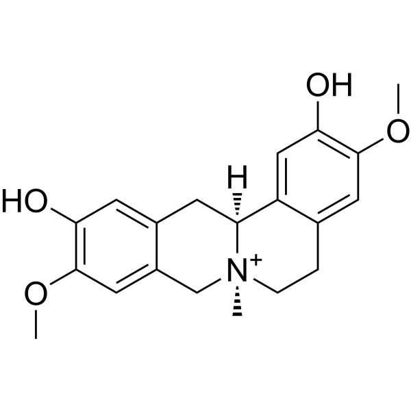
Phellodendrine
Based on 2 publication(s) in Google Scholar
Phellodendrine is an orally active plant alkaloid. Phellodendrine inhibits the proliferation of KRAS-mutated pancreatic cancer cells by suppressing macropinocytosis and glutamine metabolism, inducing ROS accumulation and mitochondrial apoptosis. Phellodendrine promotes autophagy by activating the AMPK/mTOR pathway, alleviating intestinal damage in ulcerative colitis. Phellodendrine can alleviate gouty arthritis by inhibiting the IL-6/STAT3 signaling pathway. Phellodendrine suppresses allergic reactions by altering the conformation of MRGPRB3/MRGPRX2 protein, thereby inhibiting the activation of PKC and subsequent downstream MAPK and NF-κB signaling. Phellodendrine inhibits the AKT/NF-κB pathway and down-regulates the expression of COX-2, thereby protecting zebrafish embryos from oxidative stress. Phellodendrine has an anti-major depressive disorder (MDD) effect by down-regulating CHRM1, HTR1A, and the PI3K/Akt signaling pathway.
Nos produits utilisent uniquement pour la recherche. Nous ne vendons pas aux patients.
- CAS No.: 6873-13-8
- Formule: C20H24NO4
- Masse moléculaire:342.41
-
Stockage:
4°C, protect from light
* In solvent : -80°C, 6 months; -20°C, 1 month (protect from light)

To place orders, for customer services and technical support, please contact: MedChemExpress USA
Tel: 609-228-6898 E-mail: [email protected] [email protected]


-
 Activité biologique
-
Chemical Information
-
Solvant et solubilité
- Protocole
- Pureté et documentation
- Références
-
 Help & FAQs


-
HY-15431
CapivasertibAkt Inhibitor -
HY-10425
Rizavasertibpan-Akt Inhibitor -
HY-101625
RecilisibRadioprotectant -
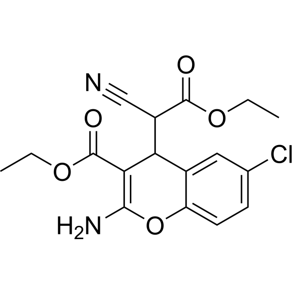
HY-18749
SC79Akt Activator -
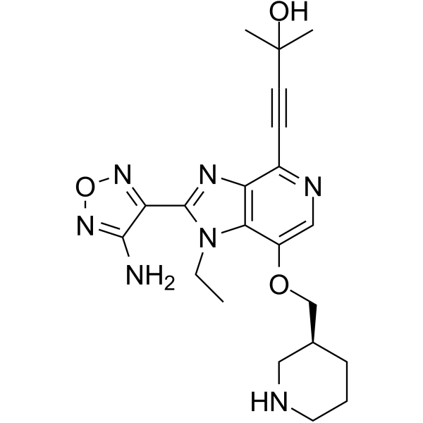
HY-10249
GSK-690693Akt Inhibitor -
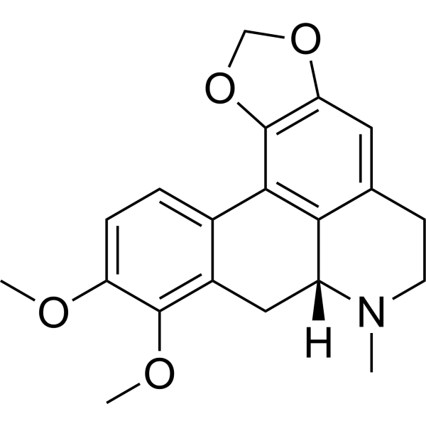
HY-N2255
CrebanineApoptosis Inducer -

HY-10425R
Rizavasertib (Standard)Reference Standard -
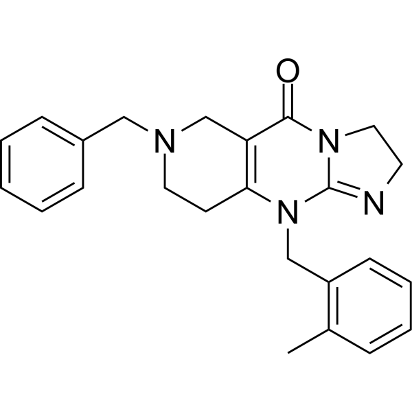
HY-15615
TIC10 isomerTIC10 Isomer -
HY-10358
MK-2206 dihydrochlorideAllosteric Akt Inhibitor -
HY-19962
PaxalisibPI3K/mTOR Inhibitor -
HY-B0094
ArtemisininParasite Inhibitor
Publications Citing Use of MedChemExpress (MCE) Phellodendrine
MoreVoir tous les produits spécifiques à Isoform AMPK
More

Activité biologique
Description
|
NF-κB |
PI3K |
mTOR |
MMP-3 |
COX-2 |
IL-1β |
STAT3 |
IL-6 |
In Vitro
Phellodendrine (0.6-80 μM, 24 h-14 d) significantly inhibits the proliferation and colony formation ability of KRAS-mutated PANC-1 and MiaPaCa-2 cells and its driven macropinocytosis, while showing no obvious toxicity to KRAS wild-type BxPC-3 cells and normal pancreatic cells HPDE6-c7[1].
Phellodendrine (10-40 μM, 12-24 h) diminishes the albumin (Alb) driven intracellular glutamine level, induces reactive oxygen species generation and causes mitochondrial membrane potential (MMP) depolarization in PANC-1 cells[1].
Phellodendrine (5-40 μM, 0-48 h) induces apoptosis cell death in PANC-1 cells through caspase-dependent mitochondrial intrinsic pathway[1].
Phellodendrine (5-20 μM) activates the p-AMPK/mTOR signalling pathway, as well as autophagy in Caco-2 cells stimulated by H2O2[2].
Phellodendrine (5 μM) attenuates Monosodium urate (MSU) (HY-B2130A)-induced MMP3 production and proteoglycan degradation by inhibiting IL-6/STAT3 pathway in rabbit primary chondrocytes and C28/I2 cells (human normal chondrocytes)[3].
Phellodendrine (0-50 μM) reduces the mRNA expression of MRGPRB3 and responsiveness of MRGPRX2 by altering its structure, is able to decrease Ca2+ levels, phosphorylation levels of CaMK, PLCβ1, PKC, ERK, JNK, p38, and p65, and inhibits the degradation of IκB-α in RBL-2H3 cells[4].
Phellodendrine (25-200 μg/mL, 37 h) increases the survival rate of zebrafish embryos and restores the abnormal heart rate levels[5].
Phellodendrine (50-200 μg/mL, 13 h) reduces the ROS level in zebrafish embryos and inhibits lipid peroxidation[5].
Phellodendrine (100 μg/mL) reverses the expression of AKT and NF-κB3, IKK, COX-2 which were abnormally changed by AAPH (HY-Y0525)-induced oxidative stress in zebrafish embryos[5].
Phellodendrine (2.5-160 μg/mL, 24-48 h) has an anti-MDD effect by regulating the mRNA levels of CHRM1, HTR1A and key targets of the PI3K/Akt signalling pathway (PI3K, Akt, mTOR) in PC12 cells[6].
MedChemExpress (MCE) has not independently confirmed the accuracy of these methods. They are for reference only.
-
Cell Line:PANC-1 cells
-
Concentration:10, 20 and 40 μM
-
Incubation Time:48 h
-
Result:Showed a higher apoptosis cells ratio both in early and late apoptosis.
Revealed a marked increase in the number of cells showing nuclear condensation and fragmentation.
-
Cell Line:PANC-1 cells
-
Concentration:5, 10 and 20 μM
-
Incubation Time:0, 6, 12, 24 h
-
Result:Induced the expressions of apoptotic proteins (cleaved caspase-3, 7, 9, PARP and Bax) and reduced the levels of Bcl-2 in time and dose dependent manner.
-
Cell Line:PC12 cells
-
Concentration:2.5, 5, 10, 20, 40, 80, 160 μg/mL
-
Incubation Time:24 and 48 h
-
Result:Reduced cells’ viability at all concentrations for 48 h.
-
Cell Line:PC12 cells
-
Concentration:5, 10, 20, 40, 80, 160 μg/mL
-
Incubation Time:24 h
-
Result:Decreased the IL-6 and IL-1β production.
-
Cell Line:PC12 cells
-
Concentration:5, 10, 20 μg/mL
-
Incubation Time:24 and 48 h
-
Result:Inhibited mRNA levels of CHRM1, HTR1A, PI3K and AKT.
Had no significant effect on the expression of mTOR.
In Vivo
Phellodendrine (30 mg/kg, p.o., once daily for 7 days) reduces the intestinal damage of ulcerative colitis (UC) in mice[2].
Phellodendrine (40 mg/kg, i.p., mice single dose or rats once daily for 5 days) alleviated Uric acid sodium (MSU) (HY-B2130A)-induced acute peritonitis in mice and arthritis in rats[3].
Phellodendrine (0.3-3 mg/kg, i.v.) protects against C48/80 (HY-130592)-induced foot swelling and Evans blue exudation in mice, and suppresses C48/80-induced RBL-2H3 rat basophilic leukemia cells degranulation, and β-HEX, HIS, IL-4, and TNF-α release[4].
MedChemExpress (MCE) has not independently confirmed the accuracy of these methods. They are for reference only.
-
Animal Model:PANC-1 xenograft model established in Balb/c nude mice (male, 7 weeks old)[1]
-
Dosage:30 and 60 mg/kg
-
Administration:Intraperitoneal injection (i.p.), once daily for 2 weeks
-
Result:Resulted in lower tumor weights and volumes.
Inhibited macropinocytosis in tumor tissues.
Reduced the proliferation activity of tumor cells.
-
Animal Model:DSS (HY-116282C) induced UC model established in female C57BL/6 mice (18-22 g)[2]
-
Dosage:30 mg/kg
-
Administration:Oral administration (p.o.), once daily for 7 days
-
Result:Significantly restored the length of the colon.
Significantly reduced the disease activity index (DAI) score and the pathological damage score.
-
Animal Model:MSU crystal-induced arthritis model established in male Sprague-Dawley (SD) rats with a mass of 240-270 g[3]
-
Dosage:40 mg/kg
-
Administration:Intraperitoneal injection (i.p.), once daily for 5 days
-
Result:Significantly reduced the degree of joint swelling in rats.
Reduced the inflammatory score and restored the loss of proteoglycans.
Significantly inhibited the expression of MMP-3.
-
Animal Model:MSU induced peritonitis model established in male C57BL/6 mice (18-20 g)[3]
-
Dosage:40 mg/kg
-
Administration:Intraperitoneal injection (i.p.), single dose
-
Result:Reduced proportion of neutrophils in the peritoneal lavage fluid.
-
Animal Model:C48/80-induced Hind paw swelling model established in adult male mice[4]
-
Dosage:0.3 and 3 mg/kg
-
Administration:Intravenously injection (i.v.)
-
Result:Was associated with reductions in both foot swelling and the degree of Evans blue exudation in mice.
suppressed C48/80-induced RBL-2H3 rat basophilic leukemia cells degranulation, and β-HEX, HIS, IL-4, and TNF-α release.
- RNA extraction experimental
- Annexin V plus membrane-impermeant dye apoptosis staining
- ROS/oxidative-stress fluorescent staining
- Research Protocol for Inflammation-related Diseases
- Cell Viability Determination by MTT Colorimetric Assay
- Collagen-Induced Arthritis
- Detection of Apoptosis
- TUNEL staining for apoptotic DNA fragmentation
- DSS-Induced Colitis
- TNBS-Induced Colitis
- Autophagy
- Apoptosis Solutions
- Lysosome and acidic-vesicle live-cell staining
- Macroautophagy Solutions


Chemical Information
-
CAS No. 6873-13-8
-
Appearance Solid
-
Masse moléculaire 342.41
-
Formule C20H24NO4
-
Color Off-white to light yellow
-
SMILES
OC1=C(OC)C=C2CC[N@@+]3(C)CC4=CC(OC)=C(O)C=C4C[C@@]3([H])C2=C1
-
Structure Classification
-
Initial Source
-
Livraison
Room temperature in continental US; may vary elsewhere.
-
Stockage
4°C, protect from light
* In solvent : -80°C, 6 months; -20°C, 1 month (protect from light)


Publications (2)
-
Journal Impact Factor
-
Most Recent
-
Int Immunopharmacol
Phellodendrine alleviates acute pancreatitis by inhibiting p38 MAPK-p47phox pathway-mediated neutrophil extracellular traps formation and ROS production. [Abstract]2025 Dec 10:167:115658. PMID: 41072078 -
Int Immunopharmacol
Phellodendrine ameliorates intestinal inflammation and protects mucosal barrier via modulating COL1A1, VCAM1 and IL-17 a. [Abstract]2025 Apr 16:152:114403. PMID: 40101527


Solvant et solubilité
In Vitro:
DMSO : 5 mg/mL (14.60 mM; Need ultrasonic; Hygroscopic DMSO has a significant impact on the solubility of product, please use newly opened DMSO)
Please refer to the solubility information to select the appropriate solvent. Once prepared, please aliquot and store the solution to prevent product inactivation from repeated freeze-thaw cycles.
Storage method and period of stock solution: -80°C, 6 months; -20°C, 1 month (protect from light). When stored at -80°C, please use it within 6 months. When stored at -20°C, please use it within 1 month.
Please refer to the solubility information to select the appropriate solvent. Once prepared, please aliquot and store the solution to prevent product inactivation from repeated freeze-thaw cycles.
Storage method and period of stock solution: -80°C, 6 months; -20°C, 1 month (protect from light). When stored at -80°C, please use it within 6 months. When stored at -20°C, please use it within 1 month.
Concentration (start) × Volume (start) = Concentration (final) × Volume (final)


Pureté et documentation
-
Fiche technique (291 KB)
-
SDS (252 KB)
- English - EN (252 KB)
- Français - FR (252 KB)
- Deutsch - DE (252 KB)
- Norwegian - NO (252 KB)
- Español - ES (252 KB)
- Swedish - SV (252 KB)
- Italian - IT (252 KB)
- Korean - KR (252 KB)
- Portuguese - PT (252 KB)
-
Instruction de manipulation (2659 KB)
Références
[1]. Thu PM, et al. Phellodendrine chloride suppresses proliferation of KRAS mutated pancreatic cancer cells through inhibition of nutrients uptake via macropinocytosis. Eur J Pharmacol. 2019 May 5;850:23-34. [Content Brief]
[2]. Su S, Wang X, Xi X, et al. Phellodendrine promotes autophagy by regulating the AMPK/mTOR pathway and treats ulcerative colitis. J Cell Mol Med. 2021;25(12):5707-5720. [Content Brief]
[3]. Qin YY, et al. Phellodendrine chloride alleviates gouty arthritis through IL-6/STAT3 signaling pathway. Immunobiology. 2025 Jul;230(4):152918. [Content Brief]
[4]. Wang J, et al. Inhibitory effect of phellodendrine on C48/80-induced allergic reaction in vitro and in vivo. Int Immunopharmacol. 2024 Jun 15;134:112256. [Content Brief]
[5]. Li L , et al. The defensive effect of phellodendrine against AAPH-induced oxidative stress through regulating the AKT/NF-κB pathway in zebrafish embryos. Life Sci. 2016 Jul 15;157:97-106. [Content Brief]
[6]. Hu L, et al. Network pharmacology combined with experimental verification for exploring the potential mechanism of phellodendrine against depression. Sci Rep. 2025 Jan 14;15(1):1958. [Content Brief]


Complete Stock Solution Preparation Table
Please refer to the solubility information to select the appropriate solvent. Once prepared, please aliquot and store the solution to prevent product inactivation from repeated freeze-thaw cycles.
Storage method and period of stock solution: -80°C, 6 months; -20°C, 1 month (protect from light). When stored at -80°C, please use it within 6 months. When stored at -20°C, please use it within 1 month.
| Optional Solvent | Concentration Solvent Mass | 1 mg | 5 mg | 10 mg | 25 mg |
|---|---|---|---|---|---|
| DMSO | 1 mM | 2.9205 mL | 14.6024 mL | 29.2048 mL | 73.0119 mL |
| 5 mM | 0.5841 mL | 2.9205 mL | 5.8410 mL | 14.6024 mL | |
| 10 mM | 0.2920 mL | 1.4602 mL | 2.9205 mL | 7.3012 mL |


Phellodendrine Related Classifications
HY-N0427 Related Classifications
 Powered by Bioz