



8-Gingerol
Based on 5 publication(s) in Google Scholar
8-Gingerol can be found in the rhizome of ginger (Z. officinale) and has oral bioactivity. It activates TRPV1, with an EC50 value of 5.0 µM. 8-Gingerol inhibits COX-2 and also suppresses the growth of H. pylori in vitro. Additionally, 8-Gingerol exhibits anticancer, antioxidant, and anti-inflammatory properties by inhibiting the epidermal growth factor receptor (EGFR) and modulating its downstream STAT3/ERK pathway to suppress the proliferation, migration, and invasion of colorectal cancer cells. 8-Gingerol also exerts immunosuppressive effects by inhibiting oxidative stress, inducing cell cycle arrest, promoting apoptosis, and regulating autophagy. Furthermore, 8-Gingerol has cardioprotective effects. 8-Gingerol is promising for research in the fields of cancer, infection, immunosuppression, and cardiovascular diseases.
For research use only. We do not sell to patients.
- Purity : 99.72%
- CAS No.: 23513-08-8
- Formula: C19H30O4
- Molecular Weight:322.44
-
Storage:
-20°C, sealed storage, away from moisture and light
* In solvent : -80°C, 6 months; -20°C, 1 month (sealed storage, away from moisture and light)

To place orders, for customer services and technical support, please contact: MedChemExpress USA
Tel: 609-228-6898 E-mail: [email protected] [email protected]


-
 Biological Activity
-
Chemical Information
-
Solvent & Solubility
- Protocols
- Purity & Documentation
- References
-
 Help & FAQs


-
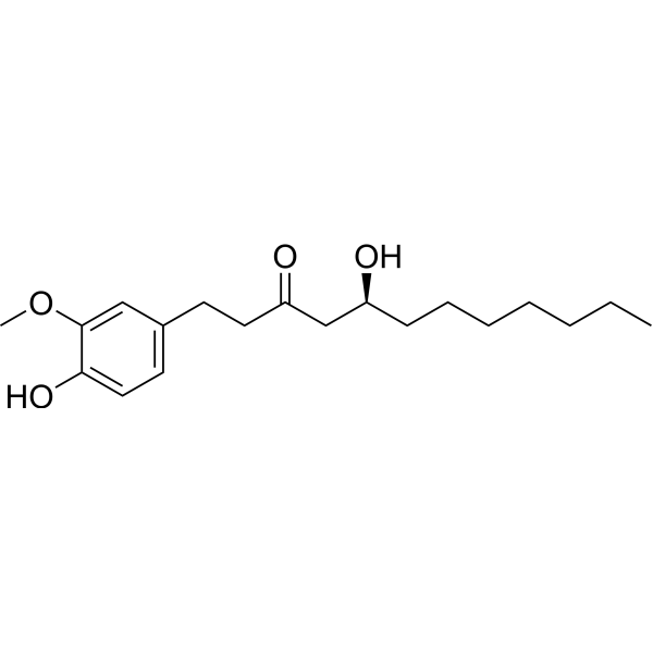
![[6]-Gingerol](https://file.medchemexpress.com/product_pic/hy-14615.gif)
HY-14615
[6]-GingerolApoptosis Inducer -
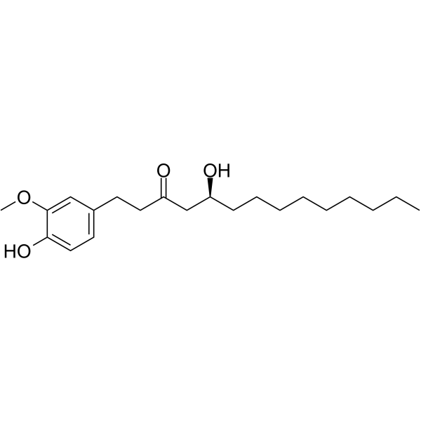
HY-N0448
10-GingerolAMPK Agonist -
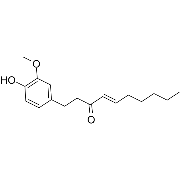
HY-14616
ShogaolAutophagy Inducer -
![[8]-Shogaol](https://file.medchemexpress.com/product_pic/hy-n2435.gif)
HY-N2435
[8]-ShogaolCOX Inhibitor -
![[10]-Shogaol](https://file.medchemexpress.com/product_pic/hy-n2434.gif)
HY-N2434
[10]-ShogaolAntioxidant -
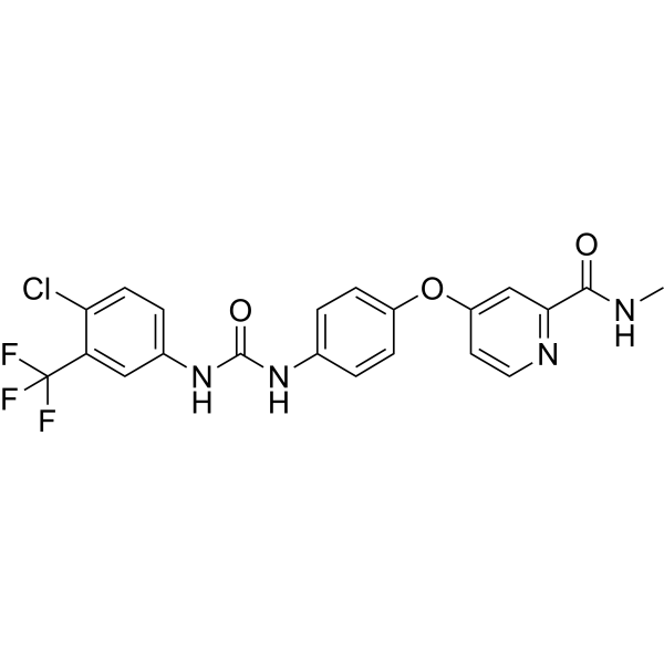
HY-10201
SorafenibMultikinase Inhibitor -
HY-15142
Doxorubicin hydrochlorideTopoisomerase Inhibitor -

HY-N0447R
8-Gingerol (Standard)Bacterial Inhibitor -
HY-111536
Mitochonic acid 5Active Compound -
HY-133865
Diacetoxy-6-gingerdiolGingerol -
HY-W020183
γ-TerpineneAntioxidant Compound -

HY-W020183R
γ-Terpinene (Standard)Antioxidant Compound
Publications Citing Use of MedChemExpress (MCE) 8-Gingerol
MoreAll EGFR Isoforms
MoreAll Caspase Isoforms
More

Biological Activity
Description
IC50 & Target
|
MMP-9 |
Caspase-9 |
In Vitro
8-Gingerol (0-70 µM, 24-72 hours) can inhibit the proliferation of HCT116 and DLD1 colorectal cancer cells, induce G0/G1 phase cell cycle arrest, and significantly promote apoptosis in HCT116 cells[3]. 8-Gingerol (0-70 µM, 48 hours) significantly inhibits the migration and invasion abilities of the cells, and its effect is dependent on the EGFR/STAT3/ERK pathway[3]. 8-Gingerol (100 µM, 48 hours) can reduce the effective concentration of 5-FU in HCT116 and DLD1 colorectal cancer cells, and in combination treatment, it may help reduce the toxicity of 5-FU[3]. 8-Gingerol (40, 80 µg/mL, 24 hours) significantly inhibits the LPS (HY-D1056) and Concanavalin A (HY-P2149) induced splenocyte proliferation, exhibiting immunosuppressive effects[4]. 8-Gingerol (10, 20 µM, 48 hours) inhibits autophagy and apoptosis in myocardial fibrosis by regulating the PI3K/Akt/mTOR signaling pathway, showing significant cardioprotective effects[5].
MedChemExpress (MCE) has not independently confirmed the accuracy of these methods. They are for reference only. Further protocols information, click here.
-
Cell Line:HCT116 (colorectal cancer), DLD1 (colorectal cancer)
-
Concentration:0, 10, 30, 50, 70 µM
-
Incubation Time:24h, 48h, 72h
-
Result:Dose-dependently inhibited HCT116 and DLD1 cell proliferation, with IC50 values of HCT116: 77.4 µM (48h), DLD1: 53.7 µM (48h)
-
Cell Line:HCT116 (colorectal cancer), DLD1 (colorectal cancer)
-
Concentration:0, 10, 30, 50, 70 µM
-
Incubation Time:48h
-
Result:Significantly induced G0/G1 cell cycle arrest in HCT116 and DLD1 cells.
-
Cell Line:HCT116 (colorectal cancer), DLD1 (colorectal cancer)
-
Concentration:0, 10, 30, 50, 70 µM
-
Incubation Time:48h
-
Result:Dose-dependently increased the apoptosis rate in HCT116 cells, but no significant change in DLD1 cells.
-
Cell Line:Cardiac cells from ISO-induced myocardial fibrosis mouse model
-
Concentration:10, 20 µM
-
Incubation Time:48h
-
Result:Significantly down-regulated the activity of MMP-9, Caspase-9, and Bax proteins, significantly up-regulated the activity of Bcl-2 protein, and inhibited apoptosis.
In Vivo
MedChemExpress (MCE) has not independently confirmed the accuracy of these methods. They are for reference only.
-
Animal Model:OVA-immunized mice (BALB/c male mice, 5 weeks old, 18-22g, immunized with ovalbumin (OVA))[4]
-
Dosage:25, 50, 100 mg/kg
-
Administration:Intraperitoneal injection (i.p.), once daily for 7 consecutive days
-
Result:Significantly suppressed LPS-, ConA-, and OVA-induced splenocyte proliferation in OVA-immunized mice and significantly reduced the percentage of CD19+ B cells and CD3+ T cells at high doses (50, 100 mg/kg)
-
Animal Model:Isoproterenol (ISO)-induced myocardial fibrosis (MF) mouse model, Kunming mice (male, 6-8 weeks old, 15-20g)[5]
-
Dosage:10 mg/kg (low dose), 20 mg/kg (high dose)
-
Administration:Intraperitoneal injection (i.p.), once daily for 14 days
-
Result:Significantly reduced the heart weight, left ventricle weight index, CK, LDH levels, ROS generation, and pathological heart damage induced by ISO.


Chemical Information
-
CAS No. 23513-08-8
-
Appearance <29°C Solid,>30°C Liquid
-
Molecular Weight 322.44
-
Formula C19H30O4
-
Color White to yellow
-
SMILES
CCCCCCC[C@H](O)CC(CCC1=CC=C(O)C(OC)=C1)=O
-
Structure Classification
-
Initial Source
-
Shipping
Room temperature in continental US; may vary elsewhere.
-
Storage
-20°C, sealed storage, away from moisture and light
* In solvent : -80°C, 6 months; -20°C, 1 month (sealed storage, away from moisture and light)


Publications (5)
-
Journal Impact Factor
-
Most Recent
-
Bioact Mater
Lipidomic analysis of plant-derived extracellular vesicles for guidance of potential anti-cancer therapy. [Abstract]2024 Dec 10:46:82-96. PMID: 39737211 -
Phytomedicine
Fangchinoline suppresses nasopharyngeal carcinoma progression by inhibiting SQLE to regulate the PI3K/AKT pathway dysregulation. [Abstract]2025 May:140:156484. PMID: 40090046 -
Food Chem
2024 Sep 15:452:139425. PMID: 38744128 -
Arch Pharm Res
Cartilage extracellular matrix regeneration with 10-gingerol via KEAP1-NRF2-ARE axis for osteoarthritis therapy. [Abstract]2025 Dec;48(11-12):1460-1481. PMID: 41212495 -
ChemMedChem
Machine Learning-Driven Discovery of Structurally Related Natural Products as Activators of the Cardiac Calcium Pump SERCA2a. [Abstract]2025 May 5;20(9):e202400913. PMID: 39853697


Solvent & Solubility
In Vitro:
DMSO : ≥ 100 mg/mL (310.14 mM; Hygroscopic DMSO has a significant impact on the solubility of product, please use newly opened DMSO)
* "≥" means soluble, but saturation unknown.
Please refer to the solubility information to select the appropriate solvent. Once prepared, please aliquot and store the solution to prevent product inactivation from repeated freeze-thaw cycles.
Storage method and period of stock solution: -80°C, 6 months; -20°C, 1 month (sealed storage, away from moisture and light). When stored at -80°C, please use it within 6 months. When stored at -20°C, please use it within 1 month.
Please refer to the solubility information to select the appropriate solvent. Once prepared, please aliquot and store the solution to prevent product inactivation from repeated freeze-thaw cycles.
Storage method and period of stock solution: -80°C, 6 months; -20°C, 1 month (sealed storage, away from moisture and light). When stored at -80°C, please use it within 6 months. When stored at -20°C, please use it within 1 month.
Concentration (start) × Volume (start) = Concentration (final) × Volume (final)
In Vivo:
Select the appropriate dissolution method based on your experimental animal and administration route.
- For the following dissolution methods, please ensure to first prepare a clear stock solution using an In Vitro approach and then sequentially add co-solvents:
- To ensure reliable experimental results, the clarified stock solution can be appropriately stored based on storage conditions. As for the working solution for In Vivo experiments, it is recommended to prepare freshly and use it on the same day.
- The percentages shown for the solvents indicate their volumetric ratio in the final prepared solution. If precipitation or phase separation occurs during preparation, heat and/or sonication can be used to aid dissolution.
Add each solvent one by one: 10% DMSO 40% PEG300 5% Tween-80 45% Saline
Solubility: ≥ 2.5 mg/mL (7.75 mM); Clear solution
This protocol yields a clear solution of ≥ 2.5 mg/mL (saturation unknown).
Taking 1 mL working solution as an example, add 100 μL DMSO stock solution (25.0 mg/mL) to 400 μL PEG300, and mix evenly; then add 50 μL Tween-80 and mix evenly; then add 450 μL Saline to adjust the volume to 1 mL.
Preparation of Saline: Dissolve 0.9 g sodium chloride in ddH₂O and dilute to 100 mL to obtain a clear Saline solution.
Add each solvent one by one: 10% DMSO 90% (20% SBE-β-CD in Saline)
Solubility: ≥ 2.5 mg/mL (7.75 mM); Clear solution
This protocol yields a clear solution of ≥ 2.5 mg/mL (saturation unknown).
Taking 1 mL working solution as an example, add 100 μL DMSO stock solution (25.0 mg/mL) to 900 μL 20% SBE-β-CD in Saline, and mix evenly.
Preparation of 20% SBE-β-CD in Saline (4°C, storage for one week): 2 g SBE-β-CD powder is dissolved in 10 mL Saline, completely dissolve until clear.
In Vivo Dissolution Calculator
Please enter the basic information of animal experiments:
-
-
-
-
Recommended: Prepare an additional quantity of animals to account for potential losses during experiments.
Please enter your animal formula composition:
-
%DMSO +
Recommended: Keep the proportion of DMSO in working solution below 2% if your animal is weak.
-
%+
-
+%Tween-80 + +
-
%Saline +
The co-solvents required include: DMSO, . All of co-solvents are available by MedChemExpress (MCE). , Tween 80. All of co-solvents are available by MedChemExpress (MCE).
Working solution concentration: 0.22 mg/mL
Method for preparing stock solution: mg drug dissolved in μL DMSO. Stock solution concentration: mg/mL. * In solvent : -80°C, 6 months; -20°C, 1 month (sealed storage, away from moisture and light)
1. Take μL DMSO stock solution;
2. Add μL .
μL , mix evenly;
3. Then add μL Tween 80, mix evenly;
4. Then add μL
Please ensure that the stock solution in the first step is dissolved to a clear state, and add co-solvents in sequence. You can use ultrasonic heating (ultrasonic cleaner, recommended frequency 20-40 kHz), vortexing, etc. to assist dissolution.

Protocols
-
Research Protocol for Infectious Diseases
Infectious-disease experiments test how pathogens interact with host barriers, innate immune receptors, inflammatory signaling, pathogen replication, and tissue injury; pattern-recognition receptors such as TLRs, RIG-I-like receptors, NOD-like receptors, and inflammasomes detect microbial molecules and activate NF-κB, interferon, and cytokine responses. The central hypothesis is that infection severity reflects the balance between pathogen burden and host response: protective inflammation restricts pathogen growth, whereas excessive or mislocalized inflammation contributes to tissue damage and disease phenotype. Unresolved questions include which host pathways are protective versus pathogenic, why some infection models fail to translate to human disease, and which combined readouts best predict clinically relevant infection outcomes.
-
Annexin V plus membrane-impermeant dye apoptosis staining
Annexin V-based apoptosis assays rely on the detection of phosphatidylserine (PS) externalization from the inner leaflet of the plasma membrane to the outer leaflet, an early biochemical hallmark of apoptosis. Fluorescently labeled Annexin V binds PS in a calcium-dependent manner, enabling identification of early apoptotic cells by flow cytometry or fluorescence microscopy. When combined with a membrane-impermeant DNA-binding dye (e. g. , propidium iodide), this approach allows discrimination between viable (Annexin V−/dye−), early apoptotic (Annexin V+/dye−), and late apoptotic or necrotic (Annexin V+/dye+) cell populations by assessing membrane integrity and PS exposure.
-
Cell migration
Cell migration is a method that plays an important role in wound healing, cell differentiation, embryonic development, etc.
-
ROS/oxidative-stress fluorescent staining
ROS/oxidative-stress fluorescent staining uses cell-permeant fluorogenic probes that become fluorescent after oxidation inside cells or tissues; commonly used examples include DCFH-DA/DCFDA for broad cellular oxidant detection, DHE for superoxide-related signal detection, MitoSOX for mitochondrial superoxide-related signal detection, and CellROX probes for oxidative-stress-associated fluorescence readouts. The assay detects probe oxidation rather than a single ROS species unless the probe and analysis method have been chemically validated for that species. DCFH-DA enters cells, is deacetylated by intracellular esterases to DCFH, and produces fluorescent DCF after oxidation, so the readout is used as an operational measure of total cellular oxidative stress rather than a species-specific ROS measurement. DHE and MitoSOX can report superoxide-related oxidation, but red fluorescence alone can include non-specific ethidium-like oxidation products; HPLC or optimized spectral approaches are
-
BrdU Incorporation Assay
Bromodeoxyuridine (BrdU) incorporation assay is based on the principle that BrdU, a thymidine analog, is incorporated into newly synthesized DNA during the S phase of the cell cycle, thereby serving as a marker of DNA replication and cellular proliferation. Incorporated BrdU can be detected using anti-BrdU antibodies following DNA denaturation, enabling visualization or quantification of proliferating cells through immunochemical detection methods such as immunofluorescence or immunohistochemistry.
-
Protocol for Cell Cycle
Cell-cycle analysis by flow cytometry measures DNA content in single cells to estimate the fraction of cells in G0/G1, S, and G2/M phases. Propidium iodide intercalates into DNA, and after RNA removal with RNase, fluorescence intensity reflects cellular DNA content: 2N cells are assigned to G0/G1, cells between 2N and 4N to S phase, and 4N cells to G2/M. DNA-content analysis alone cannot reliably separate G0 from G1 or G2 from M. Ki-67 can distinguish quiescent G0 cells from cycling cells, EdU or BrdU incorporation marks active DNA synthesis in S phase, and phospho-histone H3 staining identifies mitotic cells within the 4N population.
-
Research Protocol for Inflammation-related Diseases
The NLRP3 inflammasome is a cytosolic innate immune signaling platform that integrates priming signals and danger-signal activation to promote caspase-1 activation, maturation of IL-1β and IL-18, and gasdermin D-mediated pyroptotic cell death. The core experimental logic is to determine whether inflammatory disease phenotypes are driven by increased NLRP3 expression, ASC-containing inflammasome assembly, caspase-1 cleavage, GSDMD cleavage, and extracellular release of IL-1β/IL-18 rather than by nonspecific cell injury alone. The pathway is strongly linked to inflammation-related disease phenotypes because monosodium urate crystals activate NALP3/NLRP3 inflammasome signaling in gout-like crystal inflammation, cholesterol crystals activate NLRP3 inflammasomes in atherogenesis models, and DSS-induced intestinal inflammation has been reported to involve NLRP3 inflammasome activity. However, experimental colitis studies also show context-dependent protective effects of NLRP3 inflammasome co
-
Cell Viability Determination by MTT Colorimetric Assay
The following protocol uses the MTT colorimetric assay as a classic literature-established method for assessing cell viability/metabolic activity in cultured mammalian cells. MTT[3-(4,5-dimethylthiazol-2-yl)-2,5-diphenyltetrazolium bromide] is reduced by metabolically active cells to a colored formazan product; the amount of formazan is quantified spectrophotometrically and provides an indirect measure of metabolically active viable cells. Importantly, MTT reduction reflects cellular oxidoreductase/metabolic activity rather than an absolute direct count of living cells, so changes in cellular metabolism can alter the signal independently of cell number.
-
Apoptosis
Apoptosis, also called programmed cell death, is generally characterized by distinct morphological characteristics.
-
TUNEL staining for apoptotic DNA fragmentation
TUNEL staining detects DNA strand breaks by using terminal deoxynucleotidyl transferase to add labeled nucleotides to exposed 3′-OH DNA termini, generating either microscopic staining in fixed cells or tissue sections, or fluorescence/cytometric signal in cell suspensions. TUNEL positivity reflects DNA fragmentation but should not be interpreted alone as definitive apoptosis, because TUNEL can also label necrotic, autolytic, mechanically damaged, or DNA-repair-associated DNA breaks.
-
Flow cytometric DNA-content cell-cycle staining
Flow cytometric DNA-content cell-cycle staining measures the fluorescence intensity of DNA-bound fluorochromes in single cells or nuclei to estimate DNA content distributions, allowing assignment of populations to G0/G1, S, and G2/M phases by DNA histogram deconvolution. Propidium iodide (PI) intercalates into DNA, and PI fluorescence is proportional to cellular DNA content when staining is performed under conditions that make DNA accessible and minimize non-DNA signal. Cells with G2/M DNA content are expected to show approximately twice the fluorescence intensity of G0/G1 cells, while S-phase cells occupy intermediate fluorescence values. PI-based DNA-content analysis can also detect cells with fractional DNA content, often reported as sub-G1, when DNA fragmentation and extraction during staining reduce retained DNA signal in apoptotic cells. DAPI is an alternative DNA fluorochrome for univariate DNA-content analysis, while bivariate approaches combining DNA content with proliferation
-
Autophagy
Autophagy is a process in which eukaryotic cells use lysosomes to degrade their own cytoplasmic proteins and damaged organelles under the regulation of autophagy related gene (Atg). Microtubule-associated proteins light chain 3 (LC3) is recognized as autophagy marker, which transfers from cytoplasmic LC3 (LC3-I) to membrane type (LC3-II). LC3-II/I ratio could be detected by Western Blot and fluorescence microscopy.
-
Apoptosis Solutions
Apoptosis is a regulated, generally non-lytic cell-death pathway that removes unwanted, damaged, infected, or abnormal cells through coordinated morphological changes, caspase activation, DNA fragmentation, and membrane remodeling. The intrinsic apoptosis pathway is controlled mainly by mitochondrial outer membrane permeabilization, BCL-2 family proteins, cytochrome c release, apoptosome formation, caspase-9 activation, and downstream executioner caspase-3/7 activation. The extrinsic apoptosis pathway is initiated by death receptors such as Fas, TNFR, and TRAIL receptors, which recruit adaptor proteins and activate caspase-8 before engaging executioner caspases or mitochondrial amplification through BID cleavage. Apoptosis is linked to many phenotypes, including cancer cell killing, tissue homeostasis, immune regulation, neurodegeneration, infection response, and treatment-induced cytotoxicity; unresolved questions include how apoptosis interacts with necroptosis, pyroptosis, ferroptos
-
Cell invasion
Cell invasion is the ability of cells to migrate from one area to another via the extracellular matrix. Cell invasion is the response of normal and cancer cells to chemical and mechanical stimuli. Before migrating to a new region, the extracellular matrix is degraded by proteases within the cell. Cell invasion often occurs during wound repair, vascularization and inflammation, abnormal tissue invasion, and tumor cell metastasis.
-
Lysosome and acidic-vesicle live-cell staining
Lysosome and acidic-vesicle live-cell staining detects acidic intracellular compartments by using membrane-permeant acidotropic probes that accumulate in low-pH vesicles, including lysosomes, late endosomes, autolysosomes, and acidic phagosomes. LysoTracker staining is commonly used as an intensity-based readout of acidic lysosomal compartment abundance or enlargement, while acridine orange produces green fluorescence in less concentrated compartments and red fluorescence after concentration-dependent accumulation in acidic vesicular organelles. Loss or reduction of acridine-orange red signal can be used as a readout of lysosomal membrane permeabilization or reduced acidic-vesicle integrity. This protocol is designed for live cultured cells and can be adapted for fluorescence microscopy, high-content imaging, plate-reader readout, or flow cytometry when the selected literature supports the readout. Because these dyes report acidotropic accumulation rather than lysosome identity alone,
-
Macroautophagy Solutions
Macroautophagy is a conserved lysosome-dependent degradation pathway in which cytoplasmic material is sequestered into double-membrane autophagosomes and delivered to lysosomes for degradation and recycling. The pathway supports cellular homeostasis during nutrient limitation, organelle stress, protein-aggregate accumulation, infection, differentiation, and tissue remodeling by coupling cargo sequestration, autophagosome maturation, lysosomal fusion, and degradation of cargo-derived macromolecules. The core molecular sequence includes initiation by nutrient- and stress-regulated autophagy machinery, autophagosome nucleation, LC3/ATG8-family conjugation to autophagosomal membranes, cargo selection through receptors such as SQSTM1/p62, autophagosome-lysosome fusion, and lysosomal degradation. LC3 was identified as a mammalian homolog of yeast Atg8 that localizes to autophagosomal membranes after processing, and p62/SQSTM1 was shown to connect ubiquitinated cargo with autophagic degradati


Purity & Documentation
-
Data Sheet (280 KB)
-
SDS (394 KB)
- English - EN (394 KB)
- Français - FR (394 KB)
- Deutsch - DE (394 KB)
- Norwegian - NO (394 KB)
- Español - ES (394 KB)
- Swedish - SV (394 KB)
- Italian - IT (394 KB)
- Korean - KR (394 KB)
- Portuguese - PT (394 KB)
-
Handling Instructions (2659 KB)
References
[1]. Dedov, V.N., et al. Gingerols: a novel class of vanilloid receptor (VR1) agonists. Br. J. Pharmacol. 137(6), 793-798 (2002). [Content Brief]
[2]. Chrubasik, S., et al. Zingiberis rhizoma: A comprehensive review on the ginger effect and efficacy profiles. Phytomedicine 12(9), 684-701 (2005). [Content Brief]
[3]. Hu SM, et al. 8‑Gingerol regulates colorectal cancer cell proliferation and migration through the EGFR/STAT/ERK pathway. Int J Oncol. 2020 Jan;56(1):390-397. [Content Brief]
[4]. Lu J, et al. Immunosuppressive activity of 8-gingerol on immune responses in mice. Molecules. 2011 Mar 22;16(3):2636-45. [Content Brief]
[5]. Xue Y, et al. 8-Gingerol Ameliorates Myocardial Fibrosis by Attenuating Reactive Oxygen Species, Apoptosis, and Autophagy via the PI3K/Akt/mTOR Signaling Pathway. Front Pharmacol. 2021 Jul 28;12:711701. [Content Brief]


Complete Stock Solution Preparation Table
Please refer to the solubility information to select the appropriate solvent. Once prepared, please aliquot and store the solution to prevent product inactivation from repeated freeze-thaw cycles.
Storage method and period of stock solution: -80°C, 6 months; -20°C, 1 month (sealed storage, away from moisture and light). When stored at -80°C, please use it within 6 months. When stored at -20°C, please use it within 1 month.
| Optional Solvent | Concentration Solvent Mass | 1 mg | 5 mg | 10 mg | 25 mg |
|---|---|---|---|---|---|
| DMSO | 1 mM | 3.1014 mL | 15.5068 mL | 31.0135 mL | 77.5338 mL |
| 5 mM | 0.6203 mL | 3.1014 mL | 6.2027 mL | 15.5068 mL | |
| 10 mM | 0.3101 mL | 1.5507 mL | 3.1014 mL | 7.7534 mL | |
| 15 mM | 0.2068 mL | 1.0338 mL | 2.0676 mL | 5.1689 mL | |
| 20 mM | 0.1551 mL | 0.7753 mL | 1.5507 mL | 3.8767 mL | |
| 25 mM | 0.1241 mL | 0.6203 mL | 1.2405 mL | 3.1014 mL | |
| 30 mM | 0.1034 mL | 0.5169 mL | 1.0338 mL | 2.5845 mL | |
| 40 mM | 0.0775 mL | 0.3877 mL | 0.7753 mL | 1.9383 mL | |
| 50 mM | 0.0620 mL | 0.3101 mL | 0.6203 mL | 1.5507 mL | |
| 60 mM | 0.0517 mL | 0.2584 mL | 0.5169 mL | 1.2922 mL | |
| 80 mM | 0.0388 mL | 0.1938 mL | 0.3877 mL | 0.9692 mL | |
| 100 mM | 0.0310 mL | 0.1551 mL | 0.3101 mL | 0.7753 mL |


8-Gingerol Related Classifications
HY-N0447 Related Classifications
 Powered by Bioz