



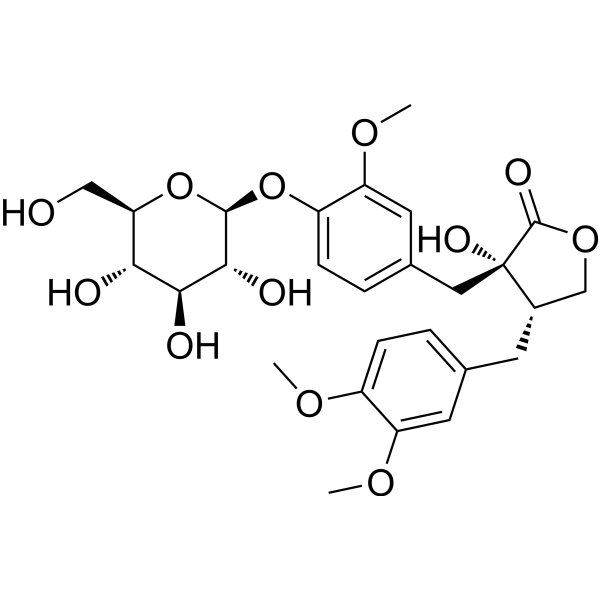
Tracheloside

Based on 1 Customer Validation
Tracheloside is an orally active anticancer agent. Tracheloside can be extracted from Trachelospermum jasminoides. Tracheloside blocks TMEM16A currents, inhibits TMEM16B, and does not inhibit Bestrophin or CFTR. Tracheloside activates AMPK, inhibits NOX4, reduces Smad3 phosphorylation/nuclear translocation, and promotes YAP1 phosphorylation/degradation. Tracheloside reduces ROS/NO production, iNOS, IL-17, JNK/p38 MAPK, MMP-2/9, Bcl-2/Bcl-xL, and cyclin D1/CDK4, while increasing Nrf2, Bax, p16, Apoptosis, and cell cycle arrest. Tracheloside can be used for research on colorectal cancer, pulmonary fibrosis, full-thickness excisional wounds, lung adenocarcinoma, and rheumatoid arthritis.
For research use only. We do not sell to patients.
- Purity : 99.91%
- CAS No.: 33464-71-0
- Formula: C27H34O12
- Molecular Weight:550.55
-
Storage:
4°C, protect from light
* In solvent : -80°C, 6 months; -20°C, 1 month (protect from light)

To place orders, for customer services and technical support, please contact: MedChemExpress USA
Tel: 609-228-6898 E-mail: [email protected] [email protected]


-
 Biological Activity
-
Chemical Information
-
Solvent & Solubility
- Protocols
- Purity & Documentation
- References
-
 Help & FAQs




Biological Activity
Description
IC50 & Target
[1]|
Bcl-2 |
Bcl-xL |
Bax |
iNOS |
IL-17 |
MMP-2 |
MMP-9 |
Cdk4/cyclin D1 |
Cellular Effect
|
Cell Line

|
Type | Value | Description | References |
|---|---|---|---|---|
| HEK293 | IC50 |
3.09 μM
|
Inhibition of TMEM16A whole-cell currents in HEK293 cells by whole-cell patch clamp.
Inhibition of TMEM16A whole-cell currents in HEK293 cells by whole-cell patch clamp.
|
39038568 |
In Vitro
Tracheloside (0.25-1 μM; 48 h) promotes E-cadherin and inhibits N-cadherin, vimentin, snail, and twist mRNA in CT26 colorectal cancer cells[1].
Tracheloside (0.25-1 μM) increases E-cadherin protein and decreases N-cadherin, vimentin, snail, and twist proteins in CT26 colorectal cancer cells[1].
Tracheloside (50 μM) maintains YFP fluorescence in TMEM16A-expressing LA795 cells by preventing channel activation-induced quenching[5].
Tracheloside (0.1-30 μM) inhibits TMEM16A currents in LA795 cells in a concentration-dependent manner, with an IC50 of 3.09 μM[5].
Tracheloside (10-100 μM; 48 h) decreases cyclin D1 and CDK4 mRNA and increases p16 mRNA in CT26 colorectal cancer cells[1].
MedChemExpress (MCE) has not independently confirmed the accuracy of these methods. They are for reference only.
-
Cell Line:RBL-2H3 rat mast cells
-
Concentration:1, 10, and 100 µM
-
Incubation Time:96 h
-
Result:No cytotoxicity was observed in RBL-2H3 cells after 96 h at 1, 10, and 100 µM.
-
Cell Line:RAW264.7 mouse macrophage cells
-
Concentration:1, 10, and 100 µM; 0.5 mg/mL (MTT)
-
Incubation Time:24 h; 4 h (MTT)
-
Result:No cytotoxicity was observed in RAW264.7 macrophage cells after 24 h at 1, 10, and 100 µM.
-
Cell Line:CT26 murine colorectal cancer cells
-
Concentration:10, 25, 50, and 100 µM
-
Incubation Time:48 h
-
Result:Early apoptosis was 2.1% and late apoptosis/dead was 7.9% at 10 µM.
Early apoptosis was 7.3% and late apoptosis/dead was 8.2% at 50 µM.
Early apoptosis was 9.2% and late apoptosis/dead was 8.9% at 100 µM.
Apoptosis of CT26 cells increased in a dose-dependent manner.
-
Cell Line:CT26 murine colorectal cancer cells
-
Concentration:10, 25, 50, and 100 µM
-
Incubation Time:48 h
-
Result:Cell cycle phase distributions were: 10 µM, G0/G1 51.9%, S 19.0%, G2/M 28.1%; 25 µM, G0/G1 48.2%, S 23.2%, G2/M 28.3%; 50 µM, G0/G1 58.8%, S 24.0%, G2/M 16.7%; 100 µM, G0/G1 58.0%, S 17.0%, G2/M 24.7%.
The G0/G1 phase increased at concentrations above 50 µM, with 58.8% at 50 µM.
-
Cell Line:CT26 murine colorectal cancer cells
-
Concentration:10, 25, 50, and 100 µM
-
Incubation Time:48 h
-
Result:mRNA expression of cyclin D1 and CDK4 decreased and p16 mRNA expression increased in CT26 cells.
Parmacokinetics
| Species | Dose | Route | T1/2α | Tmax | Cmax | AUC0-t | AUC0-∞ | Clearance (CL) | MRT |
|---|---|---|---|---|---|---|---|---|---|
| Mice[5] | 30 μM | i.g. | 6.39 h | 2.92 h | 1144.05 ng/mL | 9437.51 ng·h/mL | 10943.26 ng·h/mL | 0.0089 ng/mL | 7.56 h |
In Vivo
Tracheloside (0.02-0.2%; diet; ad libitum; during the 8-week PhIP initiation period) does not significantly inhibit mammary carcinogenesis in female SD rats[7].
Tracheloside (0.02-0.2%; diet; ad libitum; for 44 weeks after the 8-week PhIP exposure) does not show a significant inhibitory effect on PhIP-induced mammary carcinogenesis in female SD rats[7].
MedChemExpress (MCE) has not independently confirmed the accuracy of these methods. They are for reference only.
-
Animal Model:BALB/c (female, four weeks old, 16-18 g body weight)[1]
-
Dosage:25 mg/kg; 50 mg/kg
-
Administration:once daily
-
Result:Dose-dependently reduced lung weight and the number of tumor nodules in the lung.
In lung tissue, cleaved caspase-3, cleaved caspase-9, cleaved PARP, and Bax were increased, while Bcl-2 and Bcl-xL were decreased.
E-cadherin mRNA expression increased and N-cadherin, vimentin, snail, and twist mRNA expression decreased.
-
Animal Model:C57BL/6 (6-8 weeks old)[2]
-
Dosage:1 mg/kg; 5 mg/kg; 25 mg/kg
-
Administration:i.p.; daily; days 15-28
-
Result:Reduced mortality and Ashcroft fibrosis score, decreased alveolar wall thickening and extracellular matrix deposition, with optimal effects at 25 mg/kg, and lowered hydroxyproline levels.
Suppressed bleomycin-induced increases in collagen I and α-SMA protein/mRNA and CTGF/ANKRD1 mRNA.
Reduced DHE staining and MDA, counteracted bleomycin-induced changes in GSH and SOD activity, decreased NOX4, and increased CAT, SOD1, and SOD2.
-
Animal Model:C57BL/6 (6-8 weeks old)[2]
-
Dosage:25 mg/kg
-
Administration:i.p.; daily; days 15-28
-
Result:In bleomycin-induced mice, activated AMPK in vivo, increasing p-AMPK and p-ACC.
Reduced Ashcroft score, hydroxyproline, CTGF, ANKRD1, collagen I, and α-SMA, and reduced MDA and NOX4 while increasing SOD, GSH, CAT, SOD1, and SOD2.
Compound C co-administration attenuated anti-fibrotic and anti-oxidative effects, including effects on fibrosis scores, survival metrics, hydroxyproline, fibrotic gene/protein expression, and oxidative stress markers.\nProduced anti-fibrotic efficacy comparable to pirfenidone in the bleomycin-induced pulmonary fibrosis model.
-
Animal Model:Sprague-Dawley (male, 12 weeks old)[4]
-
Dosage:10 µg/mL
-
Administration:Topical; single application at day 0
-
Result:Achieved 76.51% closure at week 1 and 90.74% closure at week 2.
Relative wound area was 23.48% at week 1 and 10.25% at week 2.
At 2 weeks, histology showed complete hydrogel degradation without visible necrosis or inflammatory reaction, dense well-aligned collagen fibers, a thick continuous epidermal layer, and hair follicles and sebaceous glands.
Western blot showed higher Col-1A at weeks 1 and 2, higher elastin at week 1 with the trend persisting at week 2, and higher α-SMA at week 2.
Immunohistochemistry showed higher α-SMA, CD31, and CD34 positive area; at week 2, CCR7 was lower and CD206 was higher, fibronectin was lower, and Col-1A remained higher.
RNA-seq showed 920 uniquely expressed genes, with 1968 genes upregulated, 1333 downregulated, and 1731 unchanged, including enrichment of angiogenesis, wound healing, skin epidermis development, and ERK1/2-related genes.
ERK1/2 expression remained elevated at weeks 1 and 2, while p-ERK1/2 increased at week 1 and declined by week 2.
-
Animal Model:BALB/c (6- to 8-week-old)[5]
-
Dosage:16.5 mg/kg (about 30 μM) (alone); 8.25 mg/kg (about 15 μM) plus doxorubicin 4.1 mg/kg (about 7.5 μM) (combination)
-
Administration:intragastric; six administrations
-
Result:In the 16.5 mg/kg single-agent group, the final tumor volume inhibition rate was 52.05% and the tumor weight inhibition rate was 40.54%.
In the 8.25 mg/kg plus doxorubicin 4.1 mg/kg combination group, the final tumor volume inhibition rate was 77.81% and the tumor weight inhibition rate was 81.08%.
-
Animal Model:Crj:CD(SD) SPF (Sprague-Dawley) (female; 5 weeks old at arrival; 6 weeks old at PhIP initiation)[7]
-
Dosage:0.02% (initiation); 0.2% (initiation)
-
Administration:powdered diet; ad libitum; during the 8-week PhIP initiation period
-
Result:In group 2 (0.02%, initiation; 20 rats), fibroadenoma incidence was 3 (15%) with multiplicity 0.15 no./rat, carcinoma incidence was 6 (30%) with multiplicity 0.60 no./rat, and total mammary tumor incidence was 9 (45%) with multiplicity 0.75 no./rat.
Total tumors numbered 15, with tumor volume 5.8 cm3/rat and 7.7 cm3/tumor.
In group 3 (0.2%, initiation; 20 rats), fibroadenoma incidence was 1 (5%) with multiplicity 0.05 no./rat, carcinoma incidence was 6 (30%) with multiplicity 0.55 no./rat, and total mammary tumor incidence was 7 (35%) with multiplicity 0.60 no./rat.
Total tumors numbered 12, with tumor volume 3.1 cm3/rat and 5.2 cm3/tumor.
No significant inter-group differences were observed in incidences or multiplicities of fibroadenomas and carcinomas.
-
Animal Model:Crj:CD(SD) SPF (Sprague-Dawley) (female; 5 weeks old at arrival; 6 weeks old at PhIP initiation)[7]
-
Dosage:0.02% (post-initiation); 0.2% (post-initiation)
-
Administration:powdered diet; ad libitum; for 44 weeks after the 8-week PhIP exposure
-
Result:In group 4 (0.02%, post-initiation; 20 rats), fibroadenoma incidence was 3 (15%) with multiplicity 0.15 no./rat, carcinoma incidence was 7 (35%) with multiplicity 0.90 no./rat, and total mammary tumor incidence was 9 (45%) with multiplicity 1.05 no./rat.
Total tumors numbered 21, with tumor volume 2.0 cm3/rat and 1.9 cm3/tumor.
In group 5 (0.2%, post-initiation; 20 rats), fibroadenoma incidence was 1 (5%) with multiplicity 0.10 no./rat, carcinoma incidence was 11 (55%) with multiplicity 0.90 no./rat, and total mammary tumor incidence was 11 (55%) with multiplicity 1.00 no./rat.
Total tumors numbered 20, with tumor volume 18.3 cm3/rat and 18.3 cm3/tumor.
No significant inter-group differences were observed in incidences or multiplicities of fibroadenomas and carcinomas.
- Western Blot
- Collagen: Sirius Red Staining
- Mitochondrial membrane-potential fluorescent assay
- Annexin V plus membrane-impermeant dye apoptosis staining
- ROS/oxidative-stress fluorescent staining
- BrdU Incorporation Assay
- Connective Tissue: Masson's Trichrome/Collagen Trichrome Staining
- Protocol for Cell Cycle
- Kinase activity and phosphorylation assays
- Fibrosis/Collagen Morphometry
- Cytoplasmic-Nuclear Fractionated Protein Extraction
- Collagen-Induced Arthritis
- Detection of Apoptosis
- TUNEL staining for apoptotic DNA fragmentation
- Flow cytometric DNA-content cell-cycle staining
- Mitochondrial membrane-potential and mitochondrial mass staining
- Apoptosis Solutions
- Protocol for Kinase activity and phosphorylation assays


Chemical Information
-
CAS No. 33464-71-0
-
Appearance Solid
-
Molecular Weight 550.55
-
Formula C27H34O12
-
Color White to off-white
-
SMILES
O=C1OC[C@H](CC2=CC=C(OC)C(OC)=C2)[C@]1(CC3=CC=C(O[C@H]4[C@@H]([C@H]([C@@H]([C@@H](CO)O4)O)O)O)C(OC)=C3)O
-
Structure Classification
-
Initial Source
-
Shipping
Room temperature in continental US; may vary elsewhere.
-
Storage
4°C, protect from light
* In solvent : -80°C, 6 months; -20°C, 1 month (protect from light)


Solvent & Solubility
In Vitro:
DMSO : 100 mg/mL (181.64 mM; Need ultrasonic; Hygroscopic DMSO has a significant impact on the solubility of product, please use newly opened DMSO)
Please refer to the solubility information to select the appropriate solvent. Once prepared, please aliquot and store the solution to prevent product inactivation from repeated freeze-thaw cycles.
Storage method and period of stock solution: -80°C, 6 months; -20°C, 1 month (protect from light). When stored at -80°C, please use it within 6 months. When stored at -20°C, please use it within 1 month.
Please refer to the solubility information to select the appropriate solvent. Once prepared, please aliquot and store the solution to prevent product inactivation from repeated freeze-thaw cycles.
Storage method and period of stock solution: -80°C, 6 months; -20°C, 1 month (protect from light). When stored at -80°C, please use it within 6 months. When stored at -20°C, please use it within 1 month.
Concentration (start) × Volume (start) = Concentration (final) × Volume (final)
In Vivo:
Select the appropriate dissolution method based on your experimental animal and administration route.
- For the following dissolution methods, please ensure to first prepare a clear stock solution using an In Vitro approach and then sequentially add co-solvents:
- To ensure reliable experimental results, the clarified stock solution can be appropriately stored based on storage conditions. As for the working solution for In Vivo experiments, it is recommended to prepare freshly and use it on the same day.
- The percentages shown for the solvents indicate their volumetric ratio in the final prepared solution. If precipitation or phase separation occurs during preparation, heat and/or sonication can be used to aid dissolution.
Add each solvent one by one: 10% DMSO 40% PEG300 5% Tween-80 45% Saline
Solubility: ≥ 2.5 mg/mL (4.54 mM); Clear solution
This protocol yields a clear solution of ≥ 2.5 mg/mL (saturation unknown).
Taking 1 mL working solution as an example, add 100 μL DMSO stock solution (25.0 mg/mL) to 400 μL PEG300, and mix evenly; then add 50 μL Tween-80 and mix evenly; then add 450 μL Saline to adjust the volume to 1 mL.
Preparation of Saline: Dissolve 0.9 g sodium chloride in ddH₂O and dilute to 100 mL to obtain a clear Saline solution.
Add each solvent one by one: 10% DMSO 90% (20% SBE-β-CD in Saline)
Solubility: ≥ 2.5 mg/mL (4.54 mM); Clear solution
This protocol yields a clear solution of ≥ 2.5 mg/mL (saturation unknown).
Taking 1 mL working solution as an example, add 100 μL DMSO stock solution (25.0 mg/mL) to 900 μL 20% SBE-β-CD in Saline, and mix evenly.
Preparation of 20% SBE-β-CD in Saline (4°C, storage for one week): 2 g SBE-β-CD powder is dissolved in 10 mL Saline, completely dissolve until clear.
In Vivo Dissolution Calculator
Please enter the basic information of animal experiments:
-
-
-
-
Recommended: Prepare an additional quantity of animals to account for potential losses during experiments.
Please enter your animal formula composition:
-
%DMSO +
Recommended: Keep the proportion of DMSO in working solution below 2% if your animal is weak.
-
%+
-
+%Tween-80 + +
-
%Saline +
The co-solvents required include: DMSO, . All of co-solvents are available by MedChemExpress (MCE). , Tween 80. All of co-solvents are available by MedChemExpress (MCE).
Working solution concentration: 0.22 mg/mL
Method for preparing stock solution: mg drug dissolved in μL DMSO. Stock solution concentration: mg/mL. * In solvent : -80°C, 6 months; -20°C, 1 month (protect from light)
1. Take μL DMSO stock solution;
2. Add μL .
μL , mix evenly;
3. Then add μL Tween 80, mix evenly;
4. Then add μL
Please ensure that the stock solution in the first step is dissolved to a clear state, and add co-solvents in sequence. You can use ultrasonic heating (ultrasonic cleaner, recommended frequency 20-40 kHz), vortexing, etc. to assist dissolution.


Purity & Documentation
-
Data Sheet (317 KB)
-
SDS (393 KB)
- English - EN (393 KB)
- Français - FR (393 KB)
- Deutsch - DE (393 KB)
- Norwegian - NO (393 KB)
- Español - ES (393 KB)
- Swedish - SV (393 KB)
- Italian - IT (393 KB)
- Korean - KR (393 KB)
- Portuguese - PT (393 KB)
-
Handling Instructions (2659 KB)
References


Complete Stock Solution Preparation Table
Please refer to the solubility information to select the appropriate solvent. Once prepared, please aliquot and store the solution to prevent product inactivation from repeated freeze-thaw cycles.
Storage method and period of stock solution: -80°C, 6 months; -20°C, 1 month (protect from light). When stored at -80°C, please use it within 6 months. When stored at -20°C, please use it within 1 month.
| Optional Solvent | Concentration Solvent Mass | 1 mg | 5 mg | 10 mg | 25 mg |
|---|---|---|---|---|---|
| DMSO | 1 mM | 1.8164 mL | 9.0818 mL | 18.1637 mL | 45.4091 mL |
| 5 mM | 0.3633 mL | 1.8164 mL | 3.6327 mL | 9.0818 mL | |
| 10 mM | 0.1816 mL | 0.9082 mL | 1.8164 mL | 4.5409 mL | |
| 15 mM | 0.1211 mL | 0.6055 mL | 1.2109 mL | 3.0273 mL | |
| 20 mM | 0.0908 mL | 0.4541 mL | 0.9082 mL | 2.2705 mL | |
| 25 mM | 0.0727 mL | 0.3633 mL | 0.7265 mL | 1.8164 mL | |
| 30 mM | 0.0605 mL | 0.3027 mL | 0.6055 mL | 1.5136 mL | |
| 40 mM | 0.0454 mL | 0.2270 mL | 0.4541 mL | 1.1352 mL | |
| 50 mM | 0.0363 mL | 0.1816 mL | 0.3633 mL | 0.9082 mL | |
| 60 mM | 0.0303 mL | 0.1514 mL | 0.3027 mL | 0.7568 mL | |
| 80 mM | 0.0227 mL | 0.1135 mL | 0.2270 mL | 0.5676 mL | |
| 100 mM | 0.0182 mL | 0.0908 mL | 0.1816 mL | 0.4541 mL |


Tracheloside Related Classifications
HY-N1507 Related Classifications
 Powered by Bioz
Keywords
- Tracheloside
- 33464-71-0
- Chloride Channel
- AMPK
- NADPH Oxidase
- TGF-beta/Smad
- YAP
- Reactive Oxygen Species (ROS)
- NO Synthase
- Interleukin Related
- JNK
- p38 MAPK
- MMP
- Bcl-2 Family
- CDK
- Keap1-Nrf2
- Apoptosis
- Cl− Channels
- AMP-activated protein kinase
- NOX
- Transforming growth factor beta
- Yes-associated protein
- Nitric oxide synthases
- NOS
- IL
- c-Jun N-terminal kinase
- Matrix metalloproteinases
- Cyclin dependent kinase
- NOX4
- Smad3
- TMEM16A
- YAP1
- calcium-activated chloride channel
- colorectal cancer
- myofibroblast differentiation
- oxidative stress
- pulmonary fibrosis
- Inhibitor
- inhibitor
- inhibit