



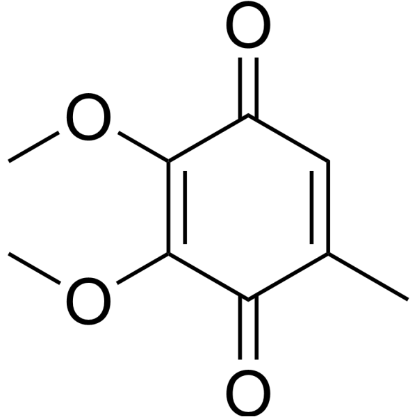
Coenzyme Q0
Based on 2 publication(s) in Google Scholar
Coenzyme Q0 (CoQ0) is a potent, oral active ubiquinone compound can be derived from Antrodia cinnamomea. Coenzyme Q0 induces apoptosis and autophagy, suppresses of HER-2/AKT/mTOR signaling to potentiate the apoptosis and autophagy mechanisms. Coenzyme Q0 regulates NFκB/AP-1 activation and enhances Nrf2 stabilization in attenuation of inflammation and redox imbalance. Coenzyme Q0 has anti-angiogenic activity through downregulation of MMP-9/NF-κB and upregulation of HO-1 signaling.
For research use only. We do not sell to patients.
- Purity : 99.79%
- CAS No.: 605-94-7
- Formula: C9H10O4
- Molecular Weight:182.18
-
Storage:Powder -20°C, 3 years , 4°C, 2 years ; In solvent -80°C, 6 months , -20°C, 1 month

To place orders, for customer services and technical support, please contact: MedChemExpress USA
Tel: 609-228-6898 E-mail: [email protected] [email protected]


-
 Biological Activity
-
Chemical Information
-
Solvent & Solubility
- Protocols
- Purity & Documentation
- References
-
 Help & FAQs


-
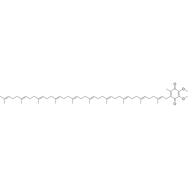
HY-N0111
Coenzyme Q10Antioxidant -
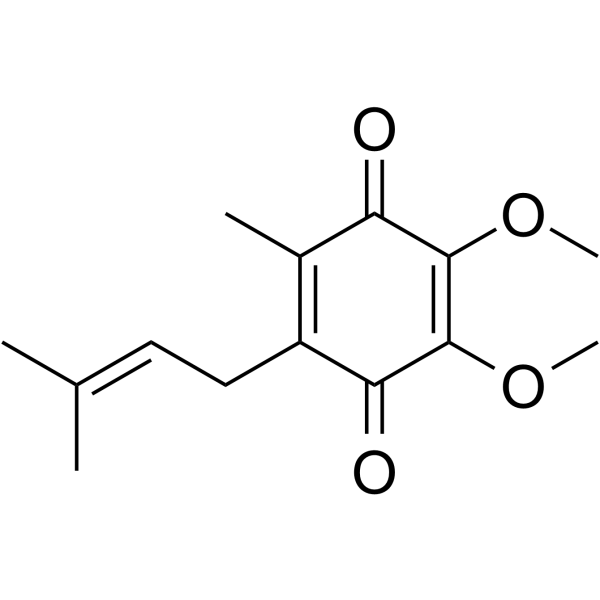
HY-113449
Ubiquinone-1Endogenous Metabolite -

HY-W050151
2,5-Di-tert-butyl-1,4-benzoquinoneAntibacterial Agent, RNA Polymerase Inhibitor -
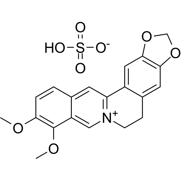
HY-N0716B
Berberine sulfateAntibiotic -
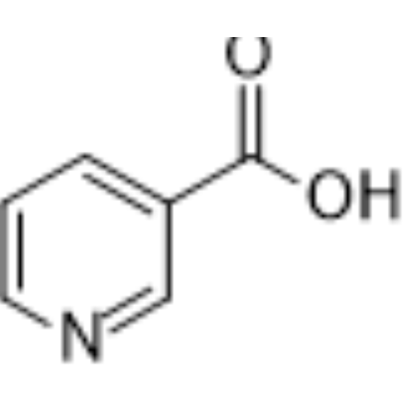
HY-B0143
NiacinB3 vitamin -

HY-W100959
DuroquinoneModel Quinone -
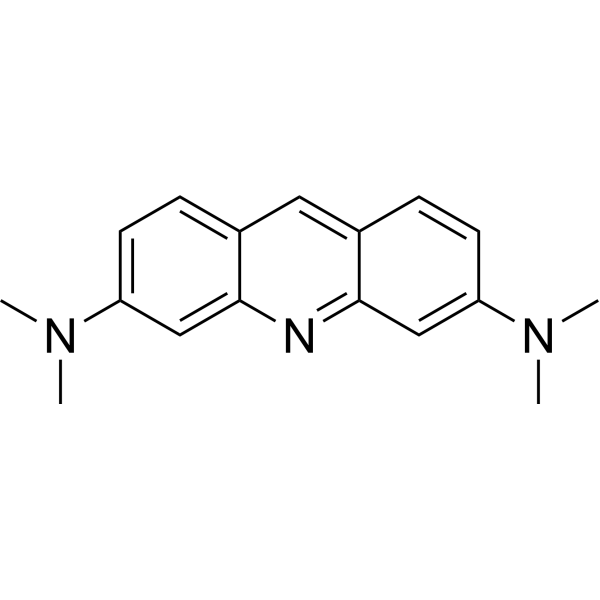
HY-D0952
Acridine Orange baseFluorescent Dye -
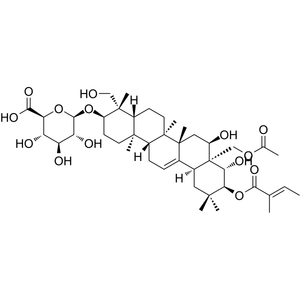
HY-N2541
Gymnemic acid IAntisweetness Agent -
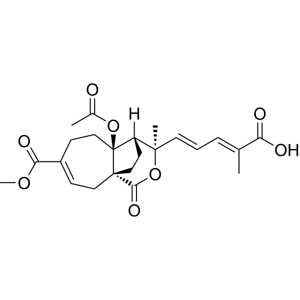
HY-N6939
Pseudolaric Acid BAutophagy Inducer -

HY-B0143R
Niacin (standard)B3 vitamin -
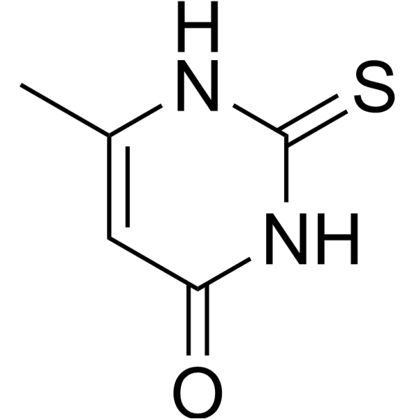
HY-B0513
MethylthiouracilTNF Receptor Inhibitor -
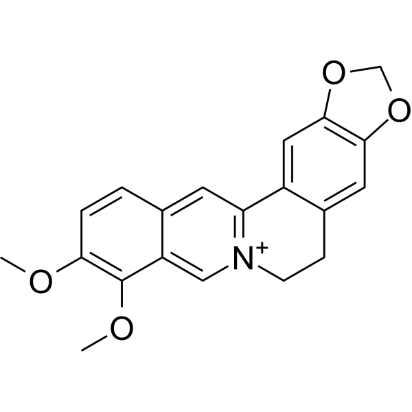
HY-N0716
BerberineAlkaloid
Publications Citing Use of MedChemExpress (MCE) Coenzyme Q0
MoreAll EGFR Isoforms
MoreAll Caspase Isoforms
More

Biological Activity
Description
In Vitro
Coenzyme Q0 (0-40 μM; 24 h) and inhibits viability and growth of human ovarian carcinoma cells[1].
Coenzyme Q0 (CoQ0) (0-30 μM; 24 h; SKOV-3 cells) has anti-proliferative activity through induction of G2/M cell-cycle arrest and reduction of cell-cycle regulatory proteins[1].
Coenzyme Q0 (CoQ0) (0-30 μM; 0-30 min; SKOV-3 cells) increases intracellular ROS levels to promote SKOV-3 cell death[1].
Coenzyme Q0 (CoQ0) (0-30 μM; 24 h; SKOV-3 cells) induces autophagy by increase accumulation of LC3-II, GFP-LC3 puncta, AVOs formation and Beclin-1/Bcl-2 dysregulation[1].
Coenzyme Q0 (CoQ0) (0-30 μM; 24 h; SKOV-3 cells) induces apoptosis by mitochondrial (caspase-3, PARP and Bax/Bcl-2 dysregulation) and ER stress (caspase-12 and Hsp70) signals[1].
Coenzyme Q0 (CoQ0) (30 μM; 24 h; SKOV-3 cells) suppresses of HER-2/AKT/mTOR signaling to potentiate the apoptosis and autophagy mechanisms[1].
Coenzyme Q0 (CoQ0) (0-10 μM; 0.5-18 h; RAW264.7 cells) regulates NFκB/AP-1 activation and enhances Nrf2 stabilization[2].
Coenzyme Q0 (CoQ0) (5 μM; 0-12 h; EA.hy 926 cells) has anti-angiogenic activity in EA.hy 926 cells[3].
MedChemExpress (MCE) has not independently confirmed the accuracy of these methods. They are for reference only. Further protocols information, click here.
-
Cell Line:SKOV-3, A2780 and A2870/CP70 cells
-
Concentration:0, 10, 20, 30 and 40 µM
-
Incubation Time:24 hours
-
Result:Decreased viability with the IC50 values of 26.6 µM, 27.3 µM and 28.4 µM for SKOV-3, A2780 and A2870/CP70 cells, respectively.
-
Cell Line:SKOV-3, A2780 and A2870/CP70 cells
-
Concentration:0, 10, 20 and 30 µM
-
Incubation Time:24 hours
-
Result:Arrested cell cycle at G2/M phase and reduced cell-cycle proteins in SKOV-3 cells.
-
Cell Line:SKOV-3, A2780 and A2870/CP70 cells
-
Concentration:0, 5, 15 and 30 µM
-
Incubation Time:24 hours
-
Result:Promoted the conversion of LC3–1 to LC3-II and increased the LC3-II accumulation. Increased Bax/Bcl-2 ratio in a dose-dependent manner.
-
Cell Line:SKOV-3 cells
-
Concentration:0, 10, 20 and 30 µM
-
Incubation Time:24 hours
-
Result:Had the percentage of early apoptotic cells are 25.1%, 34% and 36% for 10, 20 and 30 µM, respectively.
-
Cell Line:SKOV-3 cells
-
Concentration:0, 5, 15 and 30 µM
-
Incubation Time:24 hours
-
Result:Activated of caspase-3 and cleavaged of PARP. Increased the expressions of caspase-12, HSP-70 and Bax in a dose-dependent manner, decreased the expressions of Bcl-2.
-
Cell Line:SKOV-3 cells
-
Concentration:30 µM
-
Incubation Time:24 hours
-
Result:Decreased the phosphorylated HER-2 (Y1221) levels, p-AKT (Ser473) and p-mTOR (S2448) levels.
-
Cell Line:RAW264.7 cells
-
Concentration:0, 2.5, 5 and 10 µM
-
Incubation Time:0.5-18 hours
-
Result:Inhibited iNOS/COX-2 protein expressions with reductions of NO, PGE2, TNF-α and IL-1β secretions.
-
Cell Line:EA.hy 926 cells
-
Concentration:5 µM
-
Incubation Time:0, 1, 3, 6 and 12 hours
-
Result:Increased expressions of heme oxygenase-1 (HO-1) and γ-glutamylcysteine synthetase (γ-GCLC), inhibits protein expressions of matrix metalloproteinase-9 (MMP-9), reduces TNF-α-induced nuclear translocation and transcriptional activation of nuclear factor-κB (NF-κB).
In Vivo
Coenzyme Q0(CoQ0) (5 mg/kg; p.o.; for 4 h) has anti-inflammatory activities through Nrf2 activation and NFκB inhibition in liver and spleen of LPS-treated mice[2].
MedChemExpress (MCE) has not independently confirmed the accuracy of these methods. They are for reference only.
-
Animal Model:SKOV-3 xenografted nude mice[1]
-
Dosage:1.5 and 2.5 mg/kg
-
Administration:Intraperitoneal injection; Once every four days, for 52 days
-
Result:Inhibited the tumor growth at 1.5 and 2.5 mg/kg.
-
Animal Model:LPS-treated female FVB mice[2]
-
Dosage:5 mg/kg
-
Administration:Oral administration; for 4 hours
-
Result:Down-regulates inflammatory genes in liver and spleen tissues of LPS injected mice.


Chemical Information
-
CAS No. 605-94-7
-
Appearance Solid
-
Molecular Weight 182.18
-
Formula C9H10O4
-
Color Brown to red
-
SMILES
O=C1C(OC)=C(OC)C(C(C)=C1)=O
-
Synonyms
CoQ0
-
Shipping
Room temperature in continental US; may vary elsewhere.
-
Storage
Powder -20°C 3 years 4°C 2 years In solvent -80°C 6 months -20°C 1 month


Publications (2)
-
Journal Impact Factor
-
Most Recent
-
Emerg Microbes Infect
First report of Candida auris in Guangdong, China: clinical and microbiological characteristics of 7 episodes of candidemia. [Abstract]2024 Dec;13(1):2300525. PMID: 38164742 -
FASEB J
Amphiregulin improves ventricular remodeling after myocardial infarction by modulating autophagy and apoptosis. [Abstract]2024 Feb 29;38(4):e23488. PMID: 38358359


Solvent & Solubility
In Vitro:
DMSO : 50 mg/mL (274.45 mM; Need ultrasonic; Hygroscopic DMSO has a significant impact on the solubility of product, please use newly opened DMSO)
Please refer to the solubility information to select the appropriate solvent. Once prepared, please aliquot and store the solution to prevent product inactivation from repeated freeze-thaw cycles.
Storage method and period of stock solution: -80°C, 6 months; -20°C, 1 month. When stored at -80°C, please use it within 6 months. When stored at -20°C, please use it within 1 month.
Please refer to the solubility information to select the appropriate solvent. Once prepared, please aliquot and store the solution to prevent product inactivation from repeated freeze-thaw cycles.
Storage method and period of stock solution: -80°C, 6 months; -20°C, 1 month. When stored at -80°C, please use it within 6 months. When stored at -20°C, please use it within 1 month.
Concentration (start) × Volume (start) = Concentration (final) × Volume (final)
In Vivo:
Select the appropriate dissolution method based on your experimental animal and administration route.
- For the following dissolution methods, please ensure to first prepare a clear stock solution using an In Vitro approach and then sequentially add co-solvents:
- To ensure reliable experimental results, the clarified stock solution can be appropriately stored based on storage conditions. As for the working solution for In Vivo experiments, it is recommended to prepare freshly and use it on the same day.
- The percentages shown for the solvents indicate their volumetric ratio in the final prepared solution. If precipitation or phase separation occurs during preparation, heat and/or sonication can be used to aid dissolution.
Add each solvent one by one: 10% DMSO 40% PEG300 5% Tween-80 45% Saline
Solubility: ≥ 1.67 mg/mL (9.17 mM); Clear solution
This protocol yields a clear solution of ≥ 1.67 mg/mL (saturation unknown).
Taking 1 mL working solution as an example, add 100 μL DMSO stock solution (16.7 mg/mL) to 400 μL PEG300, and mix evenly; then add 50 μL Tween-80 and mix evenly; then add 450 μL Saline to adjust the volume to 1 mL.
Preparation of Saline: Dissolve 0.9 g sodium chloride in ddH₂O and dilute to 100 mL to obtain a clear Saline solution.
Add each solvent one by one: 10% DMSO 90% (20% SBE-β-CD in Saline)
Solubility: ≥ 1.67 mg/mL (9.17 mM); Clear solution
This protocol yields a clear solution of ≥ 1.67 mg/mL (saturation unknown).
Taking 1 mL working solution as an example, add 100 μL DMSO stock solution (16.7 mg/mL) to 900 μL 20% SBE-β-CD in Saline, and mix evenly.
Preparation of 20% SBE-β-CD in Saline (4°C, storage for one week): 2 g SBE-β-CD powder is dissolved in 10 mL Saline, completely dissolve until clear.
In Vivo Dissolution Calculator
Please enter the basic information of animal experiments:
-
-
-
-
Recommended: Prepare an additional quantity of animals to account for potential losses during experiments.
Please enter your animal formula composition:
-
%DMSO +
Recommended: Keep the proportion of DMSO in working solution below 2% if your animal is weak.
-
%+
-
+%Tween-80 + +
-
%Saline +
The co-solvents required include: DMSO, . All of co-solvents are available by MedChemExpress (MCE). , Tween 80. All of co-solvents are available by MedChemExpress (MCE).
Working solution concentration: 0.22 mg/mL
Method for preparing stock solution: mg drug dissolved in μL DMSO. Stock solution concentration: mg/mL.
1. Take μL DMSO stock solution;
2. Add μL .
μL , mix evenly;
3. Then add μL Tween 80, mix evenly;
4. Then add μL
Please ensure that the stock solution in the first step is dissolved to a clear state, and add co-solvents in sequence. You can use ultrasonic heating (ultrasonic cleaner, recommended frequency 20-40 kHz), vortexing, etc. to assist dissolution.

Protocols
-
Apoptosis
Apoptosis, also called programmed cell death, is generally characterized by distinct morphological characteristics.
-
TUNEL staining for apoptotic DNA fragmentation
TUNEL staining detects DNA strand breaks by using terminal deoxynucleotidyl transferase to add labeled nucleotides to exposed 3′-OH DNA termini, generating either microscopic staining in fixed cells or tissue sections, or fluorescence/cytometric signal in cell suspensions. TUNEL positivity reflects DNA fragmentation but should not be interpreted alone as definitive apoptosis, because TUNEL can also label necrotic, autolytic, mechanically damaged, or DNA-repair-associated DNA breaks.
-
Mitochondrial membrane-potential fluorescent assay
Mitochondrial membrane potential fluorescent assays estimate ΔΨm in living cells using lipophilic cationic dyes such as TMRM, TMRE, rhodamine 123, and JC-1, which accumulate in mitochondria according to membrane polarization; loss of signal after FCCP or CCCP treatment is interpreted as mitochondrial depolarization. TMRM/TMRE and rhodamine 123 are commonly used for semi-quantitative live-cell microscopy or flow cytometry, while JC-1 can report a shift from red aggregate fluorescence to green monomer fluorescence during depolarization; interpretation requires controls because dye concentration, quenching mode, cell type, dye efflux, and mitochondrial mass can affect fluorescence independently of ΔΨm.
-
Annexin V plus membrane-impermeant dye apoptosis staining
Annexin V-based apoptosis assays rely on the detection of phosphatidylserine (PS) externalization from the inner leaflet of the plasma membrane to the outer leaflet, an early biochemical hallmark of apoptosis. Fluorescently labeled Annexin V binds PS in a calcium-dependent manner, enabling identification of early apoptotic cells by flow cytometry or fluorescence microscopy. When combined with a membrane-impermeant DNA-binding dye (e. g. , propidium iodide), this approach allows discrimination between viable (Annexin V−/dye−), early apoptotic (Annexin V+/dye−), and late apoptotic or necrotic (Annexin V+/dye+) cell populations by assessing membrane integrity and PS exposure.
-
ROS/oxidative-stress fluorescent staining
ROS/oxidative-stress fluorescent staining uses cell-permeant fluorogenic probes that become fluorescent after oxidation inside cells or tissues; commonly used examples include DCFH-DA/DCFDA for broad cellular oxidant detection, DHE for superoxide-related signal detection, MitoSOX for mitochondrial superoxide-related signal detection, and CellROX probes for oxidative-stress-associated fluorescence readouts. The assay detects probe oxidation rather than a single ROS species unless the probe and analysis method have been chemically validated for that species. DCFH-DA enters cells, is deacetylated by intracellular esterases to DCFH, and produces fluorescent DCF after oxidation, so the readout is used as an operational measure of total cellular oxidative stress rather than a species-specific ROS measurement. DHE and MitoSOX can report superoxide-related oxidation, but red fluorescence alone can include non-specific ethidium-like oxidation products; HPLC or optimized spectral approaches are
-
Autophagy
Autophagy is a process in which eukaryotic cells use lysosomes to degrade their own cytoplasmic proteins and damaged organelles under the regulation of autophagy related gene (Atg). Microtubule-associated proteins light chain 3 (LC3) is recognized as autophagy marker, which transfers from cytoplasmic LC3 (LC3-I) to membrane type (LC3-II). LC3-II/I ratio could be detected by Western Blot and fluorescence microscopy.
-
Endothelial Tube Formation Assay
Endothelial tube formation assay evaluates the ability of endothelial cells to attach, migrate, align, and organize into capillary-like networks when cultured on gelled basement membrane extract or Matrigel; the readout is the morphology and quantity of tube-like networks, which reflects an in vitro endothelial morphogenesis step related to angiogenesis. Basement membrane extract/Matrigel provides laminin-rich extracellular matrix cues that support endothelial differentiation into capillary-like structures, but it can contain biologically active growth factors, so growth-factor-reduced matrix is preferred when testing defined angiogenic stimulators or inhibitors.
-
Mitochondrial membrane-potential and mitochondrial mass staining
Mitochondrial membrane potential staining measures the electrochemical polarization across the mitochondrial inner membrane in live cells using lipophilic cationic fluorescent probes; early rhodamine-based work showed that selective mitochondrial dye accumulation is lost when the mitochondrial transmembrane potential is dissipated. JC-1 reports mitochondrial polarization by shifting from green monomer fluorescence to red J-aggregate fluorescence as dye concentration increases within energized mitochondria; therefore, the red/green fluorescence ratio is used as a relative readout of mitochondrial membrane potential. TMRE or TMRM staining provides a single-channel relative readout because these cationic rhodamine esters accumulate in polarized mitochondria, and lower fluorescence indicates reduced mitochondrial polarization when acquisition and dye-loading conditions are controlled. Mitochondrial mass staining is commonly performed with MitoTracker Green FM or related MitoTracker dyes as
-
Apoptosis Solutions
Apoptosis is a regulated, generally non-lytic cell-death pathway that removes unwanted, damaged, infected, or abnormal cells through coordinated morphological changes, caspase activation, DNA fragmentation, and membrane remodeling. The intrinsic apoptosis pathway is controlled mainly by mitochondrial outer membrane permeabilization, BCL-2 family proteins, cytochrome c release, apoptosome formation, caspase-9 activation, and downstream executioner caspase-3/7 activation. The extrinsic apoptosis pathway is initiated by death receptors such as Fas, TNFR, and TRAIL receptors, which recruit adaptor proteins and activate caspase-8 before engaging executioner caspases or mitochondrial amplification through BID cleavage. Apoptosis is linked to many phenotypes, including cancer cell killing, tissue homeostasis, immune regulation, neurodegeneration, infection response, and treatment-induced cytotoxicity; unresolved questions include how apoptosis interacts with necroptosis, pyroptosis, ferroptos
-
Lysosome and acidic-vesicle live-cell staining
Lysosome and acidic-vesicle live-cell staining detects acidic intracellular compartments by using membrane-permeant acidotropic probes that accumulate in low-pH vesicles, including lysosomes, late endosomes, autolysosomes, and acidic phagosomes. LysoTracker staining is commonly used as an intensity-based readout of acidic lysosomal compartment abundance or enlargement, while acridine orange produces green fluorescence in less concentrated compartments and red fluorescence after concentration-dependent accumulation in acidic vesicular organelles. Loss or reduction of acridine-orange red signal can be used as a readout of lysosomal membrane permeabilization or reduced acidic-vesicle integrity. This protocol is designed for live cultured cells and can be adapted for fluorescence microscopy, high-content imaging, plate-reader readout, or flow cytometry when the selected literature supports the readout. Because these dyes report acidotropic accumulation rather than lysosome identity alone,
-
Research Protocol for Inflammation-related Diseases
The NLRP3 inflammasome is a cytosolic innate immune signaling platform that integrates priming signals and danger-signal activation to promote caspase-1 activation, maturation of IL-1β and IL-18, and gasdermin D-mediated pyroptotic cell death. The core experimental logic is to determine whether inflammatory disease phenotypes are driven by increased NLRP3 expression, ASC-containing inflammasome assembly, caspase-1 cleavage, GSDMD cleavage, and extracellular release of IL-1β/IL-18 rather than by nonspecific cell injury alone. The pathway is strongly linked to inflammation-related disease phenotypes because monosodium urate crystals activate NALP3/NLRP3 inflammasome signaling in gout-like crystal inflammation, cholesterol crystals activate NLRP3 inflammasomes in atherogenesis models, and DSS-induced intestinal inflammation has been reported to involve NLRP3 inflammasome activity. However, experimental colitis studies also show context-dependent protective effects of NLRP3 inflammasome co
-
Macroautophagy Solutions
Macroautophagy is a conserved lysosome-dependent degradation pathway in which cytoplasmic material is sequestered into double-membrane autophagosomes and delivered to lysosomes for degradation and recycling. The pathway supports cellular homeostasis during nutrient limitation, organelle stress, protein-aggregate accumulation, infection, differentiation, and tissue remodeling by coupling cargo sequestration, autophagosome maturation, lysosomal fusion, and degradation of cargo-derived macromolecules. The core molecular sequence includes initiation by nutrient- and stress-regulated autophagy machinery, autophagosome nucleation, LC3/ATG8-family conjugation to autophagosomal membranes, cargo selection through receptors such as SQSTM1/p62, autophagosome-lysosome fusion, and lysosomal degradation. LC3 was identified as a mammalian homolog of yeast Atg8 that localizes to autophagosomal membranes after processing, and p62/SQSTM1 was shown to connect ubiquitinated cargo with autophagic degradati


Purity & Documentation
-
Data Sheet (287 KB)
-
SDS (393 KB)
- English - EN (393 KB)
- Français - FR (393 KB)
- Deutsch - DE (393 KB)
- Norwegian - NO (393 KB)
- Español - ES (393 KB)
- Swedish - SV (393 KB)
- Italian - IT (393 KB)
- Korean - KR (393 KB)
- Portuguese - PT (393 KB)
-
Handling Instructions (2659 KB)
References
[1]. Yang HL, et, al. Coenzyme Q0 regulates NFκB/AP-1 activation and enhances Nrf2 stabilization in attenuation of LPS-induced inflammation and redox imbalance: Evidence from in vitro and in vivo studies. Biochim Biophys Acta. 2016 Feb;1859(2):246-61. [Content Brief]
[2]. Yang HL, et, al. Coenzyme Q0 regulates NFκB/AP-1 activation and enhances Nrf2 stabilization in attenuation of LPS-induced inflammation and redox imbalance: Evidence from in vitro and in vivo studies. Biochim Biophys Acta. 2016 Feb;1859(2):246-61. [Content Brief]
[3]. Yang HL, et, al. Anti-angiogenic properties of coenzyme Q0 through downregulation of MMP-9/NF-κB and upregulation of HO-1 signaling in TNF-α-activated human endothelial cells. Biochem Pharmacol. 2015 Nov 1;98(1):144-56. [Content Brief]


Complete Stock Solution Preparation Table
Please refer to the solubility information to select the appropriate solvent. Once prepared, please aliquot and store the solution to prevent product inactivation from repeated freeze-thaw cycles.
Storage method and period of stock solution: -80°C, 6 months; -20°C, 1 month. When stored at -80°C, please use it within 6 months. When stored at -20°C, please use it within 1 month.
| Optional Solvent | Concentration Solvent Mass | 1 mg | 5 mg | 10 mg | 25 mg |
|---|---|---|---|---|---|
| DMSO | 1 mM | 5.4891 mL | 27.4454 mL | 54.8908 mL | 137.2269 mL |
| 5 mM | 1.0978 mL | 5.4891 mL | 10.9782 mL | 27.4454 mL | |
| 10 mM | 0.5489 mL | 2.7445 mL | 5.4891 mL | 13.7227 mL | |
| 15 mM | 0.3659 mL | 1.8297 mL | 3.6594 mL | 9.1485 mL | |
| 20 mM | 0.2745 mL | 1.3723 mL | 2.7445 mL | 6.8613 mL | |
| 25 mM | 0.2196 mL | 1.0978 mL | 2.1956 mL | 5.4891 mL | |
| 30 mM | 0.1830 mL | 0.9148 mL | 1.8297 mL | 4.5742 mL | |
| 40 mM | 0.1372 mL | 0.6861 mL | 1.3723 mL | 3.4307 mL | |
| 50 mM | 0.1098 mL | 0.5489 mL | 1.0978 mL | 2.7445 mL | |
| 60 mM | 0.0915 mL | 0.4574 mL | 0.9148 mL | 2.2871 mL | |
| 80 mM | 0.0686 mL | 0.3431 mL | 0.6861 mL | 1.7153 mL | |
| 100 mM | 0.0549 mL | 0.2745 mL | 0.5489 mL | 1.3723 mL |


Coenzyme Q0 Related Classifications
HY-W016412 Related Classifications
 Powered by Bioz
Keywords
- Coenzyme Q0
- 605-94-7
- CoQ0
- Coenzyme Q 0
- Coenzyme Q-0
- CoQ 0
- CoQ-0
- Apoptosis
- Autophagy
- EGFR
- Akt
- mTOR
- Caspase
- Bcl-2 Family
- Reactive Oxygen Species (ROS)
- PARP
- COX
- NO Synthase
- TNF Receptor
- Interleukin Related
- MMP
- NF-κB
- anticancer
- anti-angiogenic
- anti-inflammation
- HO-1
- ubiquinone compound
- Antrodia cinnamomea
- Inhibitor
- inhibitor
- inhibit