



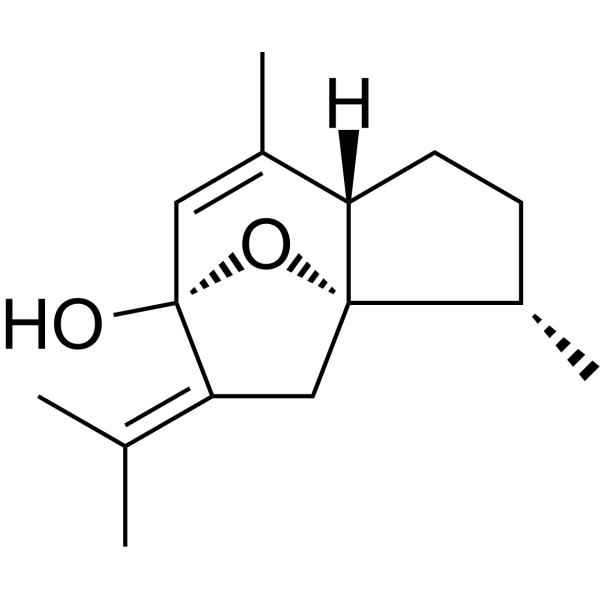
Curcumenol
Based on 5 publication(s) in Google Scholar
Curcumenol ((+)-Curcumenol) is a natural compound with oral efficacy, exhibiting an IC50 of 12.6 μM and a Ki of 10.8 μM against human CYP3A4. Curcumenol inhibits TNFα-induced phosphorylation/degradation of IκBα, phosphorylation/nuclear translocation of NF-κB p65, as well as the upregulation of MMP3, MMP9, MMP13, TRAF3, IL1RL1, TNFα and IL-1β. Curcumenol suppresses LPS-induced phosphorylation of Akt and p38 MAPK, as well as the production of pro-inflammatory mediators/proteins, and downregulates the SLC7A11/NF-κB/TGF-β pathway. Curcumenol binds to and inhibits the activation of Fyn and Lyn, blocks the function of downstream FcεRI signaling components, and reduces the release of allergic mediators/cytokines. Curcumenol upregulates the expression of KDM6B, and promotes chondrocyte proliferation and cartilage repair. Curcumenol induces ferroptosis and apoptosis, regulates the EMT process, and inhibits tumor growth and metastasis of triple-negative breast cancer. Curcumenol possesses anti-inflammatory, neuroprotective, antioxidant, antitumor, antiviral and hepatoprotective activities. Curcumenol can be used in research related to intervertebral disc degeneration, cancer, inflammation, central nervous system neurodegenerative diseases, allergic reactions and knee osteoarthritis.
For research use only. We do not sell to patients.
- Purity : 99.98%
- CAS No.: 19431-84-6
- Formula: C15H22O2
- Molecular Weight:234.33
-
Storage:Powder -20°C, 3 years , 4°C, 2 years ; In solvent -80°C, 2 years , -20°C, 1 year

To place orders, for customer services and technical support, please contact: MedChemExpress USA
Tel: 609-228-6898 E-mail: [email protected] [email protected]


-
 Biological Activity
-
Chemical Information
-
Solvent & Solubility
- Protocols
- Purity & Documentation
- References
-
 Help & FAQs


-
HY-107324
β-ElemeneApoptosis Activator -
HY-N0176
DihydroartemisininAntimalarial Agent -
HY-N0440
GermacroneAnti-viral/Cancer/Inflammatory Agent -
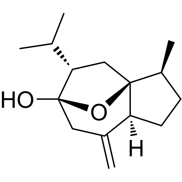
HY-N0104
CurcumolApoptosis Inducer -
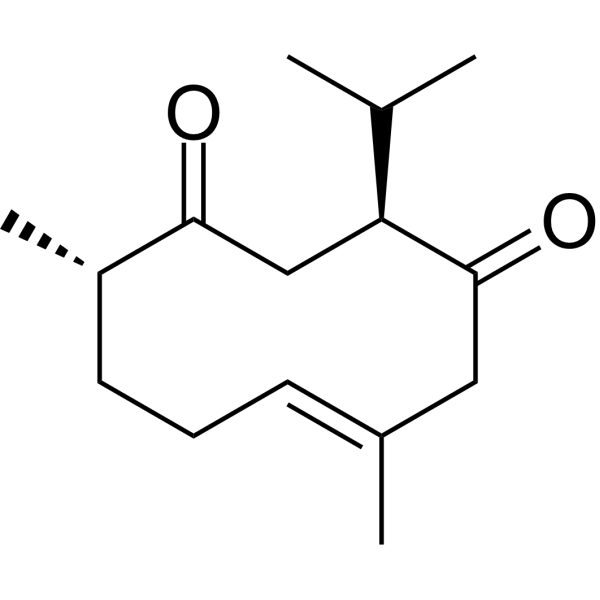
HY-N0353
CurdioneNatural Product -

HY-Y0320
Dimethyl sulfoxide, meets analytical specification of Ch.P.Aprotic Solvent -
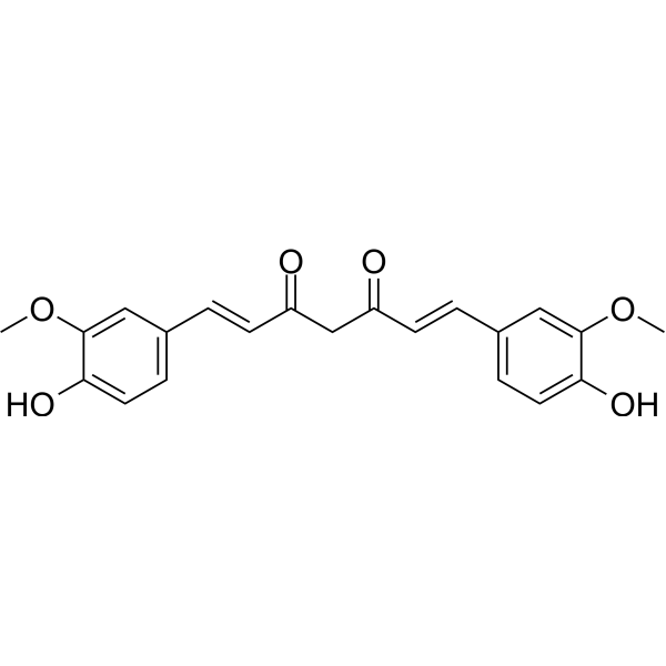
HY-N0005
Curcuminp300 Histone Acetylatransferase Inhibitor -
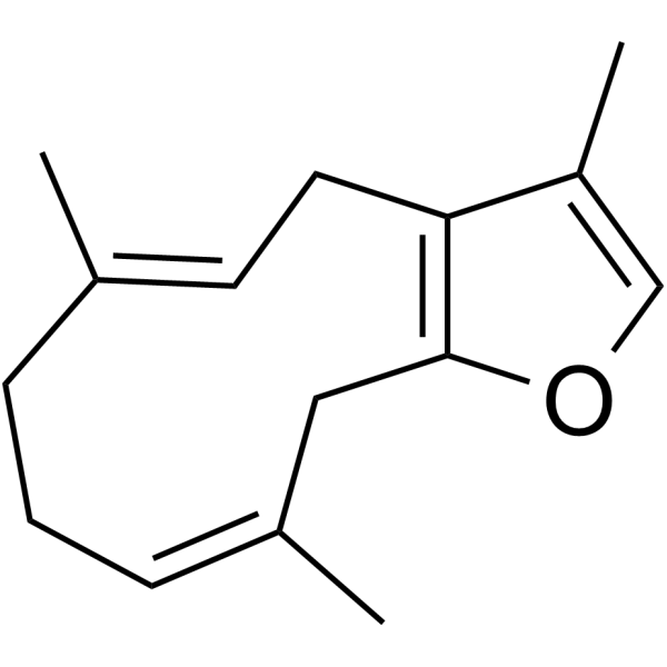
HY-126940
FuranodieneAnti-cancer Agent -
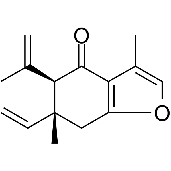
HY-N3651
CurzerenoneAntibacterial/anticancer Agent -
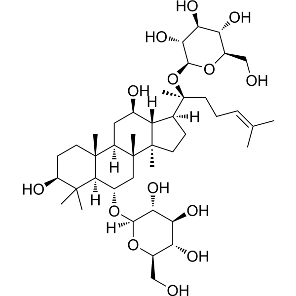
HY-N0045
Ginsenoside Rg1Apoptosis Inducer -
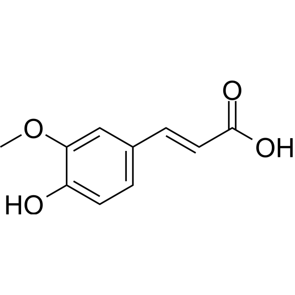
HY-N0060
Ferulic acidFGFR1 Inhibitor -
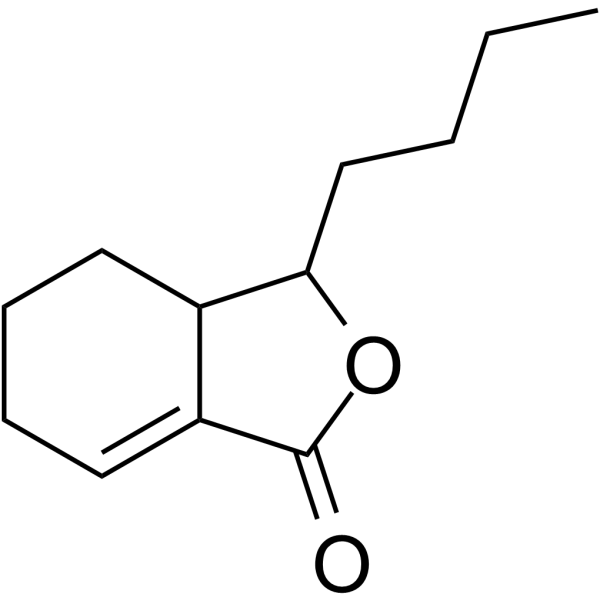
HY-N2114
SedanolideAutophagy Inducer
Publications Citing Use of MedChemExpress (MCE) Curcumenol
More Customer Validation & Images

-
IF

-
In Vivo Efficacy Study

-
Cell Proliferation/Viability Assay

-
Cell Imaging/Staining

-
WB

All Caspase Isoforms
More

Biological Activity
Description
IC50 & Target
[2]|
CYP3A4 12.6 μM (IC50) |
CYP3A4 10.8 μM (Ki) |
Cellular Effect
|
Cell Line

|
Type | Value | Description | References |
|---|---|---|---|---|
| 4T1 | IC50 |
98.76 μM
|
Reduction of cell viability against mouse 4T1 triple-negative breast cancer cells incubated for 24 hrs by Cell Counting Kit-8 (CCK-8) assay.
Reduction of cell viability against mouse 4T1 triple-negative breast cancer cells incubated for 24 hrs by Cell Counting Kit-8 (CCK-8) assay.
|
40377003 |
| MDA-MB-231 | IC50 |
190.2 μM
|
Reduction of cell viability against human MDA-MB-231 triple-negative breast cancer cells incubated for 24 hrs by Cell Counting Kit-8 (CCK-8) assay.
Reduction of cell viability against human MDA-MB-231 triple-negative breast cancer cells incubated for 24 hrs by Cell Counting Kit-8 (CCK-8) assay.
|
40377003 |
| 4T1 | IC50 |
95.11 μM
|
Reduction of cell viability against mouse 4T1 triple-negative breast cancer cells incubated for 48 hrs by Cell Counting Kit-8 (CCK-8) assay.
Reduction of cell viability against mouse 4T1 triple-negative breast cancer cells incubated for 48 hrs by Cell Counting Kit-8 (CCK-8) assay.
|
40377003 |
| MDA-MB-231 | IC50 |
169.8 μM
|
Reduction of cell viability against human MDA-MB-231 triple-negative breast cancer cells incubated for 48 hrs by Cell Counting Kit-8 (CCK-8) assay.
Reduction of cell viability against human MDA-MB-231 triple-negative breast cancer cells incubated for 48 hrs by Cell Counting Kit-8 (CCK-8) assay.
|
40377003 |
In Vitro
Curcumenol (50 μM; 24 h) regulates gene expression in immortalized rat nucleus pulposus (NP) cells (downregulating the expression of TRAF1, 2, 3, 4, 5, 6, IL1RL1, TNF and HOXA6, and upregulating the expression of CXCL1, 6, 10, 16 and Nos2), and significantly alters multiple inflammatory pathways including the TNF signaling pathway[1].
Curcumenol (6.25-50 μM; 24 h) alleviates TNFα-induced catabolic changes and inflammatory gene expression in immortalized rat nucleus pulposus (NP) cells by restoring Col2a1 levels, reducing the expression of MMP family members, and regulating genes in the TNF signaling pathway[1].
Curcumenol (50 μM; 14 days) attenuates TNFα-induced intervertebral disc inflammation and degeneration in rat in vitro models[1].
Curcumenol (50 μM) inhibits TNFα-induced activation of the NF-κB pathway and upregulation of catabolic MMP family proteins in immortalized rat nucleus pulposus cells, and regulates TRAF3 and CXCL10, two proteins involved in the TNF signaling pathway[1].
Curcumenol (50 μM) inhibits TNFα-induced nuclear translocation of phosphorylated p65 in immortalized rat nucleus pulposus (NP) cells[1].
Curcumenol (5-20 μM; 2 h pretreatment followed by 24 h LPS exposure) dose-dependently inhibits LPS-induced iNOS and COX-2 protein expression in mouse BV-2 microglia[3].
Curcumenol (5-20 μM; pretreated for 2 h followed by LPS exposure) inhibits LPS-induced NF-κB activation by reducing p65 nuclear translocation and IκBα phosphorylation in mouse BV-2 microglia[3].
Curcumenol (5-20 μM; 2 h pretreatment followed by LPS exposure) dose-dependently inhibits LPS-induced phosphorylation of Akt in mouse BV-2 microglial cells, thereby suppressing the activation of NF-κB[3].
Curcumenol (50 μM; 24 h) alleviates TNFα-induced catabolic changes in primary rat nucleus pulposus (NP) cells by restoring Col2a1 levels and reducing the expression of MMP family members[1].
Curcumenol (5-100 μM; 10-fold the concentration causing 25% reversible inhibition) competitively inhibits CYP3A4 activity in pooled human liver microsomes, with an IC50 of 12.6 μM and a Ki of 10.8 μM. In contrast, it exerts extremely weak inhibitory effects on CYP1A2, CYP2A6, CYP2C8, CYP2D6, CYP2E1 and CYP2C9, and does not act as a mechanism-based inhibitor for any of these 7 tested CYP subtypes[2].
Curcumenol (2.5-20 μM; 2 h pretreatment followed by 24 h LPS exposure) enhances the viability of LPS (HY-D1056)-stimulated mouse BV-2 microglial cells without inducing toxicity[3].
Curcumenol (5-20 μM; 2 h pretreatment, followed by 12-24 h LPS exposure) dose-dependently inhibits LPS-induced nitric oxide production in mouse BV-2 microglial cells[3].
Curcumenol (5-20 μM; 2 h pretreatment followed by LPS exposure) inhibits LPS-induced phosphorylation of p38 MAPK, but not that of JNK or ERK, in murine BV-2 microglial cells[3].
Curcumenol (250-2000 nM; 1 h pretreatment followed by 4 h DNP-BSA challenge for BMMCs; 1 h pretreatment followed by 1 h DNP-BSA challenge for RBL-2H3) dose-dependently inhibits β-hexosaminidase release from anti-DNP-IgE-sensitized, DNP-BSA-challenged mouse bone marrow-derived mast cells (BMMCs) and rat basophilic leukemia RBL-2H3 cells[5].
Curcumenol (250-2000 nM; 1 h pretreatment) dose-dependently inhibits the phosphorylation of Fyn, Lyn, Syk, PLCγ1, Akt, ERK, and p38 in mouse bone marrow-derived mast cells sensitized with anti-DNP-IgE and stimulated with DNP-BSA[5].
Curcumenol (5-20 μM; 2 h pretreatment followed by 12-24 h LPS exposure) inhibits LPS-induced production of TNF-α and IL-6 in mouse BV-2 microglial cells[3].
Curcumenol (6.25-400 μM; 24-48 h) dose-dependently inhibits the viability of 4T1 and MDA-MB-231 triple-negative breast cancer (TNBC) cells, with 24 h IC50 values of 98.76 μM and 190.2 μM, respectively, and enhances the sensitivity of these cells to Paclitaxel (HY-B0015)[4].
Curcumenol (50-100 μM; 24 h) inhibits the migration of 4T1 and MDA-MB-231 triple-negative breast cancer (TNBC) cells[4].
Curcumenol (25-100 μM; 24 h) inhibits the invasion of 4T1 and MDA-MB-231 triple-negative breast cancer (TNBC) cells[4].
Curcumenol (25-100 μM; 24 h) reduces the colony-forming ability of 4T1 and MDA-MB-231 triple-negative breast cancer (TNBC) cells[4].
Curcumenol (100 μM; 24 h) upregulates the expression of E-cadherin and downregulates the expression of Vimentin in 4T1 and MDA-MB-231 triple-negative breast cancer (TNBC) cells, indicating that it inhibits epithelial-mesenchymal transition (EMT)[4].
Curcumenol (100 μM; 24 h) regulates apoptosis-related proteins and inhibits epithelial-mesenchymal transition (EMT) in 4T1 and MDA-MB-231 triple-negative breast cancer (TNBC) cells by altering the expression of key marker proteins[4].
Curcumenol (100 μM; 24 h) reduces the mitochondrial membrane potential of 4T1 and MDA-MB-231 triple-negative breast cancer (TNBC) cells[4].
Curcumenol (100 μM; 24 h) promotes the accumulation of ROS in 4T1 and MDA-MB-231 triple-negative breast cancer (TNBC) cells[4].
Curcumenol (100 μM; 24 h) promotes lipid ROS accumulation and regulates ferroptosis-related proteins, thereby inducing ferroptosis in MDA-MB-231 triple-negative breast cancer (TNBC) cells[4].
Curcumenol (25 μM) promotes proliferation, inhibits apoptosis, and enhances extracellular matrix (ECM) synthesis of LPS-induced mouse osteoarthritis (OA) chondrocytes. It upregulates the expressions of COL2 and KDM6B, downregulates the expressions of MMP3 and H3K27me3, and enhances the binding of KDM6B to H3K27me3[6].
MedChemExpress (MCE) has not independently confirmed the accuracy of these methods. They are for reference only.
-
Cell Line:immortalized rat nucleus pulposus (NP) cells
-
Concentration:6.25, 12.5, 25, 50 μM (with 10 ng/mL TNFα)
-
Incubation Time:24 h
-
Result:Dose-dependently reversed TNFα-induced down-regulation of Col2a1 and up-regulation of MMP3, MMP9, and MMP13.
Significantly inhibited TNFα-induced up-regulation of TRAF3, CXCL1, and NOS2, and reduced expression of CXCL6, CXCL16, TRAF1, TRAF2, and TRAF6 (without statistical significance).
Inhibited TNFα-induced up-regulation of IL1RL1.
-
Cell Line:immortalized rat nucleus pulposus (NP) cells
-
Concentration:50 μM (MMP family analysis with 10 ng/mL TNFα); 50 μM (NF-κB pathway analysis with 10 ng/mL TNFα)
-
Incubation Time:24 h (MMP family analysis); 2 h pretreatment followed by 10 min stimulation (NF-κB pathway analysis)
-
Result:Significantly reduced TNFα-induced up-regulation of MMP3, MMP9, and MMP13 protein levels.
Inhibited TNFα-induced phosphorylation of p65 (Ser536) and IκBα (Ser32), and restored total IκBα protein levels that were decreased by TNFα.
Inhibited TNFα-induced up-regulation of TRAF3 protein levels, while not reducing TNFα-induced CXCL10 protein levels.
-
Cell Line:immortalized rat nucleus pulposus (NP) cells
-
Concentration:50 μM (with 10 ng/mL TNFα)
-
Incubation Time:2 h pretreatment followed by 20 min stimulation
-
Result:Effectively blocked TNFα-induced activation and nuclear translocation of p-p65.
-
Cell Line:mouse BV-2 microglial cells
-
Concentration:2.5, 5, 10, 20 μM
-
Incubation Time:2 h pretreatment, followed by 24 h LPS exposure
-
Result:Increased the viability of LPS-stimulated BV-2 cells at all tested concentrations.
Showed no toxicity up to 20 μM.
-
Cell Line:mouse BV-2 microglial cells
-
Concentration:5, 10, 20 μM
-
Incubation Time:2 h pretreatment, followed by 24 h LPS exposure
-
Result:Dose-dependently attenuated LPS-induced iNOS and COX-2 protein expression in BV-2 cells.
-
Cell Line:mouse BV-2 microglial cells
-
Concentration:5, 10, 20 μM
-
Incubation Time:2 h pretreatment, followed by LPS exposure
-
Result:Significantly decreased LPS-induced nuclear translocation of NF-κB p65 in BV-2 cells.
Attenuated LPS-induced phosphorylation of IκBα in BV-2 cells.\nDose-dependently attenuated LPS-induced phosphorylation of Akt in BV-2 cells.\nSignificantly reduced LPS-induced phosphorylation of p38 MAPK in BV-2 cells.
Did not affect phosphorylation of JNK or ERK in BV-2 cells.
-
Cell Line:4T1, MDA-MB-231
-
Concentration:6.25, 12.5, 25, 50, 100, 200, 400 μM; 25, 50, 100 μM (24 h incubation with Paclitaxel)
-
Incubation Time:24 h; 48 h
-
Result:Reduced viability of 4T1 and MDA-MB-231 cells in a dose-dependent manner.
Achieved 24 h IC50 values of 98.76 μM (4T1) and 190.2 μM (MDA-MB-231).
Achieved 48 h IC50 values of 95.11 μM (4T1) and 169.8 μM (MDA-MB-231).
Enhanced sensitivity of both cell lines to 50 nM Paclitaxel at 50 and 100 μM.
Increased sensitivity of MDA-MB-231 cells to 500 nM Paclitaxel at 50 and 100 μM.
Increased sensitivity of 4T1 cells to 500 nM Paclitaxel at 100 μM.
-
Cell Line:4T1, MDA-MB-231
-
Concentration:25, 50, 100 μM
-
Incubation Time:24 h
-
Result:Significantly reduced the migration rate of 4T1 and MDA-MB-231 cells at 50 and 100 μM after 24 h.
-
Cell Line:4T1, MDA-MB-231
-
Concentration:25, 50, 100 μM
-
Incubation Time:24 h
-
Result:Significantly reduced the number of invasive 4T1 and MDA-MB-231 cells at 25, 50, and 100 μM.
-
Cell Line:4T1, MDA-MB-231
-
Concentration:25, 50, 100 μM
-
Incubation Time:24 h
-
Result:Significantly reduced colony formation of 4T1 and MDA-MB-231 cells at 25, 50, and 100 μM.
-
Cell Line:4T1, MDA-MB-231
-
Concentration:100 μM
-
Incubation Time:24 h
-
Result:Increased protein expression of proapoptotic cleaved caspase 9, cleaved caspase 3, and BAX.
Decreased expression of antiapoptotic BCL-2.
Upregulated expression of epithelial marker E-cadherin.
Downregulated expression of mesenchymal markers N-cadherin and Vimentin.
-
Cell Line:4T1, MDA-MB-231
-
Concentration:100 μM
-
Incubation Time:24 h
-
Result:Increased fluorescent signal of E-cadherin in both cell lines.
Decreased fluorescent signal of Vimentin in both cell lines.
-
Cell Line:anti-DNP-IgE-sensitized mouse bone marrow-derived mast cells (BMMCs)
-
Concentration:250 nM, 500 nM, 1000 nM, 2000 nM
-
Incubation Time:1 h pretreatment
-
Result:Attenuated the DNP-BSA-induced phosphorylation of Fyn, Lyn, Syk, PLCγ1, Akt, ERK, and p38 in a concentration-dependent manner.
Achieved inhibition comparable to the Src family kinase inhibitor PP2 at the highest concentration of 2000 nM.
In Vivo
Curcumenol (i.p.; twice weekly; for 6 consecutive weeks at doses of 4-8 mg/kg) dose-dependently ameliorates DMM-induced knee osteoarthritis in mice by improving subchondral bone structure, reducing cartilage destruction, promoting cartilage repair, regulating the levels of key proteins and metabolites, as well as upregulating KDM6B and inhibiting H3K27me3[6].
Curcumenol (5-10 mg/kg, i.p., once every 2 days for 3 weeks) inhibits tumor growth and reduces lung metastasis of triple-negative breast cancer, regulates markers of apoptosis and epithelial-mesenchymal transition, and downregulates the SLC7A11/NF-κB/TGF-β pathway in BALB/c mice[4].
Curcumenol (10-25 mg/kg; p.o.; administered 3 times on days 9, 11, and 13) dose-dependently alleviates ovalbumin-induced systemic anaphylaxis in female BALB/c mice, reduces plasma levels of IgE, histamine, and IL-4, and attenuates hypothermia[5].
Curcumenol (10-25 mg/kg; p.o.; single administration 1 h before challenge) inhibits passive cutaneous anaphylaxis in male BALB/c mice and reduces local allergic dye extravasation[5].
MedChemExpress (MCE) has not independently confirmed the accuracy of these methods. They are for reference only.
-
Animal Model:C57/BL (male, 8-week-old, surgically induced lumbar spine instability)[1]
-
Dosage:4 mg/kg
-
Administration:i.p.; twice weekly; 1 month
-
Result:Prevented intervertebral disc height loss and osteophyte formation, restoring relative disc height index toward sham levels.
Reduced histological scores of disc degeneration.
Decreased the percentage of cells positive for inflammatory cytokines TNFα and IL-1β.
Increased the percentage of cells positive for the chondrogenic marker Col2a1 in surgically treated mice.
Confirmed reduced TNFα and IL-1β expression and restored Col2a1 expression via immunofluorescence analysis.
-
Animal Model:BALB/c (female, 6 weeks old, ~20 g, subcutaneously injected with 5×10⁴ 4T1 cells)[4]
-
Dosage:5 mg/kg; 10 mg/kg
-
Administration:i.p.; every 2 days; 3 weeks
-
Result:Suppressed tumor growth, with the 10 mg/kg dose significantly reducing tumor volume increases over 3 weeks.
Decreased Ki-67-positive proliferating cells and increased TUNEL-positive apoptotic cells in tumor tissues.
Upregulated tumor tissue expression of cleaved caspase 9, cleaved caspase 3, and BAX, while downregulating BCL-2.
Upregulated E-cadherin and downregulated N-cadherin and Vimentin in tumor tissues.
Reduced the number of lung metastatic nodules and the area of lung metastatic lesions.
Upregulated E-cadherin and downregulated Vimentin in lung metastatic tissues.
Decreased tumor tissue mRNA expression of SLC7A11, APLNR, and TGF-β, while increasing mRNA expression of IL7, HCAR2, and NF-κBID at the 10 mg/kg dose.
Downregulated tumor tissue expression of SLC7A11, phosphorylated NF-κB, and TGF-β at the 10 mg/kg dose.
-
Animal Model:BALB/c (female, 18-20 g, systemic sensitization with ovalbumin + alum adjuvant)[5]
-
Dosage:10 mg/kg; 25 mg/kg
-
Administration:p.o.; 3 doses on days 9, 11, 13
-
Result:Attenuated ovalbumin-induced rectal temperature decrease in a dose-dependent manner, with rectal temperatures at 30 minutes post-challenge similar to positive control levels.
Dose-dependently reduced ovalbumin-induced elevation of plasma IgE, histamine, and IL-4 levels.
-
Animal Model:BALB/c (male, 18-20 g, passive cutaneous anaphylaxis model via intradermal anti-DNP-IgE injection)[5]
-
Dosage:10 mg/kg; 25 mg/kg
-
Administration:p.o.; single dose 1 hour pre-challenge
-
Result:Significantly reduced Evans blue dye extravasation in mouse ears compared to untreated challenged controls.
-
Animal Model:C57BL/6 (7-week-old male, 18-22 g, DMM-induced surgical model)[6]
-
Dosage:4 mg/kg; 8 mg/kg
-
Administration:i.p.; twice weekly; 6 weeks
-
Result:Increased bone mineral density (BMD), trabecular bone volume/tissue volume (BV/TV), and trabecular number (Tb.N), while decreased trabecular separation (Tb.Sp) in a dose-dependent manner.
Reduced Osteoarthritis Research Society International (OARSI) scores compared to untreated KOA mice.
Increased glycosaminoglycan (GAG) levels in cartilage.
Downregulated d-Alanyl-d-alanine, 17a-Estradiol, Glutathione, and Succinic acid, while upregulated Sterculic acid and Azelaic acid in serum.
Upregulated type II collagen (COL2) and KDM6B protein expression, and downregulated matrix metalloproteinase 3 (MMP3) and H3K27me3 protein expression in knee joint tissue.
- Western Blot
- RNA extraction experimental
- Research Protocol for Infectious Diseases
- Annexin V plus membrane-impermeant dye apoptosis staining
- Patient-Derived Orthotopic Xenograft (PDOX)
- Research Protocol for Inflammation-related Diseases
- Breast Cancer Modeling
- Cell Viability Determination by MTT Colorimetric Assay
- Kinase activity and phosphorylation assays
- Cytoplasmic-Nuclear Fractionated Protein Extraction
- Detection of Apoptosis
- TUNEL staining for apoptotic DNA fragmentation
- Patient-Derived Xenograft (PDX)
- Apoptosis Solutions
- Protocol for Kinase activity and phosphorylation assays
- Ferroptosis Solutions
- Subcutaneous Cell-Line-Derived Xenograft
- Orthotopic Cell-Line Xenograft
- Pyroptosis Solutions


Chemical Information
-
CAS No. 19431-84-6
-
Appearance Solid
-
Molecular Weight 234.33
-
Formula C15H22O2
-
Color White to off-white
-
SMILES
C[C@@H]1[C@@](O[C@@]2(O)/C3=C(C)\C)(C3)[C@@](C(C)=C2)([H])CC1
-
Synonyms
(+)-Curcumenol
-
Structure Classification
-
Initial Source
-
Shipping
Room temperature in continental US; may vary elsewhere.
-
Storage
Powder -20°C 3 years 4°C 2 years In solvent -80°C 2 years -20°C 1 year


Publications (5)
-
Journal Impact Factor
-
Most Recent
-
-
PLoS Biol
2024 Jun 27;22(6):e3002672. PMID: 38935621 -
Int J Mol Sci
Mechanism of Curcumol Targeting the OTUB1/TGFBI Ubiquitination Pathway in the Inhibition of Angiogenesis in Colon Cancer. [Abstract]2025 May 21;26(10):4899. PMID: 40430059
Curcumenol purchased from MedChemExpress. Usage Cited in: Int J Mol Sci. 2025 May 21;26(10):4899. [Abstract]
Quantitative immunofluorescence staining was used to assess the impact of curcumol on the proliferation marker Ki67 in xenograft tumor cells treated with low-dose Curcumol (40 mg/kg, i.g.), high-dose Curcumol (80 mg/kg, i.g.).
Curcumenol purchased from MedChemExpress. Usage Cited in: Int J Mol Sci. 2025 May 21;26(10):4899. [Abstract]
Images of subcutaneous tumors in different groups of BALB/c nude mice. (Twenty-four tumor-bearing mice were randomly allocated into four groups: model control, low-dose Curcumol (40 mg/kg, i.g.), high-dose Curcumol (80 mg/kg,i.g.), and Oxaliplatin positive control (5 mg/kg), with six mice per group).
Curcumenol purchased from MedChemExpress. Usage Cited in: Int J Mol Sci. 2025 May 21;26(10):4899. [Abstract]
MTT assay demonstrated an inverse correlation between cell viability and Curcumol (40, 60, 80 μg/mL) concentration.
Curcumenol purchased from MedChemExpress. Usage Cited in: Int J Mol Sci. 2025 May 21;26(10):4899. [Abstract]
Curcumol (40, 60, 80 μg/mL)-dependent effects of curcumol on colony formation in Caco-2, HT-29, and HUVEC cells.
Curcumenol purchased from MedChemExpress. Usage Cited in: Int J Mol Sci. 2025 May 21;26(10):4899. [Abstract]
Western blot analysis of the effects of Curcumol (40, 80 μg/mL) on the expression of OTUB1, TGFBI, and VEGF in xenograft tumor cells.

Curcumenol purchased from MedChemExpress. Usage Cited in: Int J Mol Sci. 2025 May 21;26(10):4899. [Abstract]
Cell migration and invasion abilities were assessed using Transwell chambers treated with Curcumol (80 μg/mL).
-
Front Cell Dev Biol
Curdione and Schisandrin C Synergistically Reverse Hepatic Fibrosis via Modulating the TGF-β Pathway and Inhibiting Oxidative Stress. [Abstract]2021 Nov 10;9:763864. PMID: 34858986 -
bioRxiv
An efficient behavioral screening platform classifies natural products and other chemical cues according to their chemosensory valence in C. elegans. [Abstract]2024 Apr 3:2023.06.02.542933. PMID: 37333363


Solvent & Solubility
In Vitro:
DMSO : 100 mg/mL (426.75 mM; Need ultrasonic; Hygroscopic DMSO has a significant impact on the solubility of product, please use newly opened DMSO)
Please refer to the solubility information to select the appropriate solvent. Once prepared, please aliquot and store the solution to prevent product inactivation from repeated freeze-thaw cycles.
Storage method and period of stock solution: -80°C, 2 years; -20°C, 1 year. When stored at -80°C, please use it within 2 years. When stored at -20°C, please use it within 1 year.
Please refer to the solubility information to select the appropriate solvent. Once prepared, please aliquot and store the solution to prevent product inactivation from repeated freeze-thaw cycles.
Storage method and period of stock solution: -80°C, 2 years; -20°C, 1 year. When stored at -80°C, please use it within 2 years. When stored at -20°C, please use it within 1 year.
Concentration (start) × Volume (start) = Concentration (final) × Volume (final)
In Vivo:
Select the appropriate dissolution method based on your experimental animal and administration route.
- For the following dissolution methods, please ensure to first prepare a clear stock solution using an In Vitro approach and then sequentially add co-solvents:
- To ensure reliable experimental results, the clarified stock solution can be appropriately stored based on storage conditions. As for the working solution for In Vivo experiments, it is recommended to prepare freshly and use it on the same day.
- The percentages shown for the solvents indicate their volumetric ratio in the final prepared solution. If precipitation or phase separation occurs during preparation, heat and/or sonication can be used to aid dissolution.
Add each solvent one by one: 10% DMSO 40% PEG300 5% Tween-80 45% Saline
Solubility: ≥ 2.08 mg/mL (8.88 mM); Clear solution
This protocol yields a clear solution of ≥ 2.08 mg/mL (saturation unknown).
Taking 1 mL working solution as an example, add 100 μL DMSO stock solution (20.8 mg/mL) to 400 μL PEG300, and mix evenly; then add 50 μL Tween-80 and mix evenly; then add 450 μL Saline to adjust the volume to 1 mL.
Preparation of Saline: Dissolve 0.9 g sodium chloride in ddH₂O and dilute to 100 mL to obtain a clear Saline solution.
Add each solvent one by one: 10% DMSO 90% (20% SBE-β-CD in Saline)
Solubility: ≥ 2.08 mg/mL (8.88 mM); Clear solution
This protocol yields a clear solution of ≥ 2.08 mg/mL (saturation unknown).
Taking 1 mL working solution as an example, add 100 μL DMSO stock solution (20.8 mg/mL) to 900 μL 20% SBE-β-CD in Saline, and mix evenly.
Preparation of 20% SBE-β-CD in Saline (4°C, storage for one week): 2 g SBE-β-CD powder is dissolved in 10 mL Saline, completely dissolve until clear.
In Vivo Dissolution Calculator
Please enter the basic information of animal experiments:
-
-
-
-
Recommended: Prepare an additional quantity of animals to account for potential losses during experiments.
Please enter your animal formula composition:
-
%DMSO +
Recommended: Keep the proportion of DMSO in working solution below 2% if your animal is weak.
-
%+
-
+%Tween-80 + +
-
%Saline +
The co-solvents required include: DMSO, . All of co-solvents are available by MedChemExpress (MCE). , Tween 80. All of co-solvents are available by MedChemExpress (MCE).
Working solution concentration: 0.22 mg/mL
Method for preparing stock solution: mg drug dissolved in μL DMSO. Stock solution concentration: mg/mL.
1. Take μL DMSO stock solution;
2. Add μL .
μL , mix evenly;
3. Then add μL Tween 80, mix evenly;
4. Then add μL
Please ensure that the stock solution in the first step is dissolved to a clear state, and add co-solvents in sequence. You can use ultrasonic heating (ultrasonic cleaner, recommended frequency 20-40 kHz), vortexing, etc. to assist dissolution.


Purity & Documentation
-
Data Sheet (319 KB)
-
SDS (393 KB)
- English - EN (393 KB)
- Français - FR (393 KB)
- Deutsch - DE (393 KB)
- Norwegian - NO (393 KB)
- Español - ES (393 KB)
- Swedish - SV (393 KB)
- Italian - IT (393 KB)
- Korean - KR (393 KB)
- Portuguese - PT (393 KB)
-
Handling Instructions (2659 KB)
References
[2]. Sun DX, et al. Inhibitory effects of curcumenol on human liver cytochrome P450 enzymes. Phytotherapy research : PTR. 2010 Aug;24(8):1213-6. [Content Brief]
[3]. Lo JY, et al. Curcumenol isolated from Curcuma zedoaria suppresses Akt-mediated NF-κB activation and p38 MAPK signaling pathway in LPS-stimulated BV-2 microglial cells. Food & function. 2015 Nov;6(11):3550-9. [Content Brief]
[4]. Li F, et al. Curcumenol inhibits malignant progression and promotes ferroptosis via the SLC7A11/NF‑κB/TGF‑β pathway in triple‑negative breast cancer. International journal of molecular medicine. 2025 Jul;56(1):111. [Content Brief]
[6]. Chen W, et al. Curcumenol regulates Histone H3K27me3 demethylases KDM6B affecting Succinic acid metabolism to alleviate cartilage degeneration in knee osteoarthritis. Phytomedicine : international journal of phytotherapy and phytopharmacology. 2024 Oct;133:155922. [Content Brief]


Complete Stock Solution Preparation Table
Please refer to the solubility information to select the appropriate solvent. Once prepared, please aliquot and store the solution to prevent product inactivation from repeated freeze-thaw cycles.
Storage method and period of stock solution: -80°C, 2 years; -20°C, 1 year. When stored at -80°C, please use it within 2 years. When stored at -20°C, please use it within 1 year.
| Optional Solvent | Concentration Solvent Mass | 1 mg | 5 mg | 10 mg | 25 mg |
|---|---|---|---|---|---|
| DMSO | 1 mM | 4.2675 mL | 21.3374 mL | 42.6749 mL | 106.6871 mL |
| 5 mM | 0.8535 mL | 4.2675 mL | 8.5350 mL | 21.3374 mL | |
| 10 mM | 0.4267 mL | 2.1337 mL | 4.2675 mL | 10.6687 mL | |
| 15 mM | 0.2845 mL | 1.4225 mL | 2.8450 mL | 7.1125 mL | |
| 20 mM | 0.2134 mL | 1.0669 mL | 2.1337 mL | 5.3344 mL | |
| 25 mM | 0.1707 mL | 0.8535 mL | 1.7070 mL | 4.2675 mL | |
| 30 mM | 0.1422 mL | 0.7112 mL | 1.4225 mL | 3.5562 mL | |
| 40 mM | 0.1067 mL | 0.5334 mL | 1.0669 mL | 2.6672 mL | |
| 50 mM | 0.0853 mL | 0.4267 mL | 0.8535 mL | 2.1337 mL | |
| 60 mM | 0.0711 mL | 0.3556 mL | 0.7112 mL | 1.7781 mL | |
| 80 mM | 0.0533 mL | 0.2667 mL | 0.5334 mL | 1.3336 mL | |
| 100 mM | 0.0427 mL | 0.2134 mL | 0.4267 mL | 1.0669 mL |


Curcumenol Related Classifications
HY-N2259 Related Classifications
 Powered by Bioz