




 LinkedIn
 Researchgate
 Instagram
 X
X Facebook
 WhatsApp
 Reddit
 Mail
MailThe concept of ADC can be traced back to the early 1900s, It is a visionary magic bullet that could deliver a toxic drug to certain malignant cells without affecting other normal tissues. Now, it seems that a golden age of ADC drug development is coming.
Articles
All
-
All
-
Scientific Reviews
-
EDM
-
Blog
Most Recent
-
Most Recent
-
Oldest
-
 Scientific Reviews
-
 Scientific Reviews
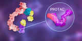
PROTAC, which exploit the ubiquitin-proteasome pathway to specifically degrade target proteins. PROTACs not only solve the problem of undruggability but they also have other advantages compared to traditional drug targeting strategies.
Scientific Reviews
PROTAC, which exploit the ubiquitin-proteasome pathway to specifically degrade target proteins. PROTACs not only solve the problem of undruggability but they also have other advantages compared to traditional drug targeting strategies. -
 Scientific Reviews
PROTAC — Target Selection and Design
Scientific Reviews
PROTAC — Target Selection and Design2022-07-08
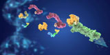
A PROTAC molecule consists of three components: a target protein binding ligand, an E3 ligase ligand, and a linker connecting these two moieties. Here, we will discuss the conventional approaches for the rational design of PROTAC molecules. -
 Scientific Reviews
Organoids represent an important bridge between 2D cultures and in vivo mouse/human models. The organoid technology exerts enormous potential in evaluation of efficacy and toxicity of drugs, regenerative medicine, and precision medicine.In this article, we will briefly introduce organoid technology.
Scientific Reviews
Organoids represent an important bridge between 2D cultures and in vivo mouse/human models. The organoid technology exerts enormous potential in evaluation of efficacy and toxicity of drugs, regenerative medicine, and precision medicine.In this article, we will briefly introduce organoid technology. -
 Scientific Reviews
Development of Intestinal Organoid
Scientific Reviews
Development of Intestinal Organoid2022-07-28
3D organoid is one of the revolutionary developments in biomedical field during the past 10 years. The establishment of intestinal organoids is an important milestone in the development of organoid technology. -
 Scientific Reviews
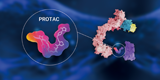
BacPROTACs is composed of a POI ligand, a chemical linker and a ClpCNTD anchor. BacPROTACs can induce in vitro and in vivo degradation of non-eukaryotic proteins in bacteria without the ubiquitin proteasome system.
Scientific Reviews
BacPROTACs is composed of a POI ligand, a chemical linker and a ClpCNTD anchor. BacPROTACs can induce in vitro and in vivo degradation of non-eukaryotic proteins in bacteria without the ubiquitin proteasome system. -
 Scientific Reviews
It's has been proved that p53, as a tumor suppressor gene and immune guardian, may become a destroyer through its own mutation. Moreover, the mechanism of p53 was found to be related to ferroptosis. This article mainly explores the mechanism between p53 and ferroptosis in detail.
Scientific Reviews
It's has been proved that p53, as a tumor suppressor gene and immune guardian, may become a destroyer through its own mutation. Moreover, the mechanism of p53 was found to be related to ferroptosis. This article mainly explores the mechanism between p53 and ferroptosis in detail. -
Scientific Reviews
Organoids have great potential in research of organ development, disease modeling, drug screening and, precision and regenerative medicin. In this article, we will expalin the origin of the organoids. Adult stem cells (ASCs) or pluripotent stem cells (PSCs) , which is the better choice.
-
 Scientific Reviews
TME (Tumor microenvironment) is considered as a complex integrated system, composed of cellular components such as tumor cells and immune cells, as well as non-cellular components such as ECM and cytokines. According to the spatial distribution of immune cells in TME, "hot" and "cold" TME will be explained in this article.
Scientific Reviews
TME (Tumor microenvironment) is considered as a complex integrated system, composed of cellular components such as tumor cells and immune cells, as well as non-cellular components such as ECM and cytokines. According to the spatial distribution of immune cells in TME, "hot" and "cold" TME will be explained in this article. -
 Scientific Reviews
PROTAC - Design Strategy for Targeting
Scientific Reviews
PROTAC - Design Strategy for Targeting2023-04-23
Protein degradation targeting chimera (PROTAC) is a technology that uses the ubiquitin proteasome pathway to silent target protein. However, PROTAC still has problems such as solubility, membrane permeability, and selectivity. In this article, we have summarized three strategies for optimization: light-controlled linker, PAC molecule, and specific E3 ligase. -
 Scientific Reviews
Necroptosis, also known as necroptosis, is a form of regulated necrotizing cell death mediated by RIP1 and RIP3 kinases. Necroptosis is a process that prevents the self-destruction of activated cells that are blocked by apoptosis. Necroptosis plays a tumor suppressor role in most cases. It may provide benefits in the researches of a variety of human diseases involving immune inflammation and cell death.
Scientific Reviews
Necroptosis, also known as necroptosis, is a form of regulated necrotizing cell death mediated by RIP1 and RIP3 kinases. Necroptosis is a process that prevents the self-destruction of activated cells that are blocked by apoptosis. Necroptosis plays a tumor suppressor role in most cases. It may provide benefits in the researches of a variety of human diseases involving immune inflammation and cell death. -
 Scientific Reviews
Stem cell classification and its application
Scientific Reviews
Stem cell classification and its application2023-05-18
Stem cells (SCs) have the unique ability to self-renew and differentiate into different cell types. SCs can differentiate into various types of tissue cells under specific conditions. Additionally, they can be further cultured to form different tissues and organs in the human body. Stem cells have numerous applications in various fields, including cell therapy, organ transplantation, neurodegenerative disease modeling, and drug screening. -
 Scientific Reviews
HLA-E, A Novel Immune Checkpoint
Scientific Reviews
HLA-E, A Novel Immune Checkpoint2023-06-29
Immune checkpoints have immunosuppressive functions. It can be used in the research of tumor immunotherapy. In this article, we introduce a new paper entitled "Immune checkpoint HLA-E: CD94 - NKG2Amediates evasion of circulating tumor cells from NK cell surveillance "research paper. -
 Scientific Reviews
Organoid Culture: Questions & Answers
Scientific Reviews
Organoid Culture: Questions & Answers2023-07-26
In the last issue, we conducted a live lecture on the theme of organoid culture. Today, we have a special topic to solve your doubts in the last live class! -
 Scientific Reviews
IHC is an indispensable technique for studying tissue morphology and in situ antigen expression, but usually only one or two antigens in tissues can be stained for analysis. It cannot judge the results more intuitively. Today, Little M will introduce you to the upgraded version mlHC.
Scientific Reviews
IHC is an indispensable technique for studying tissue morphology and in situ antigen expression, but usually only one or two antigens in tissues can be stained for analysis. It cannot judge the results more intuitively. Today, Little M will introduce you to the upgraded version mlHC. -
 Scientific Reviews
A suitable model is crucial in drug screening experiments. Organs can mimic the three-dimensional functional structure of internal organs, have similar spatial organization to corresponding organs, maintain some key characteristics, and reproduce some physiological functions. They are widely used for modeling and personalized drug screening of diseases such as cancer, infectious diseases, and rare diseases.
Scientific Reviews
A suitable model is crucial in drug screening experiments. Organs can mimic the three-dimensional functional structure of internal organs, have similar spatial organization to corresponding organs, maintain some key characteristics, and reproduce some physiological functions. They are widely used for modeling and personalized drug screening of diseases such as cancer, infectious diseases, and rare diseases. -
 Scientific Reviews
KRAS, a gene we've heard so much about, has quickly risen to fame after shedding its "undruggable" label. After reading numerous articles, it's easy to feel overwhelmed and wonder: What exactly should we know about this often-discussed but previously "undruggable" target KRAS?
Scientific Reviews
KRAS, a gene we've heard so much about, has quickly risen to fame after shedding its "undruggable" label. After reading numerous articles, it's easy to feel overwhelmed and wonder: What exactly should we know about this often-discussed but previously "undruggable" target KRAS? -
 Scientific Reviews
Wnt/β-catenin and tumor EMT
Scientific Reviews
Wnt/β-catenin and tumor EMT2024-03-18
Epithelial-Mesenchymal Transition (EMT) is closely related to the plasticity of tumor cells and is a necessary process for tumor metastasis. Wnt/β-catenin is one of the main actors involved in the EMT process. Today, we’re here to popularize the tumor EMT and Wnt/β-catenin pathway~ -
 Scientific Reviews
The 2024 AACR meeting concluded successfully in California, USA. Which antitumor drugs stole the show at this conference?
Scientific Reviews
The 2024 AACR meeting concluded successfully in California, USA. Which antitumor drugs stole the show at this conference? -
 Scientific Reviews
Science | A "new" mechanism for non-ubiquitinated Midnolin-proteasomal degradation pathway
Scientific Reviews
Science | A "new" mechanism for non-ubiquitinated Midnolin-proteasomal degradation pathway2024-04-26
“ubiquitin-mediated protein degradation” won the Nobel Prize in Chemistry in 2004! In fact, proteasomes degrade not only ubiquitinated proteins but also non-ubiquitinated ones. The mechanism remains shrouded in mystery. After reading this piece today, you might have a lightbulb moment! -
 Scientific Reviews
Exosomes, which won the Nobel Prize in 2013, are still a research hotspot in the national natural sciences, and their popularity has only increased over the past decade (in 2022, they still rank 5th in the national natural sciences hotspots!). Why have exosomes become the darling of scientific research? Let's take a look together~
Scientific Reviews
Exosomes, which won the Nobel Prize in 2013, are still a research hotspot in the national natural sciences, and their popularity has only increased over the past decade (in 2022, they still rank 5th in the national natural sciences hotspots!). Why have exosomes become the darling of scientific research? Let's take a look together~ -
 Scientific Reviews
Degrade target proteins through the autophagy-lysosome pathway including LYTAC, AUTAC, and ATTEC have gained increasing attention in recent years due to their significant research potential!
Scientific Reviews
Degrade target proteins through the autophagy-lysosome pathway including LYTAC, AUTAC, and ATTEC have gained increasing attention in recent years due to their significant research potential! -
 Scientific Reviews
Antibodies!
Scientific Reviews
Antibodies!2024-07-26
Today, We introduce antibodies for everyone! -
 Scientific Reviews
What Are Popular Anti-tumor Drug Targets?
Scientific Reviews
What Are Popular Anti-tumor Drug Targets?2024-08-20
The rapid development of targeted anti-cancer drugs has spurred diverse research across various modalities. These include small molecules, monoclonal antibodies (mAbs), cell immunotherapies, antibody-drug conjugates (ADCs), and PROTACs (proteolysis targeting chimeras). -
.jpg) Scientific Reviews
nside cells, the homeostasis and degradation of proteins is a precisely regulated process. If proteins cannot be degraded in time, it may lead to the occurrence of various diseases such as neurodegenerative diseases and cancer. This article will tell you the process of how proteins are recognized, labeled and then degraded!
Scientific Reviews
nside cells, the homeostasis and degradation of proteins is a precisely regulated process. If proteins cannot be degraded in time, it may lead to the occurrence of various diseases such as neurodegenerative diseases and cancer. This article will tell you the process of how proteins are recognized, labeled and then degraded! -
 Scientific Reviews
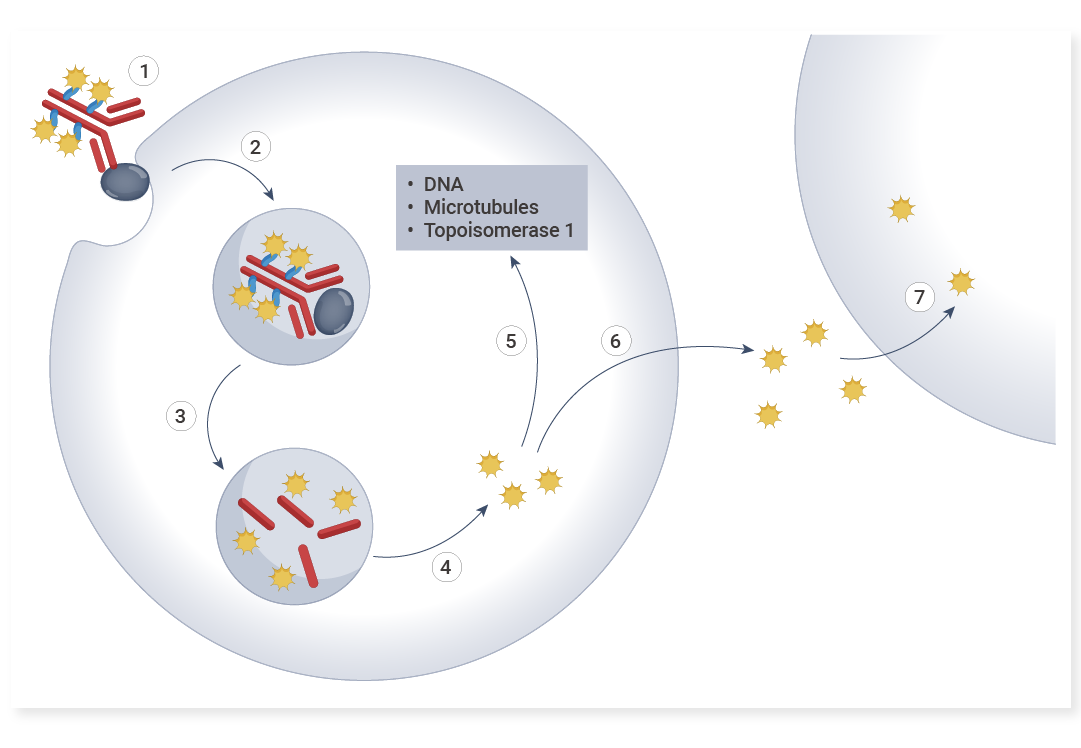
This article will walk you through the remarkable impacts of anti-payload antibodies, delving into how these molecules are revolutionizing drug development and biological research!
Scientific Reviews
This article will walk you through the remarkable impacts of anti-payload antibodies, delving into how these molecules are revolutionizing drug development and biological research! -
 Scientific Reviews
Cell Migration vs. Invasion: Differences Revealed by Scratch Assays and Transwell Experiments
Scientific Reviews
Cell Migration vs. Invasion: Differences Revealed by Scratch Assays and Transwell Experiments2025-05-30
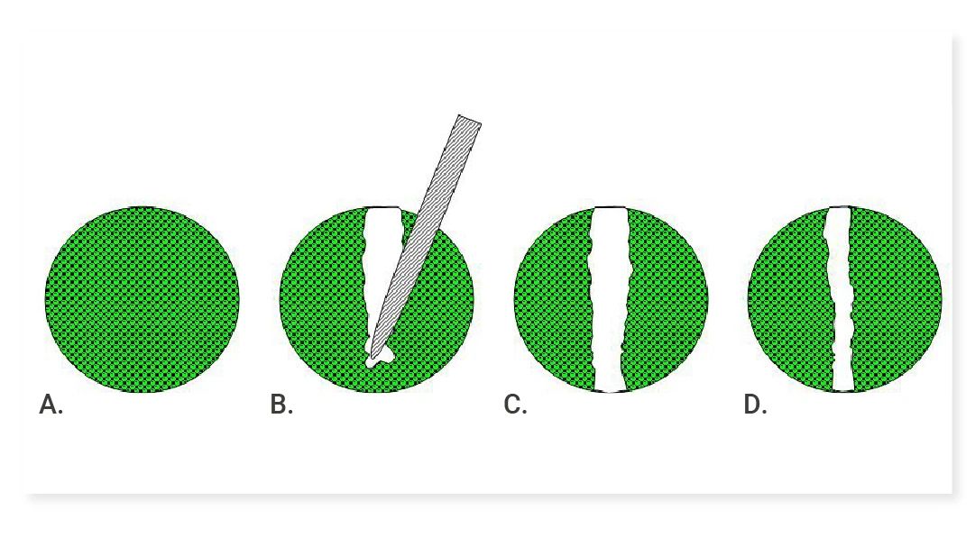
In scientific research, cell migration and invasion are crucial for understanding many important biological processes. This article delves into commonly used detection methods: the scratch assay and Transwell migration/invasion assay. -
 Scientific Reviews
In this issue, we will conduct an in-depth interpretation from the dimensions of nanoparticle design, mechanism of action, in vivo and in vitro efficacy, and immune regulation, revealing how this research brings new hope for the treatment of invasive tumors through interdisciplinary innovation!
Scientific Reviews
In this issue, we will conduct an in-depth interpretation from the dimensions of nanoparticle design, mechanism of action, in vivo and in vitro efficacy, and immune regulation, revealing how this research brings new hope for the treatment of invasive tumors through interdisciplinary innovation! -
 Scientific Reviews
IHC, ICC, IF Techniques: A Practical Guide
Scientific Reviews
IHC, ICC, IF Techniques: A Practical Guide2025-07-25
Confused About IHC/ICC/IF? Why Does Immunostaining Seem So Complicated? Read This Now! Master Immunostaining with Confidence! -
 Scientific Reviews
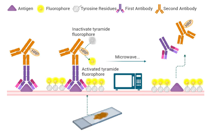
This detailed guide outlines the standardized experimental protocols for multiplex immunohistochemistry (mIHC), systematically summarizes common technical issues encountered during sample preparation, staining and imaging processes, and provides practical troubleshooting solutions to ensure reliable and reproducible results in biomedical research and clinical sample analysis.
Scientific Reviews
This detailed guide outlines the standardized experimental protocols for multiplex immunohistochemistry (mIHC), systematically summarizes common technical issues encountered during sample preparation, staining and imaging processes, and provides practical troubleshooting solutions to ensure reliable and reproducible results in biomedical research and clinical sample analysis. -
 Scientific Reviews
Exosomes—natural nanoscale carriers—are revolutionizing targeted therapy. This article uncovers the science behind their precision in drug delivery.
Scientific Reviews
Exosomes—natural nanoscale carriers—are revolutionizing targeted therapy. This article uncovers the science behind their precision in drug delivery. -
Scientific Reviews
Cracking the PROTAC Permeability Barrier: CD36-Mediated Endocytosis as a Potential Breakthrough2025-12-03
This article provides an in-depth analysis of cutting-edge literature revealing CD36 as a key mediator of cellular uptake for PROTACs and bRO5 compounds. By structurally optimizing PROTAC molecules to enhance their affinity for CD36, membrane permeability can be markedly improved, leading to significantly enhanced antitumor efficacy. -
 Scientific Reviews
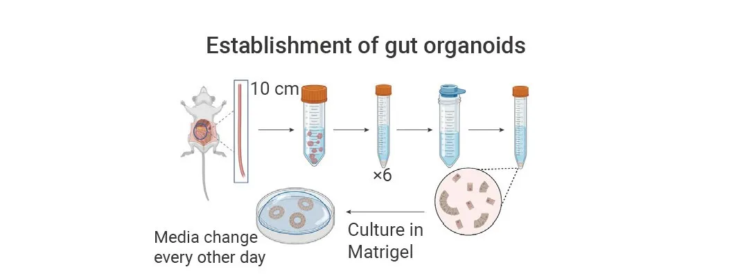
Efficient Generation of Mouse Small Intestinal Organoids: A Complete Experimental Protocol Guide
Scientific Reviews
Efficient Generation of Mouse Small Intestinal Organoids: A Complete Experimental Protocol Guide2025-12-10
How to successfully create mouse small intestine organoids? A detailed, hands-on guide to the entire process, all in one article! -
 Scientific Reviews
This article walks you through the experimental design and workflow of flow cytometry, delivering a clear, dynamic, and professional overview to elevate your research.
Scientific Reviews
This article walks you through the experimental design and workflow of flow cytometry, delivering a clear, dynamic, and professional overview to elevate your research. -
 Scientific Reviews
Molecular glue degraders have evolved from a serendipitous observation to one of the most dynamic and transformative fields in biomedical research.
Scientific Reviews
Molecular glue degraders have evolved from a serendipitous observation to one of the most dynamic and transformative fields in biomedical research. -
 EDM
Research Solution for Breast Cancer TME
EDM
Research Solution for Breast Cancer TME2025-05-21
This review examines the complex interactions within the breast cancer tumor microenvironment, emphasizing how understanding these dynamics is essential for developing effective therapies and overcoming immune resistance. -
 EDM
Targeting the ‘Undruggable’ with PROTACs
EDM
Targeting the ‘Undruggable’ with PROTACs2025-06-18
This review introduces the fundamental principles and mechanisms of PROTACs, highlights recent advances in molecular design and clinical development, and discusses emerging opportunities and remaining challenges in targeted protein degradation. -
 EDM
This review discusses the fundamentals of lipid biology and lipid metabolism, examines dysregulated lipid metabolism in cancer, and summarizes therapeutic strategies targeting lipid metabolic pathways.
EDM
This review discusses the fundamentals of lipid biology and lipid metabolism, examines dysregulated lipid metabolism in cancer, and summarizes therapeutic strategies targeting lipid metabolic pathways. -
 EDM
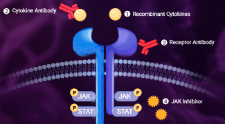
This review critically examines the role of the JAK-STAT pathway in immunity and autoimmune diseases, reviews current clinical applications of targeted therapies, and highlights emerging trends in autoimmune drug development.
EDM
This review critically examines the role of the JAK-STAT pathway in immunity and autoimmune diseases, reviews current clinical applications of targeted therapies, and highlights emerging trends in autoimmune drug development. -
 EDM
Combating Immune Evasion in Cancer
EDM
Combating Immune Evasion in Cancer2025-10-15
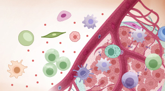
This review summarizes the mechanisms by which tumors evade immune surveillance and discusses therapeutic strategies to restore antitumor immunity. -
 EDM
This review summarizes the mechanisms of drug resistance in triple-negative breast cancer and highlights emerging therapeutic strategies.
EDM
This review summarizes the mechanisms of drug resistance in triple-negative breast cancer and highlights emerging therapeutic strategies. -
 EDM
Bispecific Antibodies in Cancer Therapy
EDM
Bispecific Antibodies in Cancer Therapy2025-10-29
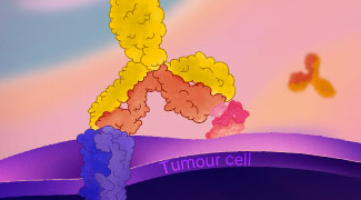
This review highlights the mechanisms, technology platforms, and clinical progress of bispecific antibodies, and discusses emerging strategies to guide future development. -
 EDM
This review provides insights into the pancreatic ductal adenocarcinoma tumor microenvironment, highlighting its cellular composition, stromal heterogeneity, and immune-targeting strategies.
EDM
This review provides insights into the pancreatic ductal adenocarcinoma tumor microenvironment, highlighting its cellular composition, stromal heterogeneity, and immune-targeting strategies. -
 EDM
This review outlines macrophage functions and classification, and provides practical experimental guidelines for macrophage differentiation, polarization, and identification.
EDM
This review outlines macrophage functions and classification, and provides practical experimental guidelines for macrophage differentiation, polarization, and identification. -
 EDM
Highlight single-cell metabolomics and stable isotope tracing (SIT) in uncovering metabolic heterogeneity and nutrient flux dynamics in tumors.
EDM
Highlight single-cell metabolomics and stable isotope tracing (SIT) in uncovering metabolic heterogeneity and nutrient flux dynamics in tumors. -
 EDM
Summarize LLPS principles, regulatory mechanisms, experimental strategies, and pathological roles to inspire mechanism-driven research and translational applications.
EDM
Summarize LLPS principles, regulatory mechanisms, experimental strategies, and pathological roles to inspire mechanism-driven research and translational applications. -
 EDM
Tumor Organoid-Immune Cell Co-Culture Models: Advances, Applications, and Future Directions
EDM
Tumor Organoid-Immune Cell Co-Culture Models: Advances, Applications, and Future Directions2026-05-07
Summarize technical strategies, application advances, and future directions of tumor organoid–immune co-culture systems. -
 EDM
Review mechanisms, major challenges, and efficacy-enhancing strategies of TCR-T therapy in solid tumors, with implications for future research and clinical translation.
EDM
Review mechanisms, major challenges, and efficacy-enhancing strategies of TCR-T therapy in solid tumors, with implications for future research and clinical translation. -
 EDM
The Dual Role of Cellular Senescence in Cancer: Mechanisms, Microenvironment, and Therapeutics
EDM
The Dual Role of Cellular Senescence in Cancer: Mechanisms, Microenvironment, and Therapeutics2026-05-21
Explore the dual role of cellular senescence in cancer, including tumor suppression, SASP-driven tumor promotion, senescence heterogeneity and plasticity, and advances in senescence-targeted therapies. -
 EDM
Breaking Immune Resistance in Colorectal Cancer: From Molecular Mechanisms to Precision Therapy
EDM
Breaking Immune Resistance in Colorectal Cancer: From Molecular Mechanisms to Precision Therapy2026-06-25
Explore CRC molecular subtypes, immune landscapes, resistance mechanisms, and emerging precision strategies for improving outcomes across distinct subtypes. -
 EDM
Explore the mechanisms driving immune tolerance breakdown in autoimmune diseases and emerging tolerance-restoring strategies, with a focus on IL-2-based immune modulation.
EDM
Explore the mechanisms driving immune tolerance breakdown in autoimmune diseases and emerging tolerance-restoring strategies, with a focus on IL-2-based immune modulation. -
 EDM
Review the evolution of molecular glues from serendipitous discovery to rational design, highlighting how emerging targets and advances in screening, proteomics, structural biology, and AI are expanding the druggable proteome.
EDM
Review the evolution of molecular glues from serendipitous discovery to rational design, highlighting how emerging targets and advances in screening, proteomics, structural biology, and AI are expanding the druggable proteome. -
 Blog
EC0489, a SMDC for Cancer Therapy
Blog
EC0489, a SMDC for Cancer Therapy2019-03-25
EC0489, a conjugate of folic acid and desacetyl vinblastine hydrazide, is a SMDC under development for the treatment of solid tumours. -
 Blog
R916562, a dual Axl/VEGF-R2 inhibitor, could be a potential anti-angiogenic and anti-metastatic drug for cancer chemotherapy.
Blog
R916562, a dual Axl/VEGF-R2 inhibitor, could be a potential anti-angiogenic and anti-metastatic drug for cancer chemotherapy. -
 Blog
Role of PRMT7 Probe SGC3027 in Cancer
Blog
Role of PRMT7 Probe SGC3027 in Cancer2019-03-27
SGC3027 is the first potent, selective and cell active chemical probe for PRMT7. SGC3027 is also a pro-drug, which converts to the active compound SGC8158 -
 Blog
Alofanib, An Allosteric Inhibitor of FGFR2
Blog
Alofanib, An Allosteric Inhibitor of FGFR22019-03-28
Alofanib is an allosteric inhibitor of FGFR2 and inhibits FGF-mediated proliferation with GI50s of 16-370 nM, showing pronounced antitumor activity. -
 Blog
A Novel and Efficacious RAF Inhibitor RAF709
Blog
A Novel and Efficacious RAF Inhibitor RAF7092019-03-30
RAF709, a novel and efficacious RAF inhibitor, activates the MAPK pathway and shows antitumor activity in tumor cells harboring BRAF or RAS mutations. -
 Blog
AZD3229 is a Potent Pan-KIT Mutant Inhibitor
Blog
AZD3229 is a Potent Pan-KIT Mutant Inhibitor2019-04-04
AZD3229 is a potent, pan-KIT mutant inhibitor with potent single digit nM growth inhibition against a diverse panel of mutant KIT driven Ba/F3 cell lines. -
 Blog
BR351 is a Brain Penetrant MMP Inhibitor
Blog
BR351 is a Brain Penetrant MMP Inhibitor2019-04-05
BR351 is a brain penetrant MMP inhibitor, and a potential tool for the molecular imaging of activated MMPs with PET, with an IC50 in the nanomolar range. -
 Blog
SGC-GAK-1 is a potent, selective, and cell-active GAK inhibitor and shows potent anti-proliferative activity in LNCaP and 22Rv1 cells.
Blog
SGC-GAK-1 is a potent, selective, and cell-active GAK inhibitor and shows potent anti-proliferative activity in LNCaP and 22Rv1 cells. -
 Blog
COH000 is an allosteric, covalent and irreversible inhibitor of SUMO-activating enzyme, with an IC50 of 0.2 μM for SUMOylation in vitro.
Blog
COH000 is an allosteric, covalent and irreversible inhibitor of SUMO-activating enzyme, with an IC50 of 0.2 μM for SUMOylation in vitro. -
 Blog
MK-0429 is An Oral Integrin (αvβ3) Inhibitor
Blog
MK-0429 is An Oral Integrin (αvβ3) Inhibitor2019-04-11
MK-0429, an orally active αvβ3 inhibitor, is a potential therapeutic agent for the prevention of kidney fibrosis, melanoma and osteoporosis. -
 Blog
AZ304, a Dual BRAF Inhibitor Against Cancer
Blog
AZ304, a Dual BRAF Inhibitor Against Cancer2019-04-12
AZ304 is a potent BRAF inhibitor, blocks both wild type BRAF and V600E mutant BRAF activity, with IC50s in the nanomolar range. -
 Blog
Erteberel is a Selective ERβ Agonist
Blog
Erteberel is a Selective ERβ Agonist2019-04-16
Erteberel (LY500307) is a synthetic, nonsteroidal estrogen which acts as a selective ERβ agonist and under development for the treatment of schizophrenia. -
 Blog
MBQ-167 is a dual Rac/Cdc42 inhibitor in in metastatic cancer, with IC50s of 103 nM for Rac 1/2/3 and 78 nM for Cdc42 in MDA-MB-231 cells, respectively.
Blog
MBQ-167 is a dual Rac/Cdc42 inhibitor in in metastatic cancer, with IC50s of 103 nM for Rac 1/2/3 and 78 nM for Cdc42 in MDA-MB-231 cells, respectively. -
 Blog
Multiple Tyrosine Kinases Inhibitor TAS-115
Blog
Multiple Tyrosine Kinases Inhibitor TAS-1152019-04-19
TAS-115 is a potent VEGFR and c-Met/HGFR-targeted kinase inhibitor with IC50s of 30 and 32 nM for rVEGFR2 and rMET, respectively. -
 Blog
JNJ-64619178 is a selective and pseudo-irreversible PRMT5 inhibitor with an IC50 of 0.14 nM. Has potent Activity In Lung Cancer.
Blog
JNJ-64619178 is a selective and pseudo-irreversible PRMT5 inhibitor with an IC50 of 0.14 nM. Has potent Activity In Lung Cancer. -
 Blog
(E)-AG 99 is an EGFR inhibitor and shows a growth-inhibition on not only serum-starved cells but also normally grown cells. Treatment for Bladder cancer.
Blog
(E)-AG 99 is an EGFR inhibitor and shows a growth-inhibition on not only serum-starved cells but also normally grown cells. Treatment for Bladder cancer. -
 Blog
Borussertib is a covalent-allosteric and first-in-class inhibitor of protein kinase Akt, with an IC50 of 0.8 nM and a Ki of 2.2 nM for Akt-wt.
Blog
Borussertib is a covalent-allosteric and first-in-class inhibitor of protein kinase Akt, with an IC50 of 0.8 nM and a Ki of 2.2 nM for Akt-wt. -
 Blog
NRX-252262 is a β-catenin:β-TrCP interaction enhancer, and its cognate E3 ligase, SCFβ-TrCP, induces mutant β-catenin degradation, with an EC50 of 3.8 nM
Blog
NRX-252262 is a β-catenin:β-TrCP interaction enhancer, and its cognate E3 ligase, SCFβ-TrCP, induces mutant β-catenin degradation, with an EC50 of 3.8 nM -
 Blog
TAK-981 is a selective inhibitor of the SUMOylation enzymatic cascade, with potential immune-activating and antineoplastic activities
Blog
TAK-981 is a selective inhibitor of the SUMOylation enzymatic cascade, with potential immune-activating and antineoplastic activities -
 Blog
SLLN-15 is an oral activ enhancer of autophagy that activates cytostatic macroautophagy/autophagy in triple-negative breast cancer (TNBC).
Blog
SLLN-15 is an oral activ enhancer of autophagy that activates cytostatic macroautophagy/autophagy in triple-negative breast cancer (TNBC). -
 Blog
CB-6644 is a selective non-ATP-competitive inhibitor of the RUVBL1/2 complex. CB-6644 significantly reduces tumor growth without obvious toxicity.
Blog
CB-6644 is a selective non-ATP-competitive inhibitor of the RUVBL1/2 complex. CB-6644 significantly reduces tumor growth without obvious toxicity. -
 Blog
NSC 228155 is a activator of EGFR and a potent inhibitor of KIX-KID interaction. NSC 228155 shows excellent anti-tumor activity.
Blog
NSC 228155 is a activator of EGFR and a potent inhibitor of KIX-KID interaction. NSC 228155 shows excellent anti-tumor activity. -
 Blog
BAY-8002 is a selective and orally active inhibitor of monocarboxylate transporter 1 (MCT1), with an IC50 of 85 nM. Has potential to treat lymphoma.
Blog
BAY-8002 is a selective and orally active inhibitor of monocarboxylate transporter 1 (MCT1), with an IC50 of 85 nM. Has potential to treat lymphoma. -
 Blog
MRTX-1257 is a selective, irreversible, covalent and oral active KRAS G12C inhibitor, with an IC50 of 900 pM for KRAS dependent ERK phosphorylation.
Blog
MRTX-1257 is a selective, irreversible, covalent and oral active KRAS G12C inhibitor, with an IC50 of 900 pM for KRAS dependent ERK phosphorylation. -
 Blog
MM-589 is an inhibitor of WDR5 and MLL protein-protein interaction. Binds to WDR5 (IC50=0.90 nM) and inhibits the MLL H3K4 methyltransferase activity.
Blog
MM-589 is an inhibitor of WDR5 and MLL protein-protein interaction. Binds to WDR5 (IC50=0.90 nM) and inhibits the MLL H3K4 methyltransferase activity. -
 Blog
USL311 is a selective CXCR4 antagonist, which prevents the binding of stromal-cell derived factor-1 (SDF-1 or CXCL12) to CXCR4. Anti-tumor activity.
Blog
USL311 is a selective CXCR4 antagonist, which prevents the binding of stromal-cell derived factor-1 (SDF-1 or CXCL12) to CXCR4. Anti-tumor activity. -
 Blog
JI051 is a stabilizer for the Hes1-PHB2 interaction, induces cell-cycle arrest by inhibiting the Notch downstream effector gene Hes1.
Blog
JI051 is a stabilizer for the Hes1-PHB2 interaction, induces cell-cycle arrest by inhibiting the Notch downstream effector gene Hes1. -
 Blog
TX1-85-1, a ATP-competitive ligand of Her3, covalent modification of Her3 to inhibit Her3 signaling
Blog
TX1-85-1, a ATP-competitive ligand of Her3, covalent modification of Her3 to inhibit Her3 signaling2019-06-01
TX1-85-1 is a Her3 (ErbB3) inhibitor with an IC50 of 23 nM. TX1-85-1 induces partial degradation of Her3 protein and attenuates Her3-dependent signaling. -
 Blog
IWP-O1, a Highly Potent Porcupine Inhibitor, Functions by Preventing the Secretion of Wnt Proteins
Blog
IWP-O1, a Highly Potent Porcupine Inhibitor, Functions by Preventing the Secretion of Wnt Proteins2019-06-03
IWP-O1 is a Porcupine (Porcn) inhibitor, with an EC50 of 80 pM in L-Wnt-STF cells. IWP-O1 functions by preventing the secretion of Wnt proteins[ -
 Blog
JMS-17-2 is a CX3CR1 Antagonist
Blog
JMS-17-2 is a CX3CR1 Antagonist2019-06-12
JMS-17-2 is a potent and selective CX3CR1 antagonist with an IC50 of 0.32 nM. Has potential to treat cancers such as breast cancer. -
 Blog
PTC299 is a dual and orally active DHODH and VEGF inhibitor, has broad and potent activity against hematological cancer cells.
Blog
PTC299 is a dual and orally active DHODH and VEGF inhibitor, has broad and potent activity against hematological cancer cells. -
 Blog
AMG 487 is an orally active and selective antagonist of CXC chemokine receptor 3 (CXCR3). AMG 487 has potential to treat metastatic cancer.
Blog
AMG 487 is an orally active and selective antagonist of CXC chemokine receptor 3 (CXCR3). AMG 487 has potential to treat metastatic cancer. -
 Blog
FGTI-2734 is a dual farnesyl and geranylgeranyl transferase-1 inhibitor. FGTI-2734 prevents membrane localization of KRAS and mutant KRAS pancreatic tumors.
Blog
FGTI-2734 is a dual farnesyl and geranylgeranyl transferase-1 inhibitor. FGTI-2734 prevents membrane localization of KRAS and mutant KRAS pancreatic tumors. -
 Blog
M-89 is a specific menin inhibitor, with a Kd of 1.4 nM. M-89 inhibits the Menin-MLL protein-protein interaction and has potential to treat MLL leukemia.
Blog
M-89 is a specific menin inhibitor, with a Kd of 1.4 nM. M-89 inhibits the Menin-MLL protein-protein interaction and has potential to treat MLL leukemia. -
 Blog
ACBI1 is a PROTAC Degrader of BAF Complex
Blog
ACBI1 is a PROTAC Degrader of BAF Complex2019-08-07
ACBI1 is a potent PROTAC degrader of BAF ATPase subunits SMARCA2, SMARCA4 and PBRM1, with DC50s of 6 nM, 11 nM and 32 nM in MV-4-11 cells, respectively. -
 Blog
BI-882370 is a potent RAF kinase inhibitor with IC50s of 0.4, 0.8, and 0.6 nM for oncogenic BRAFV600E-mutant, the WT BRAF and CRAF kinases , respectively.
Blog
BI-882370 is a potent RAF kinase inhibitor with IC50s of 0.4, 0.8, and 0.6 nM for oncogenic BRAFV600E-mutant, the WT BRAF and CRAF kinases , respectively. -
 Blog
MSX-122 is an orally active partial antagonist of CXCR4, inhibiting CXCR4/CXCL12 actions. MSX-122 has both anti-tumor and anti-metastasis activities.
Blog
MSX-122 is an orally active partial antagonist of CXCR4, inhibiting CXCR4/CXCL12 actions. MSX-122 has both anti-tumor and anti-metastasis activities. -
 Blog
ZT-12-037-01 is a ATP-competitive and specific STK19 inhibitor and inhibits oncogenic NRAS-driven melanocyte malignant transformation.
Blog
ZT-12-037-01 is a ATP-competitive and specific STK19 inhibitor and inhibits oncogenic NRAS-driven melanocyte malignant transformation. -
 Blog
PT2977 is an orally active and selective HIF-2α inhibitor with an IC50 of 9 nM. PT2977 is a potential treatment for ccRCC and VHL disease.
Blog
PT2977 is an orally active and selective HIF-2α inhibitor with an IC50 of 9 nM. PT2977 is a potential treatment for ccRCC and VHL disease. -
 Blog
BI-2852 is a potent KRAS inhibitor with nanomolar affinity and reduces pERK and pAKT levels in a dose-dependent manner in a KRAS mutant cell line NCI-H358.
Blog
BI-2852 is a potent KRAS inhibitor with nanomolar affinity and reduces pERK and pAKT levels in a dose-dependent manner in a KRAS mutant cell line NCI-H358. -
 Blog
MS31 is a highly selective spindlin 1 inhibitor, which inhibits the interactions between SPIN1 and H3K4me3. MS31 is not toxic to nontumorigenic cells.
Blog
MS31 is a highly selective spindlin 1 inhibitor, which inhibits the interactions between SPIN1 and H3K4me3. MS31 is not toxic to nontumorigenic cells. -
 Blog
BAY-293 is a potent inhibitor of Son of Sevenless 1 (SOS1) and blocks RAS activation via disruption of the KRAS-SOS1 interaction with an IC50 of 21 nM.
Blog
BAY-293 is a potent inhibitor of Son of Sevenless 1 (SOS1) and blocks RAS activation via disruption of the KRAS-SOS1 interaction with an IC50 of 21 nM. -
 Blog
WZ811 is an Orally Active CXCR4 Antagonist
Blog
WZ811 is an Orally Active CXCR4 Antagonist2019-09-07
WZ811 is an orally active, highly potent competitive antagonist of CXCR4, which inhibits chronic lymphocytic leukemia progression and tumorigenesis. -
 Blog
SGC-iMLLT is a potent, selective MLLT1/3-histone interactions inhibitor and shows high binding activity towards MLLT1 YEATS domain and MLLT3 YD.
Blog
SGC-iMLLT is a potent, selective MLLT1/3-histone interactions inhibitor and shows high binding activity towards MLLT1 YEATS domain and MLLT3 YD. -
 Blog
RU-302 is a pan-TAM inhibitor that blocks the TAM Ig1 ectodomain and the Gas6 Lg domain interaction. RU-302 blocks Gas6-inducible Axl receptor activation.
Blog
RU-302 is a pan-TAM inhibitor that blocks the TAM Ig1 ectodomain and the Gas6 Lg domain interaction. RU-302 blocks Gas6-inducible Axl receptor activation. -
 Blog
BPK-29 disrupts the NR0B1 protein-protein interactions and impairs the anchorage-independent growth of KEAP1-mutant cancer cells.
Blog
BPK-29 disrupts the NR0B1 protein-protein interactions and impairs the anchorage-independent growth of KEAP1-mutant cancer cells. -
 Blog
NV03 is a potent and selective antagonist of UHRF1-H3K9me3 interaction by binding to UHRF1 TTD with a Kd of 2.4 μM and has anticancer activity.
Blog
NV03 is a potent and selective antagonist of UHRF1-H3K9me3 interaction by binding to UHRF1 TTD with a Kd of 2.4 μM and has anticancer activity. -
 Blog
sAJM589 is a Myc inhibitor which potently disrupts the Myc-Max heterodimer in a dose dependent manner with an IC50 of 1.8 μM.
Blog
sAJM589 is a Myc inhibitor which potently disrupts the Myc-Max heterodimer in a dose dependent manner with an IC50 of 1.8 μM. -
 Blog
IPR-803 is a potent inhibitor of the uPAR•uPA protein-protein interaction, and binds directly to uPAR with sub-micromolar affinity.
Blog
IPR-803 is a potent inhibitor of the uPAR•uPA protein-protein interaction, and binds directly to uPAR with sub-micromolar affinity. -
 Blog
Vorolanib is an orally active, multikinase VEGF/PDGF receptor inhibitor with antitumor activity and is expected to disrupt tumor angiogenesis.
Blog
Vorolanib is an orally active, multikinase VEGF/PDGF receptor inhibitor with antitumor activity and is expected to disrupt tumor angiogenesis. -
 Blog
CCG-222740 is a potent and selective MRTF pathway inhibitor. It effectively reduces fibrosis in the skin and blocks melanoma metastasis.
Blog
CCG-222740 is a potent and selective MRTF pathway inhibitor. It effectively reduces fibrosis in the skin and blocks melanoma metastasis. -
 Blog
AC-73 is a first specific, orally active the cluster of differentiation 147 (CD147) inhibitor and specifically disrupts CD147 dimerization.
Blog
AC-73 is a first specific, orally active the cluster of differentiation 147 (CD147) inhibitor and specifically disrupts CD147 dimerization. -
 Blog
ADH-503 is an orally active and allosteric CD11b agonist and leads to the repolarization of tumor-associated macrophages.
Blog
ADH-503 is an orally active and allosteric CD11b agonist and leads to the repolarization of tumor-associated macrophages. -
 Blog
BI-9321 is a potent, selective and cellular active NSD3-PWWP1 domain antagonist, and specifically disrupts histone interactions of the NSD3-PWWP1 domain.
Blog
BI-9321 is a potent, selective and cellular active NSD3-PWWP1 domain antagonist, and specifically disrupts histone interactions of the NSD3-PWWP1 domain. -
 Blog
Balixafortide is a potent, selective peptidic CXCR4 antagonist with anti-cancer effects, and blocks β-arrestin recruitment and calcium flux.
Blog
Balixafortide is a potent, selective peptidic CXCR4 antagonist with anti-cancer effects, and blocks β-arrestin recruitment and calcium flux. -
 Blog
CCT365623, an Orally Active LOX Inhibitor, Supresses EGFR (pY1068) and AKT Phosphorylation
Blog
CCT365623, an Orally Active LOX Inhibitor, Supresses EGFR (pY1068) and AKT Phosphorylation2020-04-16
CCT365623 is an orally active LOX inhibitor, suppresses EGFR (pY1068) and AKT phosphorylation driven by EGF. CCT365623 has good pharmacokinetic properties. -
 Blog
SNDX-5613 is a potent, selective, small molecule inhibitor of the Menin-MLL binding interaction for targeted therapy in MLL-rearranged leukemias.
Blog
SNDX-5613 is a potent, selective, small molecule inhibitor of the Menin-MLL binding interaction for targeted therapy in MLL-rearranged leukemias. -
 Blog
MYCMI-6 is a selective MYC:MAX protein interactions inhibitor, which blocks MYC-driven transcription and binds selectively to the MYC bHLHZip domain.
Blog
MYCMI-6 is a selective MYC:MAX protein interactions inhibitor, which blocks MYC-driven transcription and binds selectively to the MYC bHLHZip domain. -
 Blog
TED-347 is a potent, irreversible, covalent and allosteric inhibitor at YAP-TEAD protein-protein interaction with antitumor activity.
Blog
TED-347 is a potent, irreversible, covalent and allosteric inhibitor at YAP-TEAD protein-protein interaction with antitumor activity. -
 Blog
M-808 is a highly potent and efficacious covalent Menin-MLL interaction inhibitor. M-808 has a binding IC50 value of 2.6 nM.
Blog
M-808 is a highly potent and efficacious covalent Menin-MLL interaction inhibitor. M-808 has a binding IC50 value of 2.6 nM. -
 Blog
EW-7195 is a potent and selective ALK5 inhibitor, and efficiently inhibits TGF-β1-induced Smad signaling, EMT and breast tumour metastasis to the lung.
Blog
EW-7195 is a potent and selective ALK5 inhibitor, and efficiently inhibits TGF-β1-induced Smad signaling, EMT and breast tumour metastasis to the lung. -
 Blog
MCP110 is an inhibitor of Ras/Raf-1 interaction in human cancer cells. The Ras family GTPases play a central role in the growth factor signaling.
Blog
MCP110 is an inhibitor of Ras/Raf-1 interaction in human cancer cells. The Ras family GTPases play a central role in the growth factor signaling. -
 Blog
SP-8356, an orally active CD147inhibitor, exerts anti-breast cancer effects by inhibiting NF-κB signaling. Anti-atherosclerotic effects.
Blog
SP-8356, an orally active CD147inhibitor, exerts anti-breast cancer effects by inhibiting NF-κB signaling. Anti-atherosclerotic effects. -
 Blog
CB-1158, a potent and orally bioavailable inhibitor of arginase, blocks myeloid cell-mediated immune suppression in the tumor microenvironment.
Blog
CB-1158, a potent and orally bioavailable inhibitor of arginase, blocks myeloid cell-mediated immune suppression in the tumor microenvironment. -
 Blog
Verucopeptin inhibits v-ATPase activity by directly targeting the v-ATPase ATP6V1G subunit but not ATP1V1B2 or ATP6V1D. Also a potent HIF-1 inhibitor.
Blog
Verucopeptin inhibits v-ATPase activity by directly targeting the v-ATPase ATP6V1G subunit but not ATP1V1B2 or ATP6V1D. Also a potent HIF-1 inhibitor. -
 Blog
BC-LI-0186 is a selective inhibitor of LeuRS and RagD interaction (IC50=46.11 nM). BC-LI-0186 suppresses the activity of cancer-associated MTOR mutants.
Blog
BC-LI-0186 is a selective inhibitor of LeuRS and RagD interaction (IC50=46.11 nM). BC-LI-0186 suppresses the activity of cancer-associated MTOR mutants. -
 Blog
E7820, sulfonamide derivative, is a unique angiogenesis inhibitor suppressing an expression of integrin alpha2 subunit on endothelium.
Blog
E7820, sulfonamide derivative, is a unique angiogenesis inhibitor suppressing an expression of integrin alpha2 subunit on endothelium. -
 Blog
Cyclo(-RGDfK) is a selective inhibitor of the αvβ3 integrin. Cyclo(-RGDfK) potently targets cancer cells through binding to the cell surface αvβ3 integrin.
Blog
Cyclo(-RGDfK) is a selective inhibitor of the αvβ3 integrin. Cyclo(-RGDfK) potently targets cancer cells through binding to the cell surface αvβ3 integrin. -
 Blog
Cilengitide is a compound targeting angiogenesis, the cornerstone of tumor growth and metastasis. ανβ3 and ανβ5 are the target of Cilengitide.
Blog
Cilengitide is a compound targeting angiogenesis, the cornerstone of tumor growth and metastasis. ανβ3 and ανβ5 are the target of Cilengitide. -
 Blog
Tetrac inhibits the cellular actions of thyroid hormone initiated at the hormone receptor on plasma membrane integrin alphavbeta3.
Blog
Tetrac inhibits the cellular actions of thyroid hormone initiated at the hormone receptor on plasma membrane integrin alphavbeta3. -
 Blog
GNE-371, a Chemical Probe for the Second Bromodomains of TFIID Subunit 1 and TFIID Subunit 1-Like
Blog
GNE-371, a Chemical Probe for the Second Bromodomains of TFIID Subunit 1 and TFIID Subunit 1-Like2020-12-09
GNE-371 is a selective chemical probe for the second bromodomains of human transcription-initiation-factor TFIID subunit 1 and TFIID subunit 1-like. -
 Blog
WL47, a high-affinity cavolin-1 (CAV1) ligand (Kd=23 nM), act as a potent and selective disrupter of CAV1 oligomers.
Blog
WL47, a high-affinity cavolin-1 (CAV1) ligand (Kd=23 nM), act as a potent and selective disrupter of CAV1 oligomers. -
 Blog
ENMD-1198 is an orally active microtubule-targeting agent with antiproliferative and antiangiogenic activity.
Blog
ENMD-1198 is an orally active microtubule-targeting agent with antiproliferative and antiangiogenic activity. -
 Blog
FAPI-46 is a quinoline-based FAP-targeted radiotracer. FAPI-46 has higher tumor uptake and prolonged tumor accumulation.
Blog
FAPI-46 is a quinoline-based FAP-targeted radiotracer. FAPI-46 has higher tumor uptake and prolonged tumor accumulation. -
 Blog
L-Moses is the first potent, selective, and cell-active PCAF Brd inhibitor. L-Moses disrupts PCAF-Brd histone H3.3 interaction.
Blog
L-Moses is the first potent, selective, and cell-active PCAF Brd inhibitor. L-Moses disrupts PCAF-Brd histone H3.3 interaction. -
 Blog
MR837 is an Inhibitor of NSD2-PWWP1
Blog
MR837 is an Inhibitor of NSD2-PWWP12021-05-11
MR837 is a potent inhibitor of NSD2 (WHSC1)-PWWP1 protein-protein interaction. MR837 can bind with human NSD2. -
 Blog
ML339 is a Selective CXCR6 Antagonist
Blog
ML339 is a Selective CXCR6 Antagonist2021-05-12
ML339 is a small molecule antagonist would block Prostate cancer cell trafficking; hence mediate a metastatic event and disease progression. -
 Blog
BI-3406 is a selective, orally bioavailable SOS1 inhibitor that binds to the catalytic domain of SOS1, preventing the interaction with KRAS.
Blog
BI-3406 is a selective, orally bioavailable SOS1 inhibitor that binds to the catalytic domain of SOS1, preventing the interaction with KRAS. -
 Blog
PHT-7.3 is a Selective Inhibitor of Connector Enhancer of Kinase Suppressor of Ras 1 (Cnk1)
Blog
PHT-7.3 is a Selective Inhibitor of Connector Enhancer of Kinase Suppressor of Ras 1 (Cnk1)2021-05-27
PHT-7.3 is a selective inhibitor of Cnk1 pleckstrin homology (PH) domain. PHT 7.3 blocks the growth of mutant KRAS cells and tumors. -
 Blog
Sitravatinib (MGCD516) is an Orally Bioavailable RTK Inhibitor with PD-1 Blockade Activity
Blog
Sitravatinib (MGCD516) is an Orally Bioavailable RTK Inhibitor with PD-1 Blockade Activity2021-06-03
Sitravatinib, a small molecule RTK inhibitor, shows potent anti-tumor activity in preclinical models of sarcoma. -
 Blog
NCGC00378430 is a potent SIX1/EYA2 interaction inhibitor and inhibits SIX1-mediated breast cancer metastasis.
Blog
NCGC00378430 is a potent SIX1/EYA2 interaction inhibitor and inhibits SIX1-mediated breast cancer metastasis. -
 Blog
Motesanib (AMG 706) is an orally active VEGFR inhibitor. Potently inhibits angiogenesis and induces regression in tumor xenografts.
Blog
Motesanib (AMG 706) is an orally active VEGFR inhibitor. Potently inhibits angiogenesis and induces regression in tumor xenografts. -
 Blog
EMD527040 is a highly selective αvβ6 antagonist with antifibrotic activities.
Blog
EMD527040 is a highly selective αvβ6 antagonist with antifibrotic activities. -
 Blog
UK122 is a potent and selective urokinase-type plasminogen activator (uPA) inhibitor with anti-cancer activity.
Blog
UK122 is a potent and selective urokinase-type plasminogen activator (uPA) inhibitor with anti-cancer activity. -
 Blog
NRX-2663 is a potent enhancer of the interaction between β-catenin SCFβ-TrCP, potentiates the ubiquitylation of mutant β-Catenin by β-TrCP.
Blog
NRX-2663 is a potent enhancer of the interaction between β-catenin SCFβ-TrCP, potentiates the ubiquitylation of mutant β-Catenin by β-TrCP. -
 Blog
Nintedanib a potent and orally active triple vascular kinase inhibitor for VEGFR1/2/3, FGFR1/2/3 and PDGFRα/β.
Blog
Nintedanib a potent and orally active triple vascular kinase inhibitor for VEGFR1/2/3, FGFR1/2/3 and PDGFRα/β. -
 Blog
Axitinib is a multi-targeted tyrosine kinase inhibitor and potently inhibitor VEGFR1, VEGFR2, VEGFR3 and PDGFRβ.
Blog
Axitinib is a multi-targeted tyrosine kinase inhibitor and potently inhibitor VEGFR1, VEGFR2, VEGFR3 and PDGFRβ. -
 Blog
CL097 is a TLR7/8 agonist. CL097 induces pro-nflammatory cytokines. CL097 induces NADPH oxidase priming and efficient diabetogenic cytotoxic T lymphocyte (CTL) function in NOD mice.
Blog
CL097 is a TLR7/8 agonist. CL097 induces pro-nflammatory cytokines. CL097 induces NADPH oxidase priming and efficient diabetogenic cytotoxic T lymphocyte (CTL) function in NOD mice. -
 Blog
Tanomastat is an orally active, non-peptidic biphenyl MMPs inhibitor, with antiangiogenic, anti-invasive and antimetastatic activities.
Blog
Tanomastat is an orally active, non-peptidic biphenyl MMPs inhibitor, with antiangiogenic, anti-invasive and antimetastatic activities. -
 Blog
BM213 is a selective C5aR1 agonist. It’s a useful research tool to study C5aR1 function
Blog
BM213 is a selective C5aR1 agonist. It’s a useful research tool to study C5aR1 function -
 Blog
Mirin is a potent MRN complex inhibitor. Mirin prevents MRN-dependent activation of ATM without affecting ATM protein kinase activity.
Blog
Mirin is a potent MRN complex inhibitor. Mirin prevents MRN-dependent activation of ATM without affecting ATM protein kinase activity. -
 Blog
Telatinib is an orally active inhibitor of VEGFR2, VEGFR3, PDGFα, and c-Kit.
Blog
Telatinib is an orally active inhibitor of VEGFR2, VEGFR3, PDGFα, and c-Kit. -
 Blog
Marimastat (BB2516) is a broad spectrum and orally bioavailable inhibitor of MMPs (Matrix metalloproteinases).
Blog
Marimastat (BB2516) is a broad spectrum and orally bioavailable inhibitor of MMPs (Matrix metalloproteinases). -
 Blog
Vatalanib is an inhibitor of VEGFR2/KDR. Vatalanib induces inhibition of the angiogenic response to VEGF and PDGF.
Blog
Vatalanib is an inhibitor of VEGFR2/KDR. Vatalanib induces inhibition of the angiogenic response to VEGF and PDGF. -
 Blog
Toceranib is a selective and orally active inhibitor of RTK and has the potential for the research of canine mast cell tumors.
Blog
Toceranib is a selective and orally active inhibitor of RTK and has the potential for the research of canine mast cell tumors. -
 Blog
Henatinib is an orally active small-molecule multikinase inhibitor that has demonstrated broad and potent antitumor activities.
Blog
Henatinib is an orally active small-molecule multikinase inhibitor that has demonstrated broad and potent antitumor activities. -
 Blog
KGP94 is a selective inhibitor of cathepsin L. KGP94 has antitumor activity and improves survival of bone metastases bearing mice.
Blog
KGP94 is a selective inhibitor of cathepsin L. KGP94 has antitumor activity and improves survival of bone metastases bearing mice. -
 Blog
MS159 is a frist-In-class nuclear receptor binding SET NSD2 PROTAC degrader for multiple myeloma research.
Blog
MS159 is a frist-In-class nuclear receptor binding SET NSD2 PROTAC degrader for multiple myeloma research. -
 Blog
Certepetide is a Tumor-penetrating Enhancer via RGD Motif Interaction with Alphav-integrins
Blog
Certepetide is a Tumor-penetrating Enhancer via RGD Motif Interaction with Alphav-integrins2022-09-01
Certepetide is a tumor-penetrating enhancer and has the potential for the research of metastatic pancreatic ductal adenocarcinoma. -
 Blog
Parsatuzumab (RG 7414) is a humanized monoclonal antibody, that acts as an immunomodulator, and binds to EGFL7.
Blog
Parsatuzumab (RG 7414) is a humanized monoclonal antibody, that acts as an immunomodulator, and binds to EGFL7. -
 Blog
CAM 833, a potent and selective inhibitor of the BRCA2-RAD51 interaction, and has the potential for the cancer research.
Blog
CAM 833, a potent and selective inhibitor of the BRCA2-RAD51 interaction, and has the potential for the cancer research. -
 Blog
ABT-510, a peptide analog of thrombospondin-1 (TSP-1), can block angiogenesis in vitro and in vivo, and slow tumor growth.
Blog
ABT-510, a peptide analog of thrombospondin-1 (TSP-1), can block angiogenesis in vitro and in vivo, and slow tumor growth. -
 Blog
GN25 is a specific inhibitor of p53-Snail binding and shows anti-tumor effect against K-Ras-mutated cancer.
Blog
GN25 is a specific inhibitor of p53-Snail binding and shows anti-tumor effect against K-Ras-mutated cancer. -
 Blog
Pegdinetanib (BMS-844203) is a selective VEGFR-2 inhibitor with antitumor activity.
Blog
Pegdinetanib (BMS-844203) is a selective VEGFR-2 inhibitor with antitumor activity.
Products
All
-
All
-
Inhibitors & Agonists
-
Isotope-Labeled Compounds
-
Fluorescent Dye
-
Peptides
-
Recombinant Proteins
-
Antibodies
-
Screening Libraries
-
Kits
-
Inhibitory Antibodies
-
Reference Standards
-
Induced Disease Models Products
-
GMP Small Molecules
-
Biochemical Assay Reagents
-
Natural Products
| Cat. No. | Product Name | Information | Application | Publication |
|---|---|---|---|---|
| HY-D0938 | CFDA-SE |
CFDA-SE is a fluorescent dye that can penetrate the cell membrane. It can react with the free amine group in the cytoskeleton protein inside the cell, and finally form a protein complex with fluorescence. After entering the cell, CFDA-SE locates in the cell membrane, cytoplasm and nucleus, and the fluorescence staining is strongest in the nucleus.
CFDA-SE dye can be uniformly inherited by the cells with cell division and proliferation, and its attenuation is proportional to the number of cell divisions. This phenomenon can be detected and analyzed by flow cytometry under the excitation light of 488 nm, and can be used to detect the proliferation of cells.
|
|
125
|
| HY-L004 | Cell Cycle/DNA Damage Compound Library |
DNA is prone to numerous forms of damage that can injure cells and impair fitness. Cells have developed an array of mechanisms to repair these injuries. Proliferating cells are especially vulnerable to DNA damage due to the added demands of cellular growth and division. Cell cycle checkpoints represent integral components of DNA repair that coordinate cooperation between the machinery of the cell cycle and several biochemical pathways that respond to damage and restore DNA structure. By delaying progression through the cell cycle, checkpoints provide more time for repair before the critical phases of DNA replication, when the genome is replicated, and of mitosis, when the genome is segregated. Loss or attenuation of checkpoint function may increase spontaneous and induced gene mutations and chromosomal aberrations by reducing the efficiency of DNA repair.
MCE owns a unique collection of 3,522 cell cycle/DNA damage-related compounds which can be used in the research of the same.
|
|
93
|
| HY-L003 | Apoptosis Compound Library |
Apoptosis is an ordered and orchestrated cellular process that occurs in physiological and pathological conditions, which is also called programmed cell death (PCD). Apoptosis plays a crucial role in developing and maintaining the health of the body by eliminating old cells, unhealthy cells and unnecessary cells. Too little or too much apoptosis contribute to many diseases. When apoptosis does not work correctly, cells that should be eliminated may persist and become immortal, for example, in cancer and leukemia. When apoptosis works overly well, it kills too many cells and inflicts grave tissue damage. This is the case in strokes and neurodegenerative disorders such as Alzheimer's, Huntington's, and Parkinson's disease.
MCE designs a unique collection of 3,650 apoptosis-related compounds mainly focusing on the key targets in the apoptosis signaling pathway and can be used in the research of apoptosis signal pathway and related diseases.
|
|
90
|
| HY-L017 | Stem Cell Signaling Compound Library |
Adult stem cells are important for tissue homeostasis and regeneration due to their ability to self-renew and generate multiple types of differentiated daughters. Self-renewal is reflected by their capacity to undergo multiple/limitless divisions. Several signaling pathways are involved in self-renewal of stem cells, that is, Notch, Wnt, and Hedgehog pathways or Polycomb family proteins. Recent studies mainly focus on cancer stem cell (CSCs), induced pluripotent stem cell (iPSCs), neural stem cell and maintenance of embryonic stem cell pluripotency. Among them, CSCs have been believed to be responsible for tumor initiation, growth, and recurrence that have implications for cancer therapy.
MCE owns a unique collection of 2,931 compounds that can be used for stem cell regulatory and signaling pathway research.
|
|
86
|
| HY-L041 | Macrocyclic Compound Library |
Macrocycles, molecules containing 12-membered or larger rings, are receiving increased attention in small-molecule drug discovery. The reasons are several, including providing access to novel chemical space, challenging new protein targets, showing favorable ADME- and PK-properties. Macrocycles have demonstrated repeated success when addressing targets that have proved to be highly challenging for standard small-molecule drug discovery, especially in modulating macromolecular processes such as protein–protein interactions (PPI). Otherwise, the size and complexity of macrocyclic compounds make possible to ensure numerous and spatially distributed binding interactions, thereby increasing both binding affinity and selectivity.
MCE offers a unique collection of 468 macrocyclic compounds which can be used for drug discovery for high throughput screening (HTS) and high content screening (HCS). MCE Macrocyclic Compound Library is a useful tool for discovering new drugs, especially for “undruggable” targets and protein–protein interactions.
|
|
83
|
| HY-L135 | Cancer Stem Cells Compound Library |
With the progress of modern cancer therapy, the life of cancer patients has been extended. However, after initial treatment and recovery, the development of secondary tumors often leads to cancer recurrence. Cancer stem cells are a small number of cells that tumor growth and reproduction depend on.
Cancer stem cells have strong self-renewal ability, which is the direct cause of tumor occurrence. In addition, cancer stem cells also have the ability to differentiate into different cell types, playing a crucial role in tumor metastasis and development. Chemotherapy and radiotherapy induced DNA damage and apoptosis are common cancer treatments. However, cancer stem cells can effectively protect cancer cells from apoptosis by activating DNA repair ability. Cancer stem cells are regarded as the key "seed" of tumor occurrence, development, metastasis and recurrence. Since its first discovery in leukemia in 1994, cancer stem cells have been considered a promising therapeutic target for cancer treatment.
MCE supplies a unique collection of 3,540 compounds targeting key proteins in cancer stem cells. MCE Cancer Stem Cells Compound Library is a useful tool for cancer stem cells related research and anti-cancer drug development.
|
|
83
|
| HY-L162 | Cell Death Library |
Cell death plays a crucial role in the development of the body and the maintenance of internal balance to prevent the development of diseases. According to the regulation of the involved processes, cell death can be defined as programmed and non-programmed death. Programmed cell death (PCD) can be divided into lytic cell death and nonlytic cell death, mainly including apoptosis, necrotic apoptosis and Pyroptosis. Non-Programmed cell death (Non-PCD) generally refers to necrosis. In stark contrast to Accidental Cell Death (ACD), Regulatory Cell Death (RCD) relies on specialized molecular mechanisms. Cell death includes internal apoptosis, external apoptosis, necrotic apoptosis, ferroptosis, pyroptosis, lysosome-dependent cell death, etc.
MCE designs a unique collection of 3,860 cell death compounds, covering multiple targets, such as Apoptosis, Ferroptosis, Pyroptosis, Necroptosis, etc. It is a useful tool for screening cell death drugs.
|
|
83
|
| HY-L162M | Cell Death Inhibitor Library Mini |
Cell death is a core biological process that maintains homeostasis in multicellular organisms, playing a dual role in life activities. On one hand, cell death participates in physiological processes such as cell renewal and damage repair through precise regulation; on the other hand, it actively eliminates damaged, infected, or cancerous cells, thereby blocking pathological progression and preserving organism health. Cell death not only ensures the normal development and growth regulation of organisms but is also closely associated with the occurrence and development of various diseases. Numerous studies have shown that specific types of programmed cell death play critical roles in disease progression, providing an important theoretical basis for developing novel therapeutic strategies by regulating cell death pathways.
MCE offers 23 types of commonly used cell death inhibitors, such as apoptosis, ferroptosis, pyroptosis, and cuproptosis, suitable for use as positive controls in the study of novel cell death mechanisms.
|
|
83
|
| HY-L170 | Allosteric Modulators (AMs) Compound Library |
An emerging drug design method is based on the secondary binding site effect, where small molecule drugs are designed to bind to secondary binding sites on target biomolecules rather than primary orthomorphic sites. Successful potential drugs (known as allosteric modulators) will be able to bind to allosteric sites and remotely alter (or modify) the conformation of the main orthosteric binding sites of biological targets. Allosteric modulators (AMs) are ligands of proteins that act through binding sites different from natural (orthosteric) ligand sites. AMs are relatively small, more lipophilic, and more rigid compounds. The binding efficacy of AMs with their targets is often slightly lower. AMs are divided into positive AMs (PAMs) and negative AMs (NAMs). AMs are ideal drug targets because they can fine-tune receptor activity while preserving the spatial and temporal signal transduction characteristics of endogenous ligands, resulting in fewer targeted side effects, improved subtype selectivity, and better promotion of biased signal transduction than normal ligands.
MCE designs a unique collection of 258 small allosteric modulators. It is a good tool to be used for research on metabolize, cancer and other diseases.
|
|
83
|
| HY-L174 | Macrophage Related Compound Library |
Macrophages are effector cells of the innate immune system, engulfing bacteria and secreting pro-inflammatory and antibacterial mediators. They are an important component of the first line defense against pathogens and tumor cells. In addition, macrophages play an important role in eliminating damaged cells through programmed cell death. Like all immune cells, macrophages originate from pluripotent hematopoietic stem cells in the bone marrow. Macrophages play key functions in many physiological processes beyond homeostasis and innate immunity, including metabolic function, cell debris clearance, tissue repair, and remodeling. In order to fulfill their different functional roles, macrophages can polarize into a series of phenotypes, including classic (pro inflammatory, M1) and alternative (anti-inflammatory, healing promoting, M2) activation states, as well as a wide range of regulatory phenotypes and subtypes. Macrophages exist in all vertebrate tissues and have a dual function in host protection and tissue damage, maintaining a good balance.
MCE designs a unique collection of 276 macrophage related compounds. It is a good tool to be used for research on Inflammation, cancer and other diseases.
|
|
83
|
| HY-L201 | Cell Proliferation Compound Library |
Cell proliferation, the increase in cell numbers resulting from cell division, is a complex and tightly regulated process. Cell proliferation is regulated by coordinated entry into the cell cycle, and changes in proliferation are closely linked to disease development. Evolutionary dynamics links tumor growth and progression with cell proliferation, cell death, and mutation rates. In addition, cell proliferation is central to degenerative diseases, the development of which is often accompanied by accelerated multiplication of cancer cells. Therefore, assays of cell proliferation levels are frequently used for laboratory research purposes and increasingly for clinical assessment of tumor aggressiveness and potentially to guide care. It has been shown that multiple key targets are collectively involved in regulating the process of cell proliferation, such as CDK, E2F, pRB, β-Catenin, and others.
MCE collects 3,628 compounds that target and regulate key targets of cell proliferation, which can be used in studies of cell proliferation mechanisms and drug discovery.
|
|
83
|
| HY-L215 | Mass Spectrometry Human Metabolite Library |
Metabolomics, positioned as the systemic characterization of small-molecule metabolites within biological systems, has emerged as an indispensable analytical platform in both fundamental research and translational applications across plant sciences, microbial biotechnology, and biomedical investigations. Functioning as a critical component in multi-omics integration, this discipline deciphers the intricate molecular networks operating downstream of genomic, transcriptomic, and proteomic regulation, thereby capturing the dynamic biochemical phenotype closest to organismal functionality. The metabolome, comprising endogenous compounds with molecular weights typically below 1500 Da, serves as the functional readout of cellular processes and environmental interactions, where perturbations in metabolic networks are frequently implicated in disease pathogenesis. Such unique attributes have propelled metabolomics into a pivotal role in pharmacological research, particularly in target deconvolution, pharmacodynamic assessment, and mechanistic elucidation of pathological processes.
MCE can provide 6,077 mass spectrometry human metabolites that can be used for metabolite identification and quantification, functional cell detection and phenotypic screening of mass spectrometry.
|
|
83
|
| HY-L216 | Polysaccharides Compound Library |
Polysaccharides are long chains of carbohydrate molecules, consisting of multiple smaller monosaccharides. Polysaccharides are found mainly in natural sources such as plants, microorganisms, algae and animals. Polysaccharides have a large number of active functional groups, different chemical compositions and different molecular weight ranges, which determines their diversity in nature and structure. Also in the field of medical research, polysaccharides act as a class of functional compounds and thus play a role. For example, nanocarrier construction, immunomodulation and vaccine development, new strategies for antitumor therapy, tissue regeneration engineering applications and disease diagnosis. With the advancement of glycomics and synthetic biotechnology, human beings are moving from “knowing polysaccharides” to “designing polysaccharides”, which will provide innovative solutions for materials science, precision medicine and sustainable development.
MCE offers 70 polysaccharides that can be used in biomedical studies.
|
|
83
|
| HY-L248 | RNA Binding Bioactive Compound Library |
The RNA-targeted bioactive compound library is a high-quality collection of small molecules specifically designed and curated to target RNA structures and functions. It is widely applied in cutting-edge drug discovery and life science research. Unlike traditional strategies that focus on protein targets, RNA-targeted compounds can directly modulate various functional RNA molecules by influencing their splicing, translation, stability, or structural conformation, thereby enabling precise intervention in key biological processes. In the field of drug development, these compounds provide a novel approach to addressing previously “undruggable” targets and have demonstrated significant potential in areas such as oncology, antiviral therapies, and neurodegenerative diseases. For example, by targeting disease-associated RNA structural domains or regulating the aberrant expression of non-coding RNAs, these compounds can effectively inhibit disease progression or restore normal cellular function. In mechanistic studies, RNA-targeted compounds serve as valuable chemical biology tools to elucidate the roles of RNA in gene expression regulation, cellular signaling pathways, and disease development.
The MCE RNA-targeted bioactive compound library contains 858 compounds, sourced from databases such as TargetRX Atlas and R-BIND. The library features excellent structural diversity and biological activity, making it suitable for high-throughput screening (HTS), target validation, phenotypic screening, and lead compound discovery. It represents a valuable resource for RNA-related research and innovative drug development.
|
|
83
|
| HY-L252 | Carbohydrate Metabolite Compound Library |
Carbohydrate metabolism serves as a central hub for energy supply and biosynthesis in living organisms and plays a critical role in the onset and progression of various diseases. In recent years, studies have shown that tumor cells reprogram their energy metabolism through aerobic glycolysis (the Warburg effect) to support rapid proliferation. Immune cells also rely on specific carbohydrate metabolic pathways to regulate their activation and differentiation states, while disorders such as diabetes and metabolic syndrome arise directly from dysregulation of carbohydrate metabolism. In addition, enzymes and key metabolic nodes involved in carbohydrate metabolism have become important targets for drug discovery, and therapeutic strategies targeting glycolysis, the pentose phosphate pathway, and energy metabolism are continuously advancing the treatment of cancer and metabolic diseases. Therefore, systematic analysis of carbohydrate metabolic networks and their associated metabolites is of great significance for elucidating disease mechanisms and developing novel therapeutic approaches.
The MCE Carbohydrate Metabolism Metabolite Library is constructed based on classical carbohydrate metabolic pathways and contains 76 metabolites. It systematically integrates key metabolic networks, including glycolysis, the pentose phosphate pathway, the tricarboxylic acid (TCA) cycle, monosaccharide metabolism, and sugar acid interconversions. The library comprehensively covers core metabolic nodes from glucose uptake and utilization to energy production and biosynthesis, while also incorporating important upstream and downstream intermediates. It enables accurate representation of intracellular metabolic flux dynamics and is well suited for applications such as metabolic flux analysis, target validation, and mechanistic studies. Furthermore, it provides robust support for multi-omics integration and the development of precision intervention strategies.
|
|
83
|
| HY-L264 | DNA Damage Repair Inhibitor Library |
DNA damage response (DDR) is a fundamental mechanism for maintaining genomic stability. When DNA damage occurs, such as single- or double-strand breaks or replication fork stalling, cells rely on key proteins including ATM, ATR, PARP, and DNA-PK to sense the damage and transmit signals, thereby regulating DNA repair, cell-cycle arrest, and cell death. Inhibition of specific DNA repair or checkpoint pathways can prevent tumor cells from effectively repairing accumulated DNA damage, ultimately leading to tumor cell death.
MCE DNA Damage Repair Inhibitor Library contains 1,544 compounds, focusing on key nodes involved in DNA damage response and DNA repair. The library covers multiple DNA repair and cell-cycle checkpoint pathways, providing a systematic compound screening tool for research on precision oncology, synthetic lethality, drug resistance mechanisms, and chemo- or radiosensitization.
|
|
83
|
| HY-L944 | MCE‑18 Novelty Focused Drug‑Like Library |
MCE 18 stands for Medicinal Chemistry Evolution 2018. This metric was established based on structural data of 28,161 patented lead molecules, 1,370 marketed innovative drugs, and nearly 30,000 investigational candidates from preclinical to Phase III stages across 23 major global pharmaceutical companies from 1950 to 2018. After scaffold clustering analysis, a scoring model was constructed by integrating five three dimensional scaffold characteristics, including aromatic rings (AR), non aromatic heterocycles (NAR), chiral centers (CHIRAL), spirocycles (SPIRO), and the sp³ carbon ratio in cyclic and acyclic moieties, enabling quantitative assessment of molecular scaffold novelty and three dimensional complexity.
According to the score distribution of patented molecules, the top 25% of the original patent dataset was defined as the high novelty region. MCE 18 high scoring compounds selected based on this criterion can effectively avoid scaffold patent conflicts and intellectual property risks from the source. Molecules in this range typically feature a high sp³ carbon ratio, abundant chiral centers, spirocycles, and fused heterocycles with prominent three dimensional conformations. Their spatial properties allow precise matching to complex non traditional undruggable target pockets such as PPI interfaces and allosteric sites, making them ideal structural types for early stage screening of First in class drugs.
MCE‑18 Novelty Focused drug‑Like library strictly selects molecules from the aforementioned high scoring range, containing more than 10,000 premium drug like molecules with highly diverse scaffolds and rich 3D diversity. It can be used for high throughput screening of well established targets such as kinases, GPCRs, and proteases, and is especially suitable for hit identification in allosteric modulation, protein–protein interactions, and various undruggable orphan targets, fully supporting early stage drug discovery for cutting edge innovat
|
|
83
|
| HY-Y1269C | Ammonium chloride, for cell culture |
Ammonium chloride, for cell culture (Salmiac, for cell culture) is a reagent that can be used in cell culture to provide a source of nitrogen. Ammonium chloride can be used as a heteropolar compound to regulate pH value, which can cause intracellular alkalination and metabolic acidosis, thus affecting the activity of enzymes and affecting the process of biological systems. Ammonium chloride acts as an autophagy inhibitor.Ammonium chloride is also a lysosome inhibitor.
|
|
62
|
| HY-P1430A | 11R-VIVIT TFA |
11R-VIVIT TFA is a cell-permeable nuclear factor of activated T cells (NFAT) inhibitor. 11R-VIVIT TFA can be used for the research of podocyte and diabetic nephropathy.
|
|
9
|
| HY-P1430 | 11R-VIVIT |
11R-VIVIT is a cell-permeable nuclear factor of activated T cells (NFAT) inhibitor. 11R-VIVIT can be used for the research of podocyte and diabetic nephropathy.
|
|
9
|
| HY-W004260 | Arachidic acid |
Arachidic acid is an orally bioavailable membrane fluidity modulator and receptor/ion channel effector. Arachidic acid does not activate arachidonic acid-sensitive K+ channels in rat midbrain and hypothalamic neurons. Arachidic acid alleviates age-related spatial cognitive impairment. Arachidic acid is applicable for research related to age-related spatial cognitive impairment.
Source: Bee Pollen |
|
3
|
| HY-D1583 | Cyanine5 DBCO hexafluorophosphate |
Cyanine5 DBCO (DBCO-Cy5) hexafluorophosphate is a low-toxicity azide reactive probe (NIR fluorescent dye), for imaging azide-labeled biomolecules via a copper-free "click-through" reaction. Cyanine5 DBCO hexafluorophosphate has no apparent cytotoxicity or animal toxicity and shows no damage to the physiological functions of cells other than the target cells (azide-labeled cells). Cyanine5 DBCO hexafluorophosphate can be used to label and track cells in vitro and in vivo (Ex=635 nm, Em=650-700 nm)
|
|
3
|
| HY-Y0344D | Sodium chloride, for cell culture |
Sodium chloride, for cell culture is an orally active salt. Sodium chloride, for cell culture induces the expression of ATP1A1. Sodium chloride, for cell culture induces the pro-inflammatory cytokines IL-2, TNFα, IL-9 and several chemokines. Sodium chloride, for cell culture enhances the anti-tumor activities of Digoxin (HY-B1049) against small cell lung cancer. Sodium chloride, for cell culture drives autoimmune disease by the induction of pathogenic Th17 cells.
|
Small Cell Lung Cancer
Experimental Autoimmune Encephalomyelitis
Experimental Autoimmune Encephalomyelitis
|
3
|
| HY-Y0406E | Calcium chloride anhydrous, for cell culture |
Calcium chloride anhydrous, for cell culture is an inorganic salt for the preparation of various buffers. Calcium chloride anhydrous, for cell culture can be used for animal modeling, to construct abdominal aortic aneurysm models.
|
|
3
|
| HY-K0353 | Human CD3/CD28 T Cell Activation Magnetic Beads |
MCE Human CD3/CD28 T Cell Activation Magnetic Beads are based on the two important co-stimulatory signals, without relying on feeder cells (antigen-presenting cells) or antigens, CD3 and CD28, without relying on feeder cells (antigen-presenting cells) or antigens, enabling simple and rapid T cell activation. |
|
3
|
| HY-P2259 | TAT-GluA2 3Y |
TAT-GluA2 3Y is a blood-brain barrier-permeable AMPA receptor inhibitory peptide that crosses cell membranes via the HIV-1 TAT protein domain. TAT-GluA2 3Y blocks the endocytosis of AMPA receptors, including the internalization of GluA1/GluA2 subunits, by disrupting interactions with the AP2, Brag2 and Syt3-GluA2 complexes, while also inhibiting long-term depression. TAT-GluA2 3Y blocks hypoxia-mediated AMPAR internalization, alleviates A1R-induced persistent synaptic inhibition, and reduces cerebral ischemic volume, neurological deficits and spatial memory deficits. TAT-GluA2 3Y blocks the effect of NLRP3 deficiency on fear generalization, inhibits amphetamine-induced behavioral/neurochemical sensitization, weakens the unconditioned stimulus-conditioned stimulus association of morphine, and promotes the extinction of morphine CPP. TAT-GluA2 3Y can be used in studies related to fear generalization, ischemic stroke, hypoxia, drug addiction and opioid addiction.
|
|
2
|
| HY-D0175 | 3-Aminopropyltriethoxysilane |
3-Aminopropyltriethoxysilane (APTES) acts as a strong glue to immobilize biomolecules such as antibodies and enzymes to silicon and silicon derivatives such as silicon nitride (Si3N4 )) on. 3-Aminopropyltriethoxysilane also acts as a spacer, providing biomolecules with more spatial freedom during immobilization for higher specific activity. 3-Aminopropyltriethoxysilane can form a more stable, sensitive, and highly biocompatible bioanalytical platform by immobilizing biomolecules onto some solid materials, electrode materials, nanomaterials, and nanocomposites.
|
|
2
|
| HY-K0310 | Mouse CD8+ Cell Positive Selection Kit |
MCE Mouse CD8+ Cells Positive Selection Kit is designed for the isolation of CD8+ cells from single cell suspensions of mouse spleen cells, lymph nodes or other tissues. |
|
2
|
| HY-P0285 | Rabies Virus Glycoprotein |
Rabies Virus Glycoprotein is a 29-amino-acid cell penetrating peptide derived from a rabies virus glycoprotein that can cross the blood-brain barrier (BBB) and enter brain cells.
Source: Rabies Virus Glycoprotein |
|
1
|
| HY-124344 | Avenanthramide C |
Avenanthramide C is an orally active polyphenolic compound that penetrates the blood-brain barrier. Avenanthramide C is derived from oats. Avenanthramide C inhibits NF-κB nuclear translocation and inhibits the phosphorylation of PI3K, PLCγ1, Lyn, Syk, and Akt. Avenanthramide C inhibits the expression and activity of MMP-9 and inhibits MAPK phosphorylation. Avenanthramide C scavenges DPPH radicals, reduces intracellular ROS, and inhibits pro-inflammatory cytokine release and adhesion molecule expression. Avenanthramide C inhibits cell degranulation. Avenanthramide C attenuates active systemic anaphylaxis and inhibits passive cutaneous anaphylaxis. Avenanthramide C ameliorates PHZ-induced blood stasis. Avenanthramide C inhibits neuroinflammation, restores long-term potentiation, and improves recognition and spatial memory in Alzheimer's disease models. Avenanthramide C can be used for research on allergic inflammation, thrombosis, atherosclerosis, and Alzheimer's disease.
|
NF-κB
PI3K
Phospholipase
Src
Syk
Akt
MMP
p38 MAPK
Free Radical Scavengers
Reactive Oxygen Species (ROS)
Blood or Cardio-cerebrovascular Disease
Allergy
Alzheimer's Disease
Atherosclerosis and Inflammation
|
1
|
| HY-P9947 | Efalizumab |
Efalizumab is a targeted T cell modulator, and is a humanized monoclonal antibody of CD11a, the α subunit of LFA-1. Efalizumab inhibits T cell activation, cutaneous T cell trafficking, and T cell adhesion to keratinocytes, can be used for plaque psoriasis research.
Species: Human |
|
1
|
| HY-Y1110I | Magnesium chloride, for insect cell culture, 97% |
Magnesium chloride, for insect cell culture, 97% is an orally active inorganic mineral salt. Magnesium chloride combined with Potassium chloride (HY-Y0537E) increases the fat body glycogen, protein, total lipids and haemolymph protein and trehalose in the silkworm, Bombyx mori L.
|
|
1
|
| HY-W019981C | Magnesium chloride hexahydrate, for cell culture |
Magnesium chloride hexahydrate (MgCl2 hexahydrate), for cell culture is the hexahydrate form of magnesium dichloride. Magnesium chloride hexahydrate, for cell culture can be used as a source of magnesium ion and a co-foactor for many enzymes. Magnesium chloride hexahydrate, for cell culture increases the activity of Ca2+ ATPase.
|
|
1
|
| HY-Y0537E | Potassium chloride, for cell culture |
Potassium chloride, for cell culture is potassium chloride that can be used for cell culture. Potassium chloride, for cell culture increases extracellular potassium ion concentration to induce cellular depolarization, thereby activating voltage-gated calcium channels and regulating intracellular calcium signaling. Potassium chloride, for cell culture upregulates Brn-5. Potassium chloride, for cell culture inhibits the proliferation of neurons and some plant cells.
|
|
1
|
| HY-Y1881B | Copper sulfate pentahydrate, for cell culture, 98% |
Copper sulfate pentahydrate, for cell culture, 98% is a biochemical reagent. Copper sulfate pentahydrate, for cell culture, 98% reduces the production of ROS and the expression levels of MyD88 as well as c-Rel genes. Copper sulfate pentahydrate, for cell culture, 98% decreases the activities of T-SOD, CAT, and GSH, increases the activities of caspase-3, caspase-8, and caspase-9. Copper sulfate pentahydrate, for cell culture, 98% is cytotoxic to various cells.
|
|
1
|
| HY-170230 | Bovine Endothelial cell growth supplement,suitable for cell culture |
Bovine Endothelial cell growth supplement, suitable for cell culture (ECGS, Bovine) is a culture medium supplement, endothelial cell growth supplement (ECGS), which promotes the proliferation of human saphenous vein endothelial cells (HSVEC) and human umbilical vein endothelial cells (HUVEC).
|
|
1
|
| HY-DY3003 | Tissue and Cell Fixative (4% Paraformaldehyde, PFA) |
Tissue and Cell Fixative (4% Paraformaldehyde, PFA) is mainly composed of paraformaldehyde, phosphate, and deionized water, with a pH of 7.0-7.4. Tissue and Cell Fixative (4% Paraformaldehyde, PFA) is suitable for fixing most tissues and cells, and can effectively protect the morphology and structure of tissues and cells as well as nucleic acids.
|
|
1
|
| HY-P7046 | IL-9 Protein, Human (CHO) |
|
1
|
|
| HY-K0306 | Mouse CD3+ T Cells Negative Selection Kit |
MCE Mouse CD3+ T Cells Negative Selection Kit is designed for the isolation of CD3+ T cells from single cell suspensions of mouse spleen cells and lymph nodes. |
|
1
|
| HY-K0307 | Mouse CD4+ T Cells Negative Selection Kit |
MCE Mouse CD4+ T Cells Negative Selection Kit is designed for the isolation of CD4+ T cells from single cell suspensions of mouse spleen cells and lymph nodes. |
|
1
|
| HY-K0350 | Mouse Neutrophil Negative Selection Kit |
MCE Mouse Neutrophil Negative Selection Kit can isolate neutrophils from single-cell suspensions of mouse bone marrow or other tissue samples. |
|
1
|
| HY-144026S | 9-PAHSA-d9 |
9-PAHSA-d9 is the deuterium labeled 9-PAHSA (HY-120657). 9-PAHSA is an orally active endogenous GPR120 agonist (EC50=18 μM). 9-PAHSA significantly inhibits LPS-induced inflammatory responses by blocking the NF-κB pathway. 9-PAHSA induces adipocyte browning, enhances glucose uptake and reduces lipid accumulation, while improving mitochondrial function and the survival rate of steatotic hepatocytes. In terms of neuroprotection, 9-PAHSA regulates the expression of REST and BDNF in the prefrontal cortex of diabetic mice, and effectively prevents spatial working memory deficits and abnormal social behaviors. 9-PAHSA does not directly regulate insulin secretion or improve systemic insulin sensitivity, and possesses specific anti-inflammatory, metabolic regulatory and neuroprotective properties. 9-PAHSA can be used in the research of diabetes-related cognitive impairment, obesity and non-alcoholic fatty liver disease.
|
|
/
|
| HY-P1987 | Mast cell degranulating peptide (28-49) |
Mast cell degranulating peptide (28-49) is a depolarizing agent from bee venom, it can raise the content of cGMP level in mouse cerebellar slices.
Source: bee venom |
|
/
|
| HY-P2004 | FFAGLDD |
FFAGLDD is MMP9 selective cleavage peptides, which used for cytosolic delivery of Doxorubi-cin (DOX) and achieve temporally and spatially controlled slow drug delivery and release.
|
|
/
|
| HY-170906S | MAT2A-IN-23 |
MAT2A-IN-23 (compound 39) is a MAT2A inhibitor, with an IC50 of 4 nM. MAT2A-IN-23 inhibits SAM in the HCT-116 (MTAP−/−) and in the HCT-116 WT cell line with IC50s of 3 and 2 nM, respectively. MAT2A-IN-23 inhibits SDMA in the HCT-116 (MTAP−/−) and in the HCT-116 WT cell line with IC50s of 2 and >3000 nM, respectively. MAT2A-IN-23 inhibits HCT-116 (MTAP−/−) cell proliferation with with an IC50 of 47 nM.
|
|
/
|
| HY-P992391 | IPH4301 |
IPH4301 is a monoclonal antibody targeting MICA/B, with dual activities of cytotoxicity and immunoregulation. IPH4301 blocks the interaction between MICA/B and NKG2D, inhibits their hydrolysis into soluble sMICA/B, and mediates antibody-dependent cellular cytotoxicity and antibody-dependent cellular phagocytosis against tumor cells expressing MICA/MICB. IPH4301 restores the expression and function of NKG2D on primary NK cells and T cells, reverses the immunosuppression induced by M2 macrophages, and enhances NK cell-mediated tumor cell apoptosis. IPH4301 can be used in research related to cancer and triple-negative breast cancer.
Species: Human |
|
/
|
| HY-W014701R | 1,4-Dihydroxy-2-naphthoic acid (Standard) |
1,4-Dihydroxy-2-naphthoic acid (Standard) is the analytical standard for 1,4-Dihydroxy-2-naphthoic acid (HY-W014701). 1,4-Dihydroxy-2-naphthoic acid is an orally active aryl hydrocarbon receptor (AhR) agonist and a bifidogenic growth stimulator. 1,4-Dihydroxy-2-naphthoic acid can improve the motor dysfunction in parkinson's disease (PD) model through AhR-dependent and -independent pathways. 1,4-Dihydroxy-2-naphthoic acid exerts anti-inflammatory effects by regulating the gut microbiota (such as promoting the proliferation of Bifidobacterium) and directly regulating the host immune system. 1,4-Dihydroxy-2-naphthoic acid induces apoptosis through G0/G1 cell cycle arrest in human keratinocyte to inhibit psoriasis.
|
Neurological, Eye or Ear Disease
Inflammation or Immune System Disease
Metabolic or Endocrine Disease
|
/
|
| HY-121034 | Acaterin |
Acaterin is a compound isolated from Pseudomonas. Its biosynthetic gene cluster has been identified, and related studies have proposed its biosynthetic pathway. Comparative transcriptomics has shown that its formation is related to the bacterial regulatory network.
Source: Pseudomonas sp. |
|
/
|
| HY-187155 | Cyclo(Gly-{Tyr(PO3H2)}-Val-Pro-Met-Leu) |
Cyclo (Gly-{Tyr (PO3H2)-Val-Pro-Met-Leu) is a binding peptide for the PI3K SH2 domain, as well as an inhibitory peptide for the mitogenic signal transduction-mediating domain. Cyclo (Gly-{Tyr (PO3H2)}-Val-Pro-Met-Leu) binds to the SH2 domain of the phosphorylated PI3K p85 subunit, and the spatial arrangement of its phosphotyrosine residue and methionine residue is associated with this interaction.
|
|
/
|
| HY-19822S2 | Elacestrant-d10 |
Elacestrant-d10 is the deuterium labeled of Elacestrant (HY-19822). Elacestrant is an orally available and selective estrogen receptor degrader (SERD) with IC50s of 48 and 870 nM for ERα and ERβ, respectively. Elacestrant also inhibits growth of ER+ breast cancer cell lines in vitro and in vivo.
|
|
/
|
| HY-P3245 | HXR9 |
HXR9 is a cell-permeable peptide and a competitive antagonist of HOX/PBX interaction. HXR9 antagonizes the interaction between HOX and a second transcrip-tion factor (PBX), which binds to HOX proteins in paralogue groups1 to 8. HXR9 selectively decreases cell proliferation and promotes apoptosis in cells with a high level of expression of the HOXA/PBX3 genes, such as MLL-rearranged leukemic cells.
|
|
/
|
| HY-P991854 | Anti-iNKT Cell Antibody (6B11) |
Anti-iNKT Cell Antibody (6B11) recognizes an epitope on T cell receptor (TCR) V alpha 24-J alpha 18. Anti-iNKT Cell Antibody is specific for cloned and primary human but not rodent iNKT and the human invariant TCR-α. Recommend Isotype Controls: Mouse IgG1 kappa, Isotype Control (HY-P99977).
Species: Human |
|
/
|
| HY-19337S1 | Ketodarolutamide-d6 |
Ketodarolutamide-d6 (BAY 1896953-d6) is the deuterium labeled Ketodarolutamide (HY-19337). Ketodarolutamide (ORM-15341) is a potent, high-affinity nonsteroidal competitive full antagonist of androgen receptor (AR). Ketodarolutamide displays a Ki value of 8 nM for rat wild-type AR and an IC50 value of 38 nM in AR-HEK293 cells. Ketodarolutamide inhibits testosterone-induced nuclear translocation of the AR and antagonizes both overexpressed and mutant ARs. Ketodarolutamide specifically suppresses the proliferation of AR-dependent prostate cancer cells and exhibits antitumor activity in models of castration-resistant prostate cancer (CRPC) . Ketodarolutamide is suitable for the mechanistic and therapeutic research of prostate cancer.
|
|
/
|
| HY-120657R | 9-PAHSA (Standard) |
Flumethrin (Standard) is the analytical standard of Flumethrin. This product is intended for research and analytical applications. 9-PAHSA is an orally active endogenous GPR120 agonist (EC50=18 μM). 9-PAHSA significantly inhibits LPS-induced inflammatory responses by blocking the NF-κB pathway. 9-PAHSA induces adipocyte browning, enhances glucose uptake and reduces lipid accumulation, while improving mitochondrial function and the survival rate of steatotic hepatocytes. In terms of neuroprotection, 9-PAHSA regulates the expression of REST and BDNF in the prefrontal cortex of diabetic mice, and effectively prevents spatial working memory deficits and abnormal social behaviors. 9-PAHSA does not directly regulate insulin secretion or improve systemic insulin sensitivity, and possesses specific anti-inflammatory, metabolic regulatory and neuroprotective properties. 9-PAHSA can be used in the research of diabetes-related cognitive impairment, obesity and non-alcoholic fatty liver disease.
|
|
/
|
| HY-135146G | GSK-3484862 (GMP) |
GSK-3484862 GMP is GSK-3484862 (HY-135146) produced by using GMP guidelines. GMP small molecules works appropriately as an auxiliary reagent for cell therapy manufacture. GSK-3484862 is a highly potent non-covalent inhibitor and demethylating agent of DNMT1. GSK-3484862 induces genome-wide DNA demethylation, including the regulatory elements of DNMT3B and the promoter region of TERT, and significantly inhibits cell viability, growth, proliferation and self-renewal. GSK-3484862 blocks the transformation of young AT2 cells, induces apoptosis, and generates transcriptomic features similar to those of senescent cells. GSK-3484862 is widely used in studies related to lung cancer, oral squamous cell carcinoma and lung adenocarcinoma.
|
|
/
|
| HY-N12209 | Neohelmanthicin A |
|
/
|
|
| HY-180885S | KRAS G12D-IN-35 |
KRAS G12D-IN-35 (example 7) is a potent and orally active KRAS G12D inhibitor. KRAS G12D-IN-35 suppresses p-ERK in AGS cells and potently inhibits the proliferation of various KRAS G12D-mutant cancer cell lines. KRAS G12D-IN-35 inhibits tumor growth in HPAC and GP2D mouse models. KRAS G12D-IN-35 can be used for cancer research, such as pancreatic and colorectal cancer.
|
|
/
|
| HY-B0114S2 | Oxcarbazepine-d8-1 |
Oxcarbazepine-d8-1 is a deuterium of Oxcarbazepine. Oxcarbazepine is a sodium channel blocker. Oxcarbazepine significantly inhibits glioblastoma cell growth and induces apoptosis or G2/M arrest in glioblastoma cell lines. Oxcarbazepine-d8-1 has anti-cancer and anticonvulsant effects.
|
|
/
|
| HY-P4139 | activable cell-penetrating peptide (ACCP) |
activable cell-penetrating peptide (ACCP) consists of a polycationic CPP (typically arg9 or r9) connected via a cleavable linker to a matching polyanion (typically glu9 or e9), which reduces the net charge to nearly zero and thereby inhibits adhesion and uptake into cells.
|
|
/
|
| HY-P2004A | FFAGLDD TFA |
FFAGLDD TFA is MMP9 selective cleavage peptides, which used for cytosolic delivery of Doxorubi-cin (DOX) and achieve temporally and spatially controlled slow drug delivery and release.
|
|
/
|
| HY-149773S | HPK1-IN-40-d2 |
HPK1-IN-40 (compound 49) is a potent and selective HPK1 inhibitor with an IC50 of 0.9 nM. HPK1-IN-40 reinvigorates T-cell receptor (TCR) signaling, promoting T-cell function and cytokine production in T cells while having anti-cancer activity.
|
|
/
|
| HY-P10156 | MAP17 |
MAP17 (Cell-penetrating peptide MAP17) is a synthetic secondary amphipathic cell-penetrating peptides, with sequence of QLALQLALQALQAALQLA.
|
|
/
|
| HY-N10188 | Nidurufin |
Nidurufin is a potent cell cycle inhibitor with antitumor activity.Nidurufin induces in vitro cell cycle arrest at G2/M transition in the K562 cell line in a concentration and time dependent manner(IC50=12.6 μM).
Source: Penicillium flavidorsum |
|
/
|
| HY-D3196 | AG2 |
AG2 is a glucose uptake tracer and two-photon fluorescent probe. AG2 is taken up by cells via glucose-specific transport systems, rather than passive diffusion; it preferentially accumulates in cancer cells and colon cancer tissues compared with normal cells and tissues; it mainly localizes to mitochondria, with a small amount also distributed in the cytoplasm and cell membrane. AG2 can be used for colon cancer research.
|
|
/
|
| HY-D0919 | NIR-Red Dead Cell-3 Dye |
NIR-Red Dead Cell-3 Dye is a multifunctional dye. Dyes are important tools in biological experiments. They can help researchers observe and analyze cell structures, track biomolecules, evaluate cell functions, distinguish cell types, detect biomolecules, study tissue pathology and monitor microorganisms. Their applications range from basic scientific research to clinical A wide range of diagnostics. Dyes are also widely used in traditional fields such as textile dyeing, as well as in emerging fields such as functional textile processing, food pigments and dye-sensitized solar cells.
|
|
/
|
| HY-N18315 | (2''S)-2'',3''-Dihydrodelicaflavone |
(2''S)-2'',3''-Dihydrodelicaflavone is a biflavonoid compound present in the whole herb of Selaginella doederleinii, which exhibits antiproliferative and apoptosis-inducing activities against non-small cell lung cancer cells. (2''S)-2'',3''-Dihydrodelicaflavone can be used in the research of non-small cell lung cancer.
|
|
/
|
| HY-P3775 | a-Bag Cell Peptide (1-7) |
a-Bag Cell Peptide (1-7) is a NH2-terminal fragment α-bag cell peptide. α-bag cell peptide can inhibit the left upper quadrant (LUQ) neurons and the depolarization of the bag cells.
α-Bag Cell Peptide Fragment
|
|
/
|
| HY-135525 | Agroclavine |
Agroclavine acts as an agonist of the D1-dopamine receptor and α1-adrenergic receptor. Agroclavine enhances the sensitivity of the brain to magnetic fields; it impairs spatial memory without affecting hippocampal long-term potentiation (LTP). Agroclavine exerts bidirectional regulatory effects on immune activity: it enhances NK cell activity with low toxicity under normal conditions, while it inhibits NK cell activity and exhibits significant cardiac and hepatic toxicity under stress conditions. Agroclavine can be used for research on neuroelectrophysiology, learning and memory, and immunoregulation.
Source: Saccharomyces cerevisiae |
|
/
|
| HY-P992388 | IO-108 |
IO-108 is a humanized IgG4 monoclonal antibody and a competitive inhibitor of LILRB2, with a KD value of 1.97 nM. IO-108 competitively blocks the binding of LILRB2 to its ligands including HLA-G, MHC-I, ANGPTL2 and SEMA4A, reprograms tumor-associated myeloid cells, drives the conversion of suppressive myeloid cells into a pro-inflammatory phenotype, and restores the cytotoxic activity of T cells and NK cells. IO-108 inhibits tumor growth in LILRB2 transgenic mouse models. IO-108 can be used for the research of solid tumors.
Species: Human |
|
/
|
| HY-136611S | ω-Hydroxy-DEET-d10 |
ω-Hydroxy-DEET-d10 is the deuterium labeled ω-Hydroxy-DEET (HY-136611). ω-Hydroxy-DEET is a major metabolite of insect repellent N-N-diethyl-meta-toluamide (DEET). ω-Hydroxy-DEET has anti-proliferative effects. DEET is a spatial repellent and an irritant that commonly used to prevent contact with mosquitoes.
|
|
/
|
| HY-P10435 | KEMPFPKYPVEP |
KEMPFPKYPVEP is a 12-amino acids neuropeptide, which upregulates levels of dopamine (DA) and norepinephrine (NE) in the prefrontal cortex, exhibits spatial and object recognition memory promoting ability in Scopolamine (HY-N0296)-induced amnesia mouse model.
|
|
/
|
| HY-N18086 | Bruceanic acid D |
Bruceanic acid D is a quassinoid compound and cytotoxic agent that can be isolated from the xylem powder of Brucea antidysenterica. Bruceanic acid D specifically inhibits P-388 lymphocytic leukemia cells but shows no activity against human lung cancer cells and human colon cancer cells. Bruceanic acid D can be used in studies related to P-388 lymphocytic leukemia.
Source: Brucea antidysenterica |
|
/
|
| HY-D3431 | CFSE-Tz |
CFSE-Tz is a fluorescent bioorthogonal probe constructed based on the IEDDA reaction, which is mainly used for visual tracing of amino acid uptake in living cells. The tetrazine group within the CFSE-Tz molecule quenches CFSE fluorescence via photoinduced electron transfer; after the probe specifically undergoes a bioorthogonal reaction with trans-cyclooctene-modified amino acids, the quenching effect is eliminated, and a green fluorescent signal is released, thereby dynamically monitoring the intracellular amino acid uptake process. CFSE-Tz can be applied to in vitro single-cell fluorescence imaging and studies related to amino acid metabolism in tumor cells (Ex = 495 nm, Em = 519 nm)[1][2]. |
|
/
|
| HY-D3172 | BD-105 |
BD-105 (Glucagon Yellow) is a glucagon-binding fluorescent probe with a KD value of 13.3 μM. BD-105 exhibits changes in fluorescence intensity upon interaction with glucagon, and colocalizes with glucagon in cells and tissues. BD-105 is a selective cell stain that labels glucagon-secreting cells without staining insulin-secreting cells or non-endocrine control cells. BD-105 serves as an imaging reagent for glucagon in live cells and tissues (Ex/Em = 540 nm/580 nm).
|
|
/
|
| HY-19337S | Ketodarolutamide-d3 |
Ketodarolutamide-d3 (BAY 1896953-d3) is the deuterium labeled Ketodarolutamide (HY-19337). Ketodarolutamide (ORM-15341) is a potent, high-affinity nonsteroidal competitive full antagonist of androgen receptor (AR). Ketodarolutamide displays a Ki value of 8 nM for rat wild-type AR and an IC50 value of 38 nM in AR-HEK293 cells. Ketodarolutamide inhibits testosterone-induced nuclear translocation of the AR and antagonizes both overexpressed and mutant ARs. Ketodarolutamide specifically suppresses the proliferation of AR-dependent prostate cancer cells and exhibits antitumor activity in models of castration-resistant prostate cancer (CRPC) . Ketodarolutamide is suitable for the mechanistic and therapeutic research of prostate cancer.
|
|
/
|
| HY-P991436 | DF6002 |
|
/
|
|
| HY-P11629 | KBP-336 |
KBP-336 is a dual amylin and calcitonin receptor agonist (DACRA). KBP-336 exhibits antidiabetic and insulin-sensitizing properties, improves glucose levels, spatial learning, and memory in diabetic rats, and reduces blood glucose. KBP-336 also alleviates pain-like symptoms in osteoarthritis rats. KBP-336 also promotes weight and fat reduction. KBP-336 is useful for research on diabetes, obesity, and arthritis.
|
Neurological, Eye or Ear Disease
Inflammation or Immune System Disease
Metabolic or Endocrine Disease
|
/
|
| HY-N12921 | (E)-8-Dodecenol |
(E)-8-Dodecenol is a pheromone that increases the frequency of electro-pulse spikes in mid-receptor neurons of codling moth (Cydia pomonella L.). (E)-8-Dodecenol is isolated from female codling moths.
Source: Cydia pomonella |
|
/
|
| HY-124329AS | BS3 Crosslinker-d4 disodium |
BS3 Crosslinker-d4 disodium is the deuterated-labeled BS3 Crosslinker disodium (HY-124329A). BS3 Crosslinker disodium is a cell-impermeable NHS ester crosslinker. BS3 Crosslinker disodium "covalently locks" two spatially adjacent proteins on the surface of living cells into a covalent complex, and the dimerization/oligomerization status is determined by observing molecular weight shifts via Western blot. BS3 Crosslinker disodium can crosslink EGFR and c-MET to assist in detecting and evaluating their dimerization status. It can be used in research related to oral cancer and glioblastoma.
|
|
/
|
| HY-120657S | 9-PAHSA-d4 |
9-PAHSA-d4 is the deuterium labeled 9-PAHSA. 9-PAHSA is an orally active endogenous GPR120 agonist (EC50=18 μM). 9-PAHSA significantly inhibits LPS-induced inflammatory responses by blocking the NF-κB pathway. 9-PAHSA induces adipocyte browning, enhances glucose uptake and reduces lipid accumulation, while improving mitochondrial function and the survival rate of steatotic hepatocytes. In terms of neuroprotection, 9-PAHSA regulates the expression of REST and BDNF in the prefrontal cortex of diabetic mice, and effectively prevents spatial working memory deficits and abnormal social behaviors. 9-PAHSA does not directly regulate insulin secretion or improve systemic insulin sensitivity, and possesses specific anti-inflammatory, metabolic regulatory and neuroprotective properties. 9-PAHSA can be used in the research of diabetes-related cognitive impairment, obesity and non-alcoholic fatty liver disease.
|
|
/
|
| HY-P990174 | Anti-Mouse Pan-endothelial Cell Antigen/MECA-32 Antibody (MECA-32) |
Anti-Mouse Pan-endothelial Cell Antigen/MECA-32 Antibody (MECA-32) is an anti-mouse Pan-endothelial Cell Antigen/MECA-32 IgG2a monoclonal antibody. Anti-Mouse Pan-endothelial Cell Antigen/MECA-32 Antibody (MECA-32) can label endothelial cells. Anti-Mouse Pan-endothelial Cell Antigen/MECA-32 Antibody (MECA-32) is often used for immunohistochemistry, delivery of siRNA and immunofluorescence.
Species: Mouse |
|
/
|
| HY-D1562 | CytoTrace Red CMTPX |
CytoTrace Red CMTPX is a cell permeable long-term cell tracer (Ex=577 nm, Em=602 nm).
|
|
/
|
| HY-120657S1 | 9-PAHSA-d31 |
9-PAHSA-d31 is the deuterium labeled 9-PAHSA. 9-PAHSA is an orally active endogenous GPR120 agonist (EC50=18 μM). 9-PAHSA significantly inhibits LPS-induced inflammatory responses by blocking the NF-κB pathway. 9-PAHSA induces adipocyte browning, enhances glucose uptake and reduces lipid accumulation, while improving mitochondrial function and the survival rate of steatotic hepatocytes. In terms of neuroprotection, 9-PAHSA regulates the expression of REST and BDNF in the prefrontal cortex of diabetic mice, and effectively prevents spatial working memory deficits and abnormal social behaviors. 9-PAHSA does not directly regulate insulin secretion or improve systemic insulin sensitivity, and possesses specific anti-inflammatory, metabolic regulatory and neuroprotective properties. 9-PAHSA can be used in the research of diabetes-related cognitive impairment, obesity and non-alcoholic fatty liver disease.
|
|
/
|
| HY-N11638 | Ganoderic acid R |
Ganoderic acid R is a potent anticancer agent. Ganoderic acid R inhibits the growth by inducing apoptosis on tumor cell line. Ganoderic acid R possesses significant cytotoxicity on a multidrug resistance (MDR) tumor cell line (KB-A-1/Dox) and a sensitive tumor cell line (KB-A-1).
|
|
/
|
| HY-184849 | Glu-Arg |
Glu-Arg is a dipeptide linked by L-glutamic acid and L-arginine. Glu-Arg contributes to the construction of the hydrophobic network and maintenance of stability of mouse cAMP-dependent protein kinase; it acts as a core hub connecting the activation segment and the GHI subdomain in eukaryotic protein kinases, and promotes signal transmission between elements and within enzymes. As a salt bridge, Glu-Arg regulates the formation and unfolding rates of α-helices, and its optimized spatial conformation is conducive to the stability and folding rate of α-helices. Glu-Arg induces concentration-dependent intracellular calcium changes in cultured human fungiform taste bud cells.
|
|
/
|
| HY-P990955 | Ebribafusp alfa |
Ebribafusp alfa (ADX-097) comprising a humanized anti-C3d monoclonal antibody linked to two moieties of the first five consensus repeats of factor H (fH1-5). Ebribafusp alfa binds C3d and related fragments, catalyzes AP convertase dissociation, acts as a factor I co-factor for C3b cleavage, and delivers fH1-5 moieties to C3d-deposited tissues for local complement inhibition without systemic blockade. Ebribafusp alfa reduces glomerular C3 deposition, proteinuria, urine albumin-creatinine ratios, and urine soluble C5b-9 levels, preserves podocyte foot-process architecture, inhibits skin complement activation, and localizes to UVB-damaged primate skin. Ebribafusp alfa can be used for the research of membranous nephropathy, bullous pemphigoid, discoid lupus erythematosus, C3 glomerulopathy, IgA nephropathy, and lupus nephritis.
Species: Human |
|
/
|
| HY-P10553A | ARF(26-44), cell-permeable acetate |
ARF(26-44), cell-permeable acetate is a cell-penetrating peptide derived from a specific amino acid sequence of the p14ARF tumor suppressor protein. As a functional inhibitor of FoxM1, ARF(26-44) cell-permeable acetate shows significant anti-tumor activity in the treatment of mouse hepatocellular carcinoma (HCC), significantly increasing tumor cell apoptosis and reducing tumor cell proliferation and angiogenesis. ARF(26-44), cell-permeable acetate can be used in research on tumor therapy.
|
|
/
|
| HY-121458 | Nemorosone |
|
/
|
|
| HY-P10553 | ARF(26-44), cell-permeable |
ARF(26-44), cell-permeable is a cell-penetrating peptide derived from a specific amino acid sequence of the p14ARF tumor suppressor protein. As a functional inhibitor of FoxM1, ARF(26-44) cell-permeable shows significant anti-tumor activity in the treatment of mouse hepatocellular carcinoma (HCC), significantly increasing tumor cell apoptosis and reducing tumor cell proliferation and angiogenesis. ARF(26-44), cell-permeable can be used in research on tumor therapy.
|
|
/
|
| HY-P991660 | ARGX-111 |
ARGX-111 is an anti-MET antibody. ARGX-111 blocks HGF-dependent and -independent signaling, downregulating MET expression on the tumor cell surface. ARGX-111 depletes MET-expressing circulating tumor cells through enhanced antibody-dependent cell-mediated cytotoxicity (ADCC), thereby inhibiting tumor metastasis. ARGX-111 depletes circulating tumor cells and inhibits bone and lung metastasis in an orthotopic mouse model of metastatic breast cancer. ARGX-111 is promising for research in breast cancer and other cancers.
Species: Human |
|
/
|
| HY-W770410 | 9-PAHSA-13C4 |
9-PAHSA-13C4 is 13C-labeled 9-PAHSA. 9-PAHSAis an orally active endogenous GPR120 agonist (EC50=18 μM). 9-PAHSAsignificantly inhibits LPS-induced inflammatory responses by blocking the NF-κB pathway. 9-PAHSAinduces adipocyte browning, enhances glucose uptake and reduces lipid accumulation, while improving mitochondrial function and the survival rate of steatotic hepatocytes. In terms of neuroprotection, 9-PAHSAregulates the expression of REST and BDNF in the prefrontal cortex of diabetic mice, and effectively prevents spatial working memory deficits and abnormal social behaviors. 9-PAHSAdoes not directly regulate insulin secretion or improve systemic insulin sensitivity, and possesses specific anti-inflammatory, metabolic regulatory and neuroprotective properties. 9-PAHSAcan be used in the research of diabetes-related cognitive impairment, obesity and non-alcoholic fatty liver disease.
|
|
/
|
| HY-N1414 | (E)-3',6-Disinapoylsucrose |
(E)-3',6-Disinapoylsucrose is an orally active, blood-brain barrier permeable neuroprotective agent that inhibits Aβ protein aggregation. (E)-3',6-Disinapoylsucrose exerts anxiolytic, anti-inflammatory and cognitive-enhancing effects. (E)-3',6-Disinapoylsucrose regulates the TrkB/BDNF signaling pathway, inhibits the expression of NF-κB p65, reduces pro-inflammatory cytokine levels, and alleviates neuronal damage. (E)-3',6-Disinapoylsucrose also enhances the functions of central 5-HT and noradrenergic systems, thereby improving spatial learning and memory abilities and reducing anxiety-like behaviors. (E)-3',6-Disinapoylsucrose can be used for the research of related diseases such as Alzheimer's disease, depression, memory impairment and anxiety disorder.
|
|
/
|
| HY-113465S | Leukotriene E4-d5 |
Leukotriene E4-d5 (LTE4-d5) is the d-5-labeled Leukotriene E4 (HY-113465). Leukotriene E4 is a stable final metabolite of cysteinyl leukotrienes, produced by immune cells such as mast cells and eosinophils. Leukotriene E4 serves as a key biomarker for evaluating asthma severity, Aspirin (HY-14654) sensitivity, and mast cell activation disorders. Leukotriene E4 is a full functional agonist of CysLT1R, which induces sustained intracellular calcium mobilization and Erk phosphorylation. Leukotriene E4 mediates inflammation in a P2Y12 receptor-dependent manner. Leukotriene E4 can be used in the research of asthma and non-steroidal anti-inflammatory drug-exacerbated respiratory diseases.
|
Isotope-Labeled Compounds
Endogenous Metabolite
Leukotriene Receptor
Calcium Channel
ERK
P2Y Receptor
|
/
|
| HY-70002S | Deutenzalutamide |
Deutenzalutamide (Enzalutamide-d3) is a developed deuterium labeled Enzalutamide (MDV3100). Enzalutamide is an androgen receptor (AR) antagonist with an IC50 of 36 nM in LNCaP prostate cells.
|
|
/
|
| HY-DY1009 | CFDA-SE (solution) |
CFDA-SE (solution) is a fluorescent dye that can penetrate the cell membrane. It can react with the free amine group in the cytoskeleton protein inside the cell, and finally form a protein complex with fluorescence. After entering the cell, CFDA-SE locates in the cell membrane, cytoplasm and nucleus, and the fluorescence staining is strongest in the nucleus. CFDA-SE dye can be uniformly inherited by the cells with cell division and proliferation, and its attenuation is proportional to the number of cell divisions. This phenomenon can be detected and analyzed by flow cytometry under the excitation light of 488 nm, and can be used to detect the proliferation of cells.
Solvent and concentration: DMSO: 5 mM |
|
/
|
| HY-118622 | Isogentisin |
Isogentisin is a cell protector. Isogentisin can shield human umbilical vein endothelial cells (HUVEC) from cell death caused by H202 and UV exposure. Isogentisin can also prevent endothelial damage triggered by smoking by activating cell repair functions to promote cell survival.
|
|
/
|
| HY-N10889 | Sominone |
Sominone is the active metabolite of Withanoside IV (HY-N8693). Sominone enhances neuronal morphological plasticity by activating the RET pathway. Sominone can also induce axon/dendrite regeneration and synaptic reconstruction, thereby improving spatial memory. Sominone can be used in the research of neurodegenerative diseases such as Alzheimer's disease.
|
|
/
|
| HY-D0917 | NIR-Red Dead Cell-1 Dye |
NIR-Red Dead Cell-1 Dye is a DNA-binding fluorescent dye for non-living cells (Ex/Em=515 nm/531 nm). NIR-Red Dead Cell-1 Dye can intercalate into base pairs of double-stranded DNA and produce stronger fluorescence. NIR-Red Dead Cell-1 Dye is suitable for necrotic cells or late apoptotic cells with damaged cell membranes, showing green fluorescence under fluorescence microscopy or flow cytometry. NIR-Red Dead Cell-1 Dye can be used to distinguish live cells from dead cells and distinguish cell membrane integrity. NIR-Red Dead Cell-1 Dye can be attached to the surface of Feraheme (FH) nanoparticles (NPs) to obtain fluorescent dye-functionalized NPs for drug delivery studies.
|
|
/
|
| HY-P990981 | Clesitamig |
Clesitamig (ALPS-12) is a trispecific T cell engager with two CD3/CD137 dual-specific Fabs and one DLL3 Fab. Clesitamig engages CD3 and CD137 on T cells to activate and co-stimulate T cells, promoting their proliferation, survival, and antitumor activity. Clesitamig binds to DLL3 on tumor cells for tumor-specific recognition. Clesitamig can be used for the research of small cell lung cancer (SCLC).
Species: Human |
|
/
|
| HY-P99523 | Vepsitamab |
Vepsitamab (AMG 199) is a bispecific T-cell engager (HLE BiTE) that binds to MUC17 and CD3. Vepsitamab directs CD3-positive cells to target MUC17-positive cells. Vepsitamab induces cell activation and proliferation. Vepsitamab is applicable to research related to gastric cancer and gastroesophageal junction (g/gej) cancer.
Species: Human |
|
/
|
| HY-P1341 | OXA(17-33) |
OXA (17-33) (Orexin A (17-33) (human, mouse, rat, bovine)) is the shortest active orexin peptide that selectively targets OX1 (EC50=8.29 nM), with 23-fold selectivity for the OX1 receptor over the OX2 receptor. The activity of OXA (17-33) depends on the Tyr17, Leu20, Asn25, His26 residues and the spatial conformation of the α-helix. OXA (17-33) activates signaling pathways involving inositol-1,4,5-trisphosphate receptor (IP3R), phospholipase D (PL-D) and choline-Sigma-1R, thereby increasing the cytoplasmic Ca2+ level in nucleus accumbens neurons, an effect that is blocked by Sigma-1R antagonists. OXA (17-33) serves as an important biological probe for investigating the function of the OX1 receptor. OXA (17-33) can be modified via incorporation of mixed disulfide bonds of homocysteine and cysteamine, and is widely used in studies related to insomnia and narcolepsy.
|
|
/
|
| HY-P2193 | TAT-amide |
TAT-amide is a cell penetrating peptide. Cell-penetrating peptides (CPPs) are short amino acid sequences able to enter different cells.
|
|
/
|
| HY-P991155 | Ramantamig |
Ramantamig (JNJ-79635322) is a humanized monoclonal antibody targeting human CD3ε, GPRC5D, and TNFRSF17 (BCMA). Ramantamig binds to BCMA and GPRC5D on multiple myeloma cells, binds to CD3ε on T cells, forms immunological synapses, and enables T-cell-mediated cytotoxicity. Ramantamig activates T cells concomitantly with inducing myeloma cell cytotoxicity, with no nonspecific T-cell activation in the absence of target myeloma cells. Ramantamig carries mutations to reduce interaction with Fc receptors and disrupt protein A binding of monomeric and homodimerized chains. Ramantamig can be used for the research of multiple myeloma.
Species: Human |
|
/
|
| HY-70002S1 | Enzalutamide-d6 |
Enzalutamide-d6 (MDV3100-d6) is a deuterium labeled Enzalutamide (MDV3100). Enzalutamide is an androgen receptor (AR) antagonist with an IC50 of 36 nM in LNCaP prostate cells.
|
|
/
|
| HY-P3245A | HXR9 hydrochloride |
HXR9 hydrochloride is a cell-permeable peptide and a competitive antagonist of HOX/PBX interaction. HXR9 hydrochloride antagonizes the interaction between HOX and a second transcrip-tion factor (PBX), which binds to HOX proteins in paralogue groups1 to 8. HXR9 hydrochloride selectively decreases cell proliferation and promotes apoptosis in cells with a high level of expression of the HOXA/PBX3 genes, such as MLL-rearranged leukemic cells.
|
|
/
|
| HY-131131 | 5-CFDA-AM |
5-CFDA-AM is a cell-permeable esterase substrate that can be used as an active probe to measure enzyme activity and cell membrane integrity. 5-CFDA-AM is electroneutral and can enter the cell at a lower concentration than CFDA, where it is hydrolysed by intracellular esterases to produce carboxyfluorescein, which contains an additional negative charge and can be better retained in the cell. 5-CFDA-AM can be used to detect cell viability.
|
|
/
|
| HY-P0246 | Mastoparan |
Mastoparan, a tetradecapeptide which is a component of wasp venom, stimulates release of prolactin from cultured rat anterior pituitary cells.
Source: a wasp venom peptide |
|
/
|
| HY-120657 | 9-PAHSA |
9-PAHSA is an orally active endogenous GPR120 agonist (EC50=18 μM). 9-PAHSA significantly inhibits LPS-induced inflammatory responses by blocking the NF-κB pathway. 9-PAHSA induces adipocyte browning, enhances glucose uptake and reduces lipid accumulation, while improving mitochondrial function and the survival rate of steatotic hepatocytes. In terms of neuroprotection, 9-PAHSA regulates the expression of REST and BDNF in the prefrontal cortex of diabetic mice, and effectively prevents spatial working memory deficits and abnormal social behaviors. 9-PAHSA does not directly regulate insulin secretion or improve systemic insulin sensitivity, and possesses specific anti-inflammatory, metabolic regulatory and neuroprotective properties. 9-PAHSA can be used in the research of diabetes-related cognitive impairment, obesity and non-alcoholic fatty liver disease.
|
Neurological, Eye or Ear Disease
Digestive System Disease
Digestive System Inflammation
Glucose Metabolism
Obesity
|
/
|
| HY-NP0204 | MSA Protein |
MSA Protein (Mouse Serum Albumin) is most abundant protein in plasma, which leaks into the brain parenchyma when the blood-brain barrier (BBB) is impaired. Mouse Serum Albumin induces astrocytes to A1 phenotype to remarkably increase levels of Elovl1. Mouse Serum Albumin promotes VLSFAs secretion and causes neuronal lippoapoptosis through endoplasmic reticulum stress response pathway. MSA-activated microglia triggeres remarkable tau phosphorylation at multiple sites (Ser202/Thr205) through NLRP3 inflammasome pathway. Mouse Serum Albumin decreases the spatial learning and memory abilities in mice. Mouse Serum Albumin can be used for the study of neurodegenerative diseases like Alzheimer’s disease (AD) and frontotemporal dementia (FTD).
|
|
/
|
| HY-W134423 | Agar, microbiology tested, for cell culture (powder) |
Agar, microbiology tested, for cell culture (powder) is a jelly like substance that contains agarose and agar gel. Agar, microbiology tested, for cell culture (powder) can be isolated from the cell walls of red algae species such as Gelidium and Gracilaria (Ogonori). The gelation and melting of Agar, microbiology tested, for cell culture (powder) is based on the hydrogen bridge (physical gels), so the gelation is reversible. Agar, microbiology tested, for cell culture (powder) is widely used in food additives, plant tissue culture, microorganisms culture media, fingerprint recognition, and medical fields.
|
|
/
|
| HY-W251598C | Sodium bicarbonate, for cell culture |
Sodium bicarbonate, for cell culture (Sodium hydrogen carbonate for cell culture; Soda bicarbonate for cell culture) is an inorganic salt that is neutral to slightly alkaline and easily decomposes when exposed to moisture in the air. Sodium bicarbonate, for cell culture can maintain the pH of the culture medium, thereby affecting the expression of inducible nitric oxide synthase (iNOS) and nitric oxide (NO) production in macrophages and reversing the acidosis of the tumor microenvironment. Sodium bicarbonate is widely used in the fields of food, medicine, cosmetics, etc. Its main uses include buffers, flavoring agents, disinfectants, pharmaceuticals, and proton gradient regulators. Sodium bicarbonate is also commonly used as an antacid to inhibit gastrointestinal diseases, neutralize gastric acid, and reduce gastric discomfort.
|
|
/
|
| HY-P70539 | IL-9 Protein, Human (HEK293, His) |
IL-9 protein, secreted by T-helper 2 lymphocytes, mast cells, or NKT cells, regulates immune response against parasites, intestinal permeability, and adaptive immunity. It induces differentiation of TH17 cells and mast cell proliferation through IL9R and IL2RG receptor stimulation, activating JAK1, JAK3, STAT1, STAT3, and STAT5. IL-9's diverse effects are mediated by its interaction with IL9R subunit and IL2RG. IL-9 Protein, Human (HEK293, His) is the recombinant human-derived IL-9 protein, expressed by HEK293 , with C-6*His labeled tag.
Species: Human; Source: HEK293 |
|
/
|
| HY-P7083 | IL-9 Protein, Mouse (CHO, His) |
|
/
|
|
| HY-P71823 | KIR2DS1 Protein, Human (P.pastoris, His) |
KIR2DS1, on NK cells, acts as a receptor for specific HLA-C alleles, like w6, without inhibiting NK cell activity. Interacting with the adapter protein TYROBP/DAP12, their collaboration enhances KIR2DS1 stability at the cell surface. This dynamic interplay highlights the intricate regulatory mechanisms associated with KIR2DS1, contributing to the modulation of NK cell responses. KIR2DS1 Protein, Human (P.pastoris, His) is the recombinant human-derived KIR2DS1 protein, expressed by P. pastoris , with N-6*His labeled tag.
Species: Human; Source: P. pastoris |
|
/
|
| HY-P72274 | Islet cell autoantigen 1/ICA1, Human (P.pastoris, His) |
ICA1 may be involved in neurotransmitter secretion, suggesting involvement in synaptic transmission. It may help regulate the release of neurotransmitters, a key aspect of neuronal communication. Islet cell autoantigen 1/ICA1, Human (P.pastoris, His) is the recombinant human-derived Islet cell autoantigen 1/ICA1, expressed by P. pastoris , with N-6*His labeled tag.
Species: Human; Source: P. pastoris |
|
/
|
| HY-P73230 | IL-9 Protein, Rat (sf9, His) |
IL-9 Protein, with cytokine and receptor binding activities, positively regulates cell growth. Active in the extracellular space, its human orthologs are linked to asthma and respiratory syncytial virus disease. IL-9 expression is biased, notably in testes (RPKM 6.4) and thymus (RPKM 1.0), suggesting involvement in immune and respiratory processes. IL-9 Protein, Rat (sf9, His) is the recombinant rat-derived IL-9 protein, expressed by Sf9 insect cells , with C-His labeled tag.
Species: Rat; Source: Sf9 insect cells |
|
/
|
| HY-P73229 | IL-9 Protein, Mouse (sf9, His) |
IL-9 is secreted by T helper 2 lymphocytes, mast cells, and NKT cells and plays an important role in antiparasitic immune responses, affecting intestinal permeability, adaptive immunity, and T cell subset differentiation.It promotes TH17 cell and mast cell proliferation through IL9R and IL2RG receptor activation, triggering JAK1 and JAK3 kinases and subsequent STAT-mediated transcription.IL-9 Protein, Mouse (sf9, His) is the recombinant mouse-derived IL-9 protein, expressed by Sf9 insect cells , with C-His labeled tag.
Species: Mouse; Source: Sf9 insect cells |
|
/
|
| HY-P705930 | ITK Protein, Human (Active, 269a.a, sf9, His, GST) |
ITK protein is an important tyrosine kinase that regulates adaptive immune responses by affecting the development and function of T cells and NKT cells. Upon T cell receptor (TCR) activation, ITK undergoes LCK-mediated phosphorylation, leading to its activation. ITK Protein, Human (Active, 269a.a, sf9, His, GST) is the recombinant human-derived ITK, expressed by Sf9 insect cells, with His and GST labeled tag.
Species: Human; Source: Sf9 insect cells |
|
/
|
| HY-P74795 | ITK Protein, Mouse (sf9, His-GST) |
ITK protein is a tyrosine kinase that plays a crucial role in directing adaptive immune responses by controlling the development, function, and differentiation of conventional T cells and unconventional NKT cells.After T cell receptor (TCR) activation by antigen-presenting cells (APC), ITK is recruited to the cell membrane and phosphorylated by LCK, thereby making it fully activated.ITK Protein, Mouse (sf9, His-GST) is the recombinant mouse-derived ITK protein, expressed by Sf9 insect cells , with N-His, N-GST labeled tag.
Species: Mouse; Source: Sf9 insect cells |
|
/
|
| HY-P73228 | IL-9 Protein, Human (sf9, His) |
IL-9 protein, secreted by T-helper 2 lymphocytes, mast cells, or NKT cells, regulates immune response against parasites, intestinal permeability, and adaptive immunity. It induces differentiation of TH17 cells and mast cell proliferation through IL9R and IL2RG receptor stimulation, activating JAK1, JAK3, STAT1, STAT3, and STAT5. IL-9's diverse effects are mediated by its interaction with IL9R subunit and IL2RG. IL-9 Protein, Human (sf9, His) is the recombinant human-derived IL-9 protein, expressed by Sf9 insect cells , with C-His labeled tag.
Species: Human; Source: Sf9 insect cells |
|
/
|
| HY-P87311 | CXCL13 Antibody (YA6994) |
CXCL13 Antibody (YA6994) is a Rabbit-derived and non-conjugated IgG monoclonal antibody, targeting to CXCL13.
Host: Rabbit; Reactivity: Human |
|
/
|
| HY-P811002 | CD200R Antibody |
|
/
|
|
| HY-P811875 | ICA1 Antibody(YA10346) |
ICA1 Antibody(YA10346) is a Mouse-derived and non-conjugated IgG multiclonal antibody, targeting to ICA1.
Host: Mouse; Reactivity: Human, M |
|
/
|
| HY-P811875A | ICA1 Antibody(YA10346) (PBS only) |
ICA1 Antibody(YA10346) is a Mouse-derived and non-conjugated IgG multiclonal antibody, targeting to ICA1.
Host: Mouse; Reactivity: Human, M |
|
/
|
| HY-P81660 | CD7 Antibody (YA1405) |
|
/
|
|
| HY-P81660A | CD7 Antibody (YA1405)(PBS only) |
|
/
|
|
| HY-P85013 | CD96 Antibody (YA4705) |
|
/
|
|
| HY-P85897 | CD7 Antibody (YA5589) |
|
/
|
|
| HY-P85998 | Mast cell tryptase Antibody (YA5690) |
Mast cell tryptase Antibody (YA5690) is a Mouse-derived and non-conjugated IgG1 monoclonal antibody, targeting to Mast cell tryptase.
Host: Mouse; Reactivity: Human |
|
/
|
| HY-P86015 | Mast cell tryptase Antibody (YA5707) |
Mast cell tryptase Antibody (YA5707) is a Mouse-derived and non-conjugated IgG1 monoclonal antibody, targeting to Mast cell tryptase.
Host: Mouse; Reactivity: Human |
|
/
|
| HY-K0308 | Mouse CD4+ Cell Positive Selection Kit |
MCE Mouse CD4+ Cells Positive Selection Kit is designed for the isolation of CD4+ cells from single cell suspensions of mouse spleen cells, lymph nodes or other tissues. |
|
/
|
| HY-K0309 | Mouse CD8+ T Cells Negative Selection Kit |
MCE Mouse CD8+ T Cells Negative Selection Kit is designed for the isolation of CD8+ T cells from single cell suspensions of mouse spleen cells and lymph nodes. |
|
/
|
| HY-K0354 | Mouse CD3/CD28 T Cell Activation Magnetic Beads |
MCE Mouse CD3/CD28 T Cell Activation Magnetic Beads are based on the two important co-stimulatory signals, without relying on feeder cells (antigen-presenting cells) or antigens, CD3 and CD28, without relying on feeder cells (antigen-presenting cells) or antigens, enabling simple and rapid T cell activation. |
|
/
|
| HY-K0355 | Mouse CD3 Cell Depletion Magnetic Beads (Positive Selection) |
MCE Mouse CD3 Cell Depletion Magnetic Beads (Positive Selection) are magnetic beads covalently coupled with anti-mouse CD3 antibodies, enabling the specific binding and magnetic removal of CD3+ cells from single-cell suspensions derived from mouse spleen and other tissues. This allows efficient enrichment of the non-CD3+ cell population. |
|
/
|
| HY-K0356 | Human CD3 Cell Depletion Magnetic Beads (Positive Selection) |
MCE Human CD3 Cell Depletion Magnetic Beads (Positive Selection) are magnetic beads covalently coupled with anti-human CD3 antibodies, enabling the specific binding and magnetic removal of CD3+ cells from single-cell suspensions derived from human peripheral blood mononuclear cells (PBMCs) or human peripheral blood leukocytes (WBCs). This allows efficient enrichment of the non-CD3+ cell population. |
|
/
|
| HY-K0357 | Mouse Mature B Cell Negative Selection Kit |
MCE Mouse Mature B Cell Negative Selection Kit enables the efficient isolation of mature B cells from single-cell suspensions prepared from mouse spleen, lymph nodes, or bone marrow. The principle involves using biotin-labeled monoclonal antibodies to label non-target cells (non-B cells), followed by the removal of these non-target cells through streptavidin-labeled magnetic beads, achieving the selective isolation of mouse mature B cells. |
|
/
|
| HY-K0358 | Human CD66b+ Cell Positive Selection Kit |
MCE Human CD66b+ Cell Positive Selection Kit is designed for the efficient isolation of CD66b+ cells from human peripheral blood single-cell suspensions. CD66b+ cells are first specifically labeled with the CD66b Capture Antibody and subsequently captured using Releasable Magnetic Beads. Following magnetic separation, the beads are detached from the cell surface using Magnetic Beads Release Buffer, resulting in highly purified CD66b+ cells free of residual magnetic bead labeling. |
|
/
|
| HY-K0360 | Mouse CD4+ CD25+ Regulatory T Cell Positive Selection Kit |
MCE Mouse CD4+ CD25+ Regulatory T Cell Positive Selection Kit is designed for the isolation of CD4+ CD25+ regulatory T cells (Tregs) from mouse splenic single-cell suspensions. The kit utilizes a two-step isolation strategy. First, CD4 Enrichment Beads are used to deplete non-target cells and enrich CD4+ T cells. Subsequently, CD25 Capture Antibody specifically binds to CD25+ cells, which are then captured by Releasable Magnetic Beads. Following isolation, the magnetic beads are gently detached from the cell surface using Magnetic Beads Release Buffer, yielding label-free CD4+ CD25+ regulatory T cells with no residual magnetic beads. |
|
/
|
| HY-K1043A | Polyamine Supplement (1000×) |
MCE Polyamine Supplement (1000×) can be used as a component of the CEPT Cocktail in combination with Emricasan, Chroman 1, and trans-ISRIB to enhance the survival of pluripotent stem cells (PSCs) and promote single-cell clonal expansion. In addition, this supplement improves the viability of PSCs and their differentiated derivatives during cryopreservation, organoid culture, single-cell cloning, and genome editing, thereby enhancing the robustness and reproducibility of downstream applications. |
|
/
|
| HY-K2034 | OptiLNP Antibody-ctLNP RNA Transfection Reagent (Human Primary T Cells) |
MCE OptiLNP Antibody-ctLNP pDNA Transfection Reagent (Jurkat Cells) utilizes antibody-conjugated cell-targeted lipid nanoparticle (Cell-targeted Lipid Nanoparticle, ctLNP) technology and is specifically optimized for human primary T cells. It enables efficient and targeted RNA delivery, improving transfection efficiency in difficult-to-transfect primary T cells. The kit is ideally suited for applications including CAR-T and TCR-T cell engineering, cancer immunology research, immune cell engineering, and in vitro T cell functional studies. |
|
/
|
| HY-K3030 | Ham's F-12K (Kaighn's) |
Ham’s F-12K Medium was originally designed for the culture of primary human hepatocytes and differentiated rat and chicken cells. It is particularly suitable for single-cell cloning of Chinese hamster ovary (CHO) cells under serum-free conditions. When supplemented with serum, it can also be used for the culture of various other mammalian cell types, including chondrocytes and rat prostate epithelial cells. |
|
/
|
| HY-K3037 | McCoy’s 5A (Modified) Medium |
MCE McCoy's 5A Modified Medium is a nutrient-rich medium designed for culturing a wide range of cell types, particularly suitable for supporting the growth of primary cells. It is widely used for the culture of various mammalian cells, including normal and tumor-derived cells, epithelial cells, fibroblasts, bone marrow cells, and other cell types. The product specifications are based on the 500 mL format, and larger volume packages are provided as multiples of this standard specification. |
|
/
|
| HY-K6023 | Cardiomyocyte Dissociation Solution |
MCE Cardiomyocyte Dissociation Solution is specifically designed for the dissociation of cardiomyocytes following PSC differentiation and selection. It contains highly active collagenase, dispase, and neutral protease, which work synergistically with a low concentration of recombinant serine protease to efficiently and gently dissociate densely adherent cardiomyocytes into a single-cell suspension. |
|
/
|
