




 LinkedIn
 Researchgate
 Instagram
 X
X Facebook
 WhatsApp
 Reddit
 Mail
MailSARS-CoV-2 contains four main structural proteins: spike (S), membrane (M), envelope (E), and nucleocapsid (N) proteins. All the proteins and subcellular structures of CoVs are promising targets for SARS-CoV-2 research.
Articles
All
-
All
-
Scientific Reviews
-
EDM
-
Blog
Most Recent
-
Most Recent
-
Oldest
-
 Scientific Reviews
-
 Scientific Reviews
The concept of ADC can be traced back to the early 1900s, It is a visionary magic bullet that could deliver a toxic drug to certain malignant cells without affecting other normal tissues. Now, it seems that a golden age of ADC drug development is coming.
Scientific Reviews
The concept of ADC can be traced back to the early 1900s, It is a visionary magic bullet that could deliver a toxic drug to certain malignant cells without affecting other normal tissues. Now, it seems that a golden age of ADC drug development is coming. -
 Scientific Reviews
A comprehensive explanation of ferroptosis
Scientific Reviews
A comprehensive explanation of ferroptosis2022-09-15
Ferroptosis is a new type of RCD that depends on iron and characterized by the accumulation of lipid peroxides. In this article, we will pay our attention on ferroptosis and briefly discusses its mechanism. -
 Scientific Reviews
It's has been proved that p53, as a tumor suppressor gene and immune guardian, may become a destroyer through its own mutation. Moreover, the mechanism of p53 was found to be related to ferroptosis. This article mainly explores the mechanism between p53 and ferroptosis in detail.
Scientific Reviews
It's has been proved that p53, as a tumor suppressor gene and immune guardian, may become a destroyer through its own mutation. Moreover, the mechanism of p53 was found to be related to ferroptosis. This article mainly explores the mechanism between p53 and ferroptosis in detail. -
 Scientific Reviews
TME (Tumor microenvironment) is considered as a complex integrated system, composed of cellular components such as tumor cells and immune cells, as well as non-cellular components such as ECM and cytokines. According to the spatial distribution of immune cells in TME, "hot" and "cold" TME will be explained in this article.
Scientific Reviews
TME (Tumor microenvironment) is considered as a complex integrated system, composed of cellular components such as tumor cells and immune cells, as well as non-cellular components such as ECM and cytokines. According to the spatial distribution of immune cells in TME, "hot" and "cold" TME will be explained in this article. -
 Scientific Reviews
Necroptosis, also known as necroptosis, is a form of regulated necrotizing cell death mediated by RIP1 and RIP3 kinases. Necroptosis is a process that prevents the self-destruction of activated cells that are blocked by apoptosis. Necroptosis plays a tumor suppressor role in most cases. It may provide benefits in the researches of a variety of human diseases involving immune inflammation and cell death.
Scientific Reviews
Necroptosis, also known as necroptosis, is a form of regulated necrotizing cell death mediated by RIP1 and RIP3 kinases. Necroptosis is a process that prevents the self-destruction of activated cells that are blocked by apoptosis. Necroptosis plays a tumor suppressor role in most cases. It may provide benefits in the researches of a variety of human diseases involving immune inflammation and cell death. -
 Scientific Reviews
HLA-E, A Novel Immune Checkpoint
Scientific Reviews
HLA-E, A Novel Immune Checkpoint2023-06-29
Immune checkpoints have immunosuppressive functions. It can be used in the research of tumor immunotherapy. In this article, we introduce a new paper entitled "Immune checkpoint HLA-E: CD94 - NKG2Amediates evasion of circulating tumor cells from NK cell surveillance "research paper. -
 Scientific Reviews
WHO's Q2 drug list has been updated. Let's take you through the list of the most noteworthy small molecule drugs that we should pay attention to.
Scientific Reviews
WHO's Q2 drug list has been updated. Let's take you through the list of the most noteworthy small molecule drugs that we should pay attention to. -
 Scientific Reviews
FDA Approved Drug List!
Scientific Reviews
FDA Approved Drug List!2023-09-14
In the first half of 2023 (as of June 27), the FDA approved 26 new drugs, let's take you learn about it through the article. -
 Scientific Reviews
IHC is an indispensable technique for studying tissue morphology and in situ antigen expression, but usually only one or two antigens in tissues can be stained for analysis. It cannot judge the results more intuitively. Today, Little M will introduce you to the upgraded version mlHC.
Scientific Reviews
IHC is an indispensable technique for studying tissue morphology and in situ antigen expression, but usually only one or two antigens in tissues can be stained for analysis. It cannot judge the results more intuitively. Today, Little M will introduce you to the upgraded version mlHC. -
 Scientific Reviews
A suitable model is crucial in drug screening experiments. Organs can mimic the three-dimensional functional structure of internal organs, have similar spatial organization to corresponding organs, maintain some key characteristics, and reproduce some physiological functions. They are widely used for modeling and personalized drug screening of diseases such as cancer, infectious diseases, and rare diseases.
Scientific Reviews
A suitable model is crucial in drug screening experiments. Organs can mimic the three-dimensional functional structure of internal organs, have similar spatial organization to corresponding organs, maintain some key characteristics, and reproduce some physiological functions. They are widely used for modeling and personalized drug screening of diseases such as cancer, infectious diseases, and rare diseases. -
 Scientific Reviews
FDA Annual Review | Record-breaking number of new drug approvals in 2023!
Scientific Reviews
FDA Annual Review | Record-breaking number of new drug approvals in 2023! -
 Scientific Reviews
KRAS, a gene we've heard so much about, has quickly risen to fame after shedding its "undruggable" label. After reading numerous articles, it's easy to feel overwhelmed and wonder: What exactly should we know about this often-discussed but previously "undruggable" target KRAS?
Scientific Reviews
KRAS, a gene we've heard so much about, has quickly risen to fame after shedding its "undruggable" label. After reading numerous articles, it's easy to feel overwhelmed and wonder: What exactly should we know about this often-discussed but previously "undruggable" target KRAS? -
 Scientific Reviews
Compound Screening Guide!
Scientific Reviews
Compound Screening Guide!2024-03-15
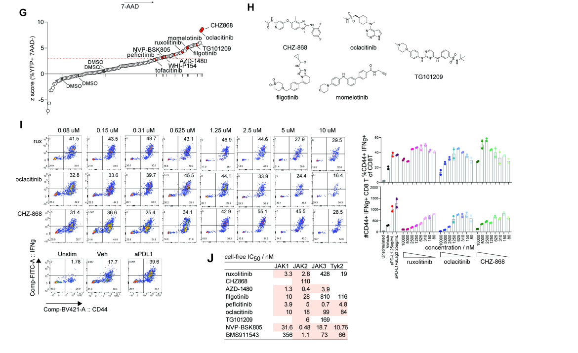
How to use compound library? How to design an experiment if you buy a compound library? Want a specific experimental protocol? This article will introduce popular experimental techniques and provide new ideas for publishing high level literature. -
 Scientific Reviews
Wnt/β-catenin and tumor EMT
Scientific Reviews
Wnt/β-catenin and tumor EMT2024-03-18
Epithelial-Mesenchymal Transition (EMT) is closely related to the plasticity of tumor cells and is a necessary process for tumor metastasis. Wnt/β-catenin is one of the main actors involved in the EMT process. Today, we’re here to popularize the tumor EMT and Wnt/β-catenin pathway~ -
 Scientific Reviews
The 2024 AACR meeting concluded successfully in California, USA. Which antitumor drugs stole the show at this conference?
Scientific Reviews
The 2024 AACR meeting concluded successfully in California, USA. Which antitumor drugs stole the show at this conference? -
 Scientific Reviews
Katalin Karikó and Drew Weissman were awarded the Nobel Prize in Physiology or Medicine in 2023 for their groundbreaking work in nucleoside modification, which paved the way for the creation of successful mRNA vaccines to fight against COVID-19. Let's now delve into the complete process of mRNA vaccine development.
Scientific Reviews
Katalin Karikó and Drew Weissman were awarded the Nobel Prize in Physiology or Medicine in 2023 for their groundbreaking work in nucleoside modification, which paved the way for the creation of successful mRNA vaccines to fight against COVID-19. Let's now delve into the complete process of mRNA vaccine development. -
 Scientific Reviews
Exosomes, which won the Nobel Prize in 2013, are still a research hotspot in the national natural sciences, and their popularity has only increased over the past decade (in 2022, they still rank 5th in the national natural sciences hotspots!). Why have exosomes become the darling of scientific research? Let's take a look together~
Scientific Reviews
Exosomes, which won the Nobel Prize in 2013, are still a research hotspot in the national natural sciences, and their popularity has only increased over the past decade (in 2022, they still rank 5th in the national natural sciences hotspots!). Why have exosomes become the darling of scientific research? Let's take a look together~ -
 Scientific Reviews
How important is the compound library? It connects to drug screening on one end and leads to lead compound modifications on the other, serving as one of the sources of new drug development.
Scientific Reviews
How important is the compound library? It connects to drug screening on one end and leads to lead compound modifications on the other, serving as one of the sources of new drug development. -
 Scientific Reviews
Antibodies!
Scientific Reviews
Antibodies!2024-07-26
Today, We introduce antibodies for everyone! -
 Scientific Reviews
What Are Popular Anti-tumor Drug Targets?
Scientific Reviews
What Are Popular Anti-tumor Drug Targets?2024-08-20
The rapid development of targeted anti-cancer drugs has spurred diverse research across various modalities. These include small molecules, monoclonal antibodies (mAbs), cell immunotherapies, antibody-drug conjugates (ADCs), and PROTACs (proteolysis targeting chimeras). -
 Scientific Reviews
Virtual Screening and New Uses for Old Drugs
Scientific Reviews
Virtual Screening and New Uses for Old Drugs2024-09-17
With the advancement of medical science, drug screening against various disease targets has become the fundamental strategy for drug development. Currently, computer-based virtual screening techniques are emerging in the field of new drug research due to their efficiency and low cost. Let's explore it today! -
 Scientific Reviews
Recently, Mol Cell reported the discovery of the first ferroptosis marker, Hyperoxidized PRDX3! Let’s take a look together~
Scientific Reviews
Recently, Mol Cell reported the discovery of the first ferroptosis marker, Hyperoxidized PRDX3! Let’s take a look together~ -
 Scientific Reviews
2024 Nobel Prize Announcements! Curious about the details? Click to dive into the exciting developments!
Scientific Reviews
2024 Nobel Prize Announcements! Curious about the details? Click to dive into the exciting developments! -
 Scientific Reviews
We are thrilled to share the latest advancements in AI technology as highlighted in this insightful article. From groundbreaking innovations to transformative applications, the future of AI is brighter than ever!
Scientific Reviews
We are thrilled to share the latest advancements in AI technology as highlighted in this insightful article. From groundbreaking innovations to transformative applications, the future of AI is brighter than ever! -
 Scientific Reviews
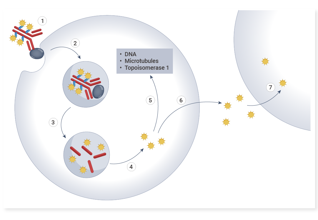
This article will walk you through the remarkable impacts of anti-payload antibodies, delving into how these molecules are revolutionizing drug development and biological research!
Scientific Reviews
This article will walk you through the remarkable impacts of anti-payload antibodies, delving into how these molecules are revolutionizing drug development and biological research! -
 Scientific Reviews
Cell Migration vs. Invasion: Differences Revealed by Scratch Assays and Transwell Experiments
Scientific Reviews
Cell Migration vs. Invasion: Differences Revealed by Scratch Assays and Transwell Experiments2025-05-30
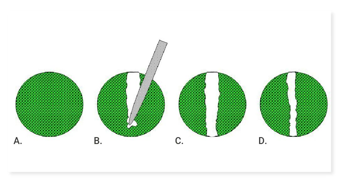
In scientific research, cell migration and invasion are crucial for understanding many important biological processes. This article delves into commonly used detection methods: the scratch assay and Transwell migration/invasion assay. -
 Scientific Reviews
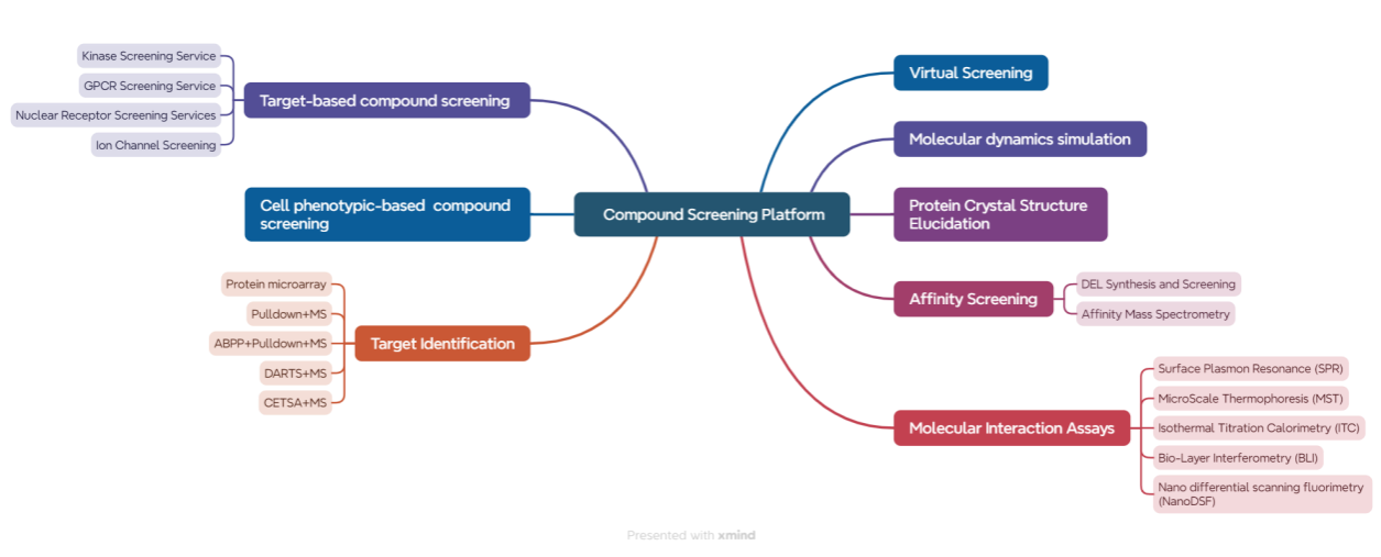
How should drug screening experiments be conducted? How can we ensure the accuracy of the lead compounds identified? This article will take you through how MCE's clients conduct drug screening experiments.
Scientific Reviews
How should drug screening experiments be conducted? How can we ensure the accuracy of the lead compounds identified? This article will take you through how MCE's clients conduct drug screening experiments. -
 Scientific Reviews
In this issue, we will conduct an in-depth interpretation from the dimensions of nanoparticle design, mechanism of action, in vivo and in vitro efficacy, and immune regulation, revealing how this research brings new hope for the treatment of invasive tumors through interdisciplinary innovation!
Scientific Reviews
In this issue, we will conduct an in-depth interpretation from the dimensions of nanoparticle design, mechanism of action, in vivo and in vitro efficacy, and immune regulation, revealing how this research brings new hope for the treatment of invasive tumors through interdisciplinary innovation! -
 Scientific Reviews
IHC, ICC, IF Techniques: A Practical Guide
Scientific Reviews
IHC, ICC, IF Techniques: A Practical Guide2025-07-25
Confused About IHC/ICC/IF? Why Does Immunostaining Seem So Complicated? Read This Now! Master Immunostaining with Confidence! -
 Scientific Reviews
HTS Breakthroughs Powered by MCE Libraries
Scientific Reviews
HTS Breakthroughs Powered by MCE Libraries2025-08-13
Key High-Throughput Screening Breakthroughs of 2024 Featuring MCE -
 Scientific Reviews
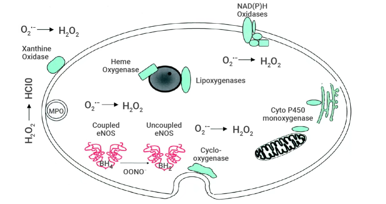
The article introduces the mechanisms of ROS generation and the methods for their detection.
Scientific Reviews
The article introduces the mechanisms of ROS generation and the methods for their detection. -
 Scientific Reviews
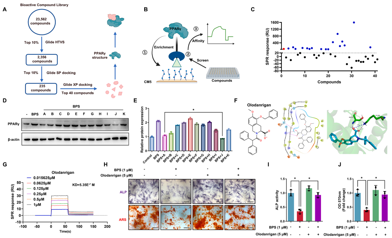
Encountering challenges with the high costs and long timelines of drug screening? Have a defined target but remain uncertain how to efficiently identify active molecules? Unsure how to validate hits generated from virtual screening? The ‘Winning Combination’ of drug screening offers a powerful solution to address these critical obstacles.
Scientific Reviews
Encountering challenges with the high costs and long timelines of drug screening? Have a defined target but remain uncertain how to efficiently identify active molecules? Unsure how to validate hits generated from virtual screening? The ‘Winning Combination’ of drug screening offers a powerful solution to address these critical obstacles. -
 Scientific Reviews
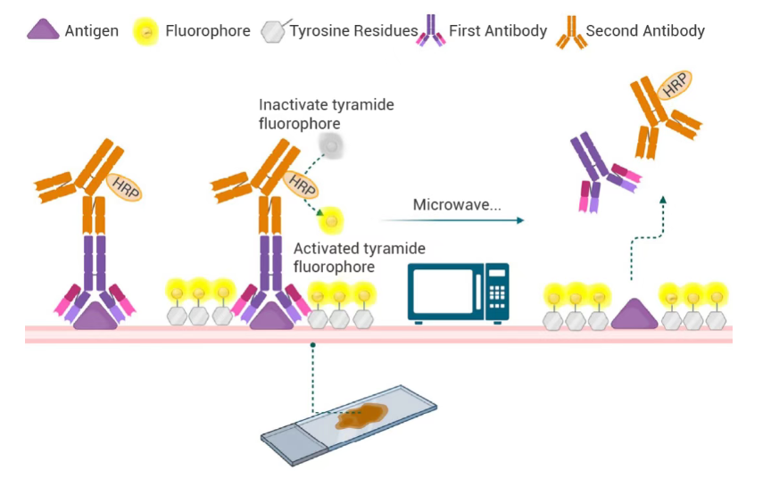
This detailed guide outlines the standardized experimental protocols for multiplex immunohistochemistry (mIHC), systematically summarizes common technical issues encountered during sample preparation, staining and imaging processes, and provides practical troubleshooting solutions to ensure reliable and reproducible results in biomedical research and clinical sample analysis.
Scientific Reviews
This detailed guide outlines the standardized experimental protocols for multiplex immunohistochemistry (mIHC), systematically summarizes common technical issues encountered during sample preparation, staining and imaging processes, and provides practical troubleshooting solutions to ensure reliable and reproducible results in biomedical research and clinical sample analysis. -
 Scientific Reviews
Exosomes—natural nanoscale carriers—are revolutionizing targeted therapy. This article uncovers the science behind their precision in drug delivery.
Scientific Reviews
Exosomes—natural nanoscale carriers—are revolutionizing targeted therapy. This article uncovers the science behind their precision in drug delivery. -
Scientific Reviews
Cracking the PROTAC Permeability Barrier: CD36-Mediated Endocytosis as a Potential Breakthrough2025-12-03
This article provides an in-depth analysis of cutting-edge literature revealing CD36 as a key mediator of cellular uptake for PROTACs and bRO5 compounds. By structurally optimizing PROTAC molecules to enhance their affinity for CD36, membrane permeability can be markedly improved, leading to significantly enhanced antitumor efficacy. -
 Scientific Reviews
This paper elaborates on cytokines for culturing major immune cells, their regulatory roles, recombinant cytokines' merits and MCE’s related high-quality products.
Scientific Reviews
This paper elaborates on cytokines for culturing major immune cells, their regulatory roles, recombinant cytokines' merits and MCE’s related high-quality products. -
 Scientific Reviews
This article walks you through the experimental design and workflow of flow cytometry, delivering a clear, dynamic, and professional overview to elevate your research.
Scientific Reviews
This article walks you through the experimental design and workflow of flow cytometry, delivering a clear, dynamic, and professional overview to elevate your research. -
 Scientific Reviews
Molecular glue degraders have evolved from a serendipitous observation to one of the most dynamic and transformative fields in biomedical research.
Scientific Reviews
Molecular glue degraders have evolved from a serendipitous observation to one of the most dynamic and transformative fields in biomedical research. -
 EDM
Research Solution for Breast Cancer TME
EDM
Research Solution for Breast Cancer TME2025-05-21
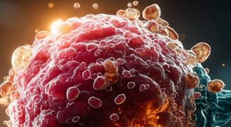
This review examines the complex interactions within the breast cancer tumor microenvironment, emphasizing how understanding these dynamics is essential for developing effective therapies and overcoming immune resistance. -
 EDM
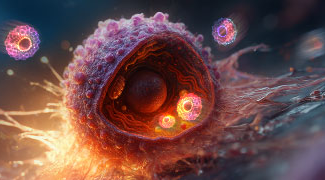
This review discusses the fundamentals of lipid biology and lipid metabolism, examines dysregulated lipid metabolism in cancer, and summarizes therapeutic strategies targeting lipid metabolic pathways.
EDM
This review discusses the fundamentals of lipid biology and lipid metabolism, examines dysregulated lipid metabolism in cancer, and summarizes therapeutic strategies targeting lipid metabolic pathways. -
 EDM
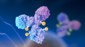
This review presents an overview of antibody–drug conjugates from design principles and antitumor mechanisms to structural innovations, clinical progress, and future development prospects.
EDM
This review presents an overview of antibody–drug conjugates from design principles and antitumor mechanisms to structural innovations, clinical progress, and future development prospects. -
 EDM
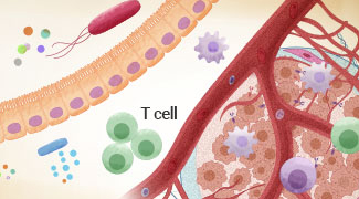
This review explores mechanisms and strategies for modulating the gut microbiota to enhance cancer immunotherapy, providing insights to improve therapeutic efficacy.
EDM
This review explores mechanisms and strategies for modulating the gut microbiota to enhance cancer immunotherapy, providing insights to improve therapeutic efficacy. -
 EDM
Combating Immune Evasion in Cancer
EDM
Combating Immune Evasion in Cancer2025-10-15
This review summarizes the mechanisms by which tumors evade immune surveillance and discusses therapeutic strategies to restore antitumor immunity. -
 EDM
This review summarizes the mechanisms of drug resistance in triple-negative breast cancer and highlights emerging therapeutic strategies.
EDM
This review summarizes the mechanisms of drug resistance in triple-negative breast cancer and highlights emerging therapeutic strategies. -
 EDM
Bispecific Antibodies in Cancer Therapy
EDM
Bispecific Antibodies in Cancer Therapy2025-10-29
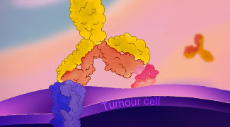
This review highlights the mechanisms, technology platforms, and clinical progress of bispecific antibodies, and discusses emerging strategies to guide future development. -
 EDM
This review provides insights into the pancreatic ductal adenocarcinoma tumor microenvironment, highlighting its cellular composition, stromal heterogeneity, and immune-targeting strategies.
EDM
This review provides insights into the pancreatic ductal adenocarcinoma tumor microenvironment, highlighting its cellular composition, stromal heterogeneity, and immune-targeting strategies. -
 EDM
Understanding In Vivo CAR-T Cell Therapy
EDM
Understanding In Vivo CAR-T Cell Therapy2026-01-22
This review provides a comprehensive overview of in vivo CAR-T therapy, covering technical platforms, clinical translation, and key challenges such as gene delivery and immunogenicity. -
 EDM
Highlight the overview of engineered EVs, their construction methods, and applications in cancer immunotherapy.
EDM
Highlight the overview of engineered EVs, their construction methods, and applications in cancer immunotherapy. -
 EDM
Highlight single-cell metabolomics and stable isotope tracing (SIT) in uncovering metabolic heterogeneity and nutrient flux dynamics in tumors.
EDM
Highlight single-cell metabolomics and stable isotope tracing (SIT) in uncovering metabolic heterogeneity and nutrient flux dynamics in tumors. -
 EDM
Explore ADC evolution, immune combination strategies, and clinical advances at the intersection of targeted therapy and immunotherapy.
EDM
Explore ADC evolution, immune combination strategies, and clinical advances at the intersection of targeted therapy and immunotherapy. -
 EDM
Tumor Organoid-Immune Cell Co-Culture Models: Advances, Applications, and Future Directions
EDM
Tumor Organoid-Immune Cell Co-Culture Models: Advances, Applications, and Future Directions2026-05-07
Summarize technical strategies, application advances, and future directions of tumor organoid–immune co-culture systems. -
 EDM
Review mechanisms, major challenges, and efficacy-enhancing strategies of TCR-T therapy in solid tumors, with implications for future research and clinical translation.
EDM
Review mechanisms, major challenges, and efficacy-enhancing strategies of TCR-T therapy in solid tumors, with implications for future research and clinical translation. -
 EDM
The Dual Role of Cellular Senescence in Cancer: Mechanisms, Microenvironment, and Therapeutics
EDM
The Dual Role of Cellular Senescence in Cancer: Mechanisms, Microenvironment, and Therapeutics2026-05-21
Explore the dual role of cellular senescence in cancer, including tumor suppression, SASP-driven tumor promotion, senescence heterogeneity and plasticity, and advances in senescence-targeted therapies. -
 EDM
Breaking Immune Resistance in Colorectal Cancer: From Molecular Mechanisms to Precision Therapy
EDM
Breaking Immune Resistance in Colorectal Cancer: From Molecular Mechanisms to Precision Therapy2026-06-25
Explore CRC molecular subtypes, immune landscapes, resistance mechanisms, and emerging precision strategies for improving outcomes across distinct subtypes. -
 EDM
Explore the mechanisms of tumor immune evasion, dynamic checkpoint regulation, and the evolution of mechanism-driven combinations and patient stratification.
EDM
Explore the mechanisms of tumor immune evasion, dynamic checkpoint regulation, and the evolution of mechanism-driven combinations and patient stratification. -
 Blog
EC0489, a SMDC for Cancer Therapy
Blog
EC0489, a SMDC for Cancer Therapy2019-03-25
EC0489, a conjugate of folic acid and desacetyl vinblastine hydrazide, is a SMDC under development for the treatment of solid tumours. -
 Blog
R916562, a dual Axl/VEGF-R2 inhibitor, could be a potential anti-angiogenic and anti-metastatic drug for cancer chemotherapy.
Blog
R916562, a dual Axl/VEGF-R2 inhibitor, could be a potential anti-angiogenic and anti-metastatic drug for cancer chemotherapy. -
 Blog
Role of PRMT7 Probe SGC3027 in Cancer
Blog
Role of PRMT7 Probe SGC3027 in Cancer2019-03-27
SGC3027 is the first potent, selective and cell active chemical probe for PRMT7. SGC3027 is also a pro-drug, which converts to the active compound SGC8158 -
 Blog
Alofanib, An Allosteric Inhibitor of FGFR2
Blog
Alofanib, An Allosteric Inhibitor of FGFR22019-03-28
Alofanib is an allosteric inhibitor of FGFR2 and inhibits FGF-mediated proliferation with GI50s of 16-370 nM, showing pronounced antitumor activity. -
 Blog
A Novel and Efficacious RAF Inhibitor RAF709
Blog
A Novel and Efficacious RAF Inhibitor RAF7092019-03-30
RAF709, a novel and efficacious RAF inhibitor, activates the MAPK pathway and shows antitumor activity in tumor cells harboring BRAF or RAS mutations. -
 Blog
CF53 is a highly potent, selective and orally active inhibitor of BET protein, with anti-tumor activity in acute leukemia and breast cancer cell lines.
Blog
CF53 is a highly potent, selective and orally active inhibitor of BET protein, with anti-tumor activity in acute leukemia and breast cancer cell lines. -
 Blog
AZD3229 is a Potent Pan-KIT Mutant Inhibitor
Blog
AZD3229 is a Potent Pan-KIT Mutant Inhibitor2019-04-04
AZD3229 is a potent, pan-KIT mutant inhibitor with potent single digit nM growth inhibition against a diverse panel of mutant KIT driven Ba/F3 cell lines. -
 Blog
BR351 is a Brain Penetrant MMP Inhibitor
Blog
BR351 is a Brain Penetrant MMP Inhibitor2019-04-05
BR351 is a brain penetrant MMP inhibitor, and a potential tool for the molecular imaging of activated MMPs with PET, with an IC50 in the nanomolar range. -
 Blog
A Lead PROTAC BRD9 Chemical Degrader
Blog
A Lead PROTAC BRD9 Chemical Degrader2019-04-06
PROTAC BRD9 Degrader-1 is a lead PROTAC BRD9 chemical degrader and a selective probe useful for the study of BAF complex biology. -
 Blog
SGC-GAK-1 is a potent, selective, and cell-active GAK inhibitor and shows potent anti-proliferative activity in LNCaP and 22Rv1 cells.
Blog
SGC-GAK-1 is a potent, selective, and cell-active GAK inhibitor and shows potent anti-proliferative activity in LNCaP and 22Rv1 cells. -
 Blog
COH000 is an allosteric, covalent and irreversible inhibitor of SUMO-activating enzyme, with an IC50 of 0.2 μM for SUMOylation in vitro.
Blog
COH000 is an allosteric, covalent and irreversible inhibitor of SUMO-activating enzyme, with an IC50 of 0.2 μM for SUMOylation in vitro. -
 Blog
MK-0429 is An Oral Integrin (αvβ3) Inhibitor
Blog
MK-0429 is An Oral Integrin (αvβ3) Inhibitor2019-04-11
MK-0429, an orally active αvβ3 inhibitor, is a potential therapeutic agent for the prevention of kidney fibrosis, melanoma and osteoporosis. -
 Blog
AZ304, a Dual BRAF Inhibitor Against Cancer
Blog
AZ304, a Dual BRAF Inhibitor Against Cancer2019-04-12
AZ304 is a potent BRAF inhibitor, blocks both wild type BRAF and V600E mutant BRAF activity, with IC50s in the nanomolar range. -
 Blog
Erteberel is a Selective ERβ Agonist
Blog
Erteberel is a Selective ERβ Agonist2019-04-16
Erteberel (LY500307) is a synthetic, nonsteroidal estrogen which acts as a selective ERβ agonist and under development for the treatment of schizophrenia. -
 Blog
MBQ-167 is a dual Rac/Cdc42 inhibitor in in metastatic cancer, with IC50s of 103 nM for Rac 1/2/3 and 78 nM for Cdc42 in MDA-MB-231 cells, respectively.
Blog
MBQ-167 is a dual Rac/Cdc42 inhibitor in in metastatic cancer, with IC50s of 103 nM for Rac 1/2/3 and 78 nM for Cdc42 in MDA-MB-231 cells, respectively. -
 Blog
PRN1008 is a Reversible Covalent and Oral Active Inhibitor of Bruton’s Tyrosine Kinase (BTK)
Blog
PRN1008 is a Reversible Covalent and Oral Active Inhibitor of Bruton’s Tyrosine Kinase (BTK)2019-04-18
PRN1008 is a selective, reversible covalent and oral active inhibitor of Bruton’s Tyrosine Kinase (BTK), with an IC50 of 1.3 nM. -
 Blog
Multiple Tyrosine Kinases Inhibitor TAS-115
Blog
Multiple Tyrosine Kinases Inhibitor TAS-1152019-04-19
TAS-115 is a potent VEGFR and c-Met/HGFR-targeted kinase inhibitor with IC50s of 30 and 32 nM for rVEGFR2 and rMET, respectively. -
 Blog
Y06036 is a potent and selective BET inhibitor for potential treatment of castration-resistant prostate cancer. With nanomolar inhibition.
Blog
Y06036 is a potent and selective BET inhibitor for potential treatment of castration-resistant prostate cancer. With nanomolar inhibition. -
 Blog
JNJ-64619178 is a selective and pseudo-irreversible PRMT5 inhibitor with an IC50 of 0.14 nM. Has potent Activity In Lung Cancer.
Blog
JNJ-64619178 is a selective and pseudo-irreversible PRMT5 inhibitor with an IC50 of 0.14 nM. Has potent Activity In Lung Cancer. -
 Blog
(E)-AG 99 is an EGFR inhibitor and shows a growth-inhibition on not only serum-starved cells but also normally grown cells. Treatment for Bladder cancer.
Blog
(E)-AG 99 is an EGFR inhibitor and shows a growth-inhibition on not only serum-starved cells but also normally grown cells. Treatment for Bladder cancer. -
 Blog
TAK-828F is a potent, selective and orally active retinoic acid receptor-related orphan receptor γt (RORγt) inverse agonist. TAK-828F inhibits IL-17A cytokine expression and reduces symptoms of autoimmune encephalomyelitis mice.
Blog
TAK-828F is a potent, selective and orally active retinoic acid receptor-related orphan receptor γt (RORγt) inverse agonist. TAK-828F inhibits IL-17A cytokine expression and reduces symptoms of autoimmune encephalomyelitis mice. -
 Blog
Y06137 is a potent and selective BET inhibitor, which binds to the BRD4(1) bromodomain with a Kd of 81 nM. Antitumor activity.
Blog
Y06137 is a potent and selective BET inhibitor, which binds to the BRD4(1) bromodomain with a Kd of 81 nM. Antitumor activity. -
 Blog
Borussertib is a covalent-allosteric and first-in-class inhibitor of protein kinase Akt, with an IC50 of 0.8 nM and a Ki of 2.2 nM for Akt-wt.
Blog
Borussertib is a covalent-allosteric and first-in-class inhibitor of protein kinase Akt, with an IC50 of 0.8 nM and a Ki of 2.2 nM for Akt-wt. -
 Blog
NRX-252262 is a β-catenin:β-TrCP interaction enhancer, and its cognate E3 ligase, SCFβ-TrCP, induces mutant β-catenin degradation, with an EC50 of 3.8 nM
Blog
NRX-252262 is a β-catenin:β-TrCP interaction enhancer, and its cognate E3 ligase, SCFβ-TrCP, induces mutant β-catenin degradation, with an EC50 of 3.8 nM -
 Blog
TAK-981 is a selective inhibitor of the SUMOylation enzymatic cascade, with potential immune-activating and antineoplastic activities
Blog
TAK-981 is a selective inhibitor of the SUMOylation enzymatic cascade, with potential immune-activating and antineoplastic activities -
 Blog
TD-428 is a Highly Specific BRD4 Degrader
Blog
TD-428 is a Highly Specific BRD4 Degrader2019-04-29
TD-428, a immunomodulatory drug analog, is a highly specific BRD4 degrader with a DC50 of 0.32 nM. TD-428 reduces c-Myc levels more efficiently than JQ1. -
 Blog
SLLN-15 is an oral activ enhancer of autophagy that activates cytostatic macroautophagy/autophagy in triple-negative breast cancer (TNBC).
Blog
SLLN-15 is an oral activ enhancer of autophagy that activates cytostatic macroautophagy/autophagy in triple-negative breast cancer (TNBC). -
 Blog
A1874 is a nutlin-based and BRD4-degrading PROTAC with a DC50 of 32 nM. Effective in inhibiting many cancer cell lines proliferation
Blog
A1874 is a nutlin-based and BRD4-degrading PROTAC with a DC50 of 32 nM. Effective in inhibiting many cancer cell lines proliferation -
 Blog
BAY-8002 is a selective and orally active inhibitor of monocarboxylate transporter 1 (MCT1), with an IC50 of 85 nM. Has potential to treat lymphoma.
Blog
BAY-8002 is a selective and orally active inhibitor of monocarboxylate transporter 1 (MCT1), with an IC50 of 85 nM. Has potential to treat lymphoma. -
 Blog
MRTX-1257 is a selective, irreversible, covalent and oral active KRAS G12C inhibitor, with an IC50 of 900 pM for KRAS dependent ERK phosphorylation.
Blog
MRTX-1257 is a selective, irreversible, covalent and oral active KRAS G12C inhibitor, with an IC50 of 900 pM for KRAS dependent ERK phosphorylation. -
 Blog
USL311 is a selective CXCR4 antagonist, which prevents the binding of stromal-cell derived factor-1 (SDF-1 or CXCL12) to CXCR4. Anti-tumor activity.
Blog
USL311 is a selective CXCR4 antagonist, which prevents the binding of stromal-cell derived factor-1 (SDF-1 or CXCL12) to CXCR4. Anti-tumor activity. -
 Blog
TX1-85-1, a ATP-competitive ligand of Her3, covalent modification of Her3 to inhibit Her3 signaling
Blog
TX1-85-1, a ATP-competitive ligand of Her3, covalent modification of Her3 to inhibit Her3 signaling2019-06-01
TX1-85-1 is a Her3 (ErbB3) inhibitor with an IC50 of 23 nM. TX1-85-1 induces partial degradation of Her3 protein and attenuates Her3-dependent signaling. -
 Blog
IWP-O1, a Highly Potent Porcupine Inhibitor, Functions by Preventing the Secretion of Wnt Proteins
Blog
IWP-O1, a Highly Potent Porcupine Inhibitor, Functions by Preventing the Secretion of Wnt Proteins2019-06-03
IWP-O1 is a Porcupine (Porcn) inhibitor, with an EC50 of 80 pM in L-Wnt-STF cells. IWP-O1 functions by preventing the secretion of Wnt proteins[ -
 Blog
PF-06465469 is a Covalent Inhibitor of ITK
Blog
PF-06465469 is a Covalent Inhibitor of ITK2019-06-08
PF-06465469 is a potent and covalent inhibitor of ITK with an IC50 of 2 nM. PF-06465469 inhibits MEK1/2 or AKT phosphorylation. -
 Blog
JMS-17-2 is a CX3CR1 Antagonist
Blog
JMS-17-2 is a CX3CR1 Antagonist2019-06-12
JMS-17-2 is a potent and selective CX3CR1 antagonist with an IC50 of 0.32 nM. Has potential to treat cancers such as breast cancer. -
 Blog
S18-000003 is a potent, selective and orally active inhibitor of retinoic acid receptor-related orphan receptor-gamma-t (RORγt). S18-000003 ameliorates psoriasis-like lesions in vivo. S18-000003 can be used for the research of skin inflammatory diseases.
Blog
S18-000003 is a potent, selective and orally active inhibitor of retinoic acid receptor-related orphan receptor-gamma-t (RORγt). S18-000003 ameliorates psoriasis-like lesions in vivo. S18-000003 can be used for the research of skin inflammatory diseases. -
 Blog
PTC299 is a dual and orally active DHODH and VEGF inhibitor, has broad and potent activity against hematological cancer cells.
Blog
PTC299 is a dual and orally active DHODH and VEGF inhibitor, has broad and potent activity against hematological cancer cells. -
 Blog
AMG 487 is an orally active and selective antagonist of CXC chemokine receptor 3 (CXCR3). AMG 487 has potential to treat metastatic cancer.
Blog
AMG 487 is an orally active and selective antagonist of CXC chemokine receptor 3 (CXCR3). AMG 487 has potential to treat metastatic cancer. -
 Blog
FGTI-2734 is a dual farnesyl and geranylgeranyl transferase-1 inhibitor. FGTI-2734 prevents membrane localization of KRAS and mutant KRAS pancreatic tumors.
Blog
FGTI-2734 is a dual farnesyl and geranylgeranyl transferase-1 inhibitor. FGTI-2734 prevents membrane localization of KRAS and mutant KRAS pancreatic tumors. -
 Blog
BI-882370 is a potent RAF kinase inhibitor with IC50s of 0.4, 0.8, and 0.6 nM for oncogenic BRAFV600E-mutant, the WT BRAF and CRAF kinases , respectively.
Blog
BI-882370 is a potent RAF kinase inhibitor with IC50s of 0.4, 0.8, and 0.6 nM for oncogenic BRAFV600E-mutant, the WT BRAF and CRAF kinases , respectively. -
 Blog
MSX-122 is an orally active partial antagonist of CXCR4, inhibiting CXCR4/CXCL12 actions. MSX-122 has both anti-tumor and anti-metastasis activities.
Blog
MSX-122 is an orally active partial antagonist of CXCR4, inhibiting CXCR4/CXCL12 actions. MSX-122 has both anti-tumor and anti-metastasis activities. -
 Blog
ZT-12-037-01 is a ATP-competitive and specific STK19 inhibitor and inhibits oncogenic NRAS-driven melanocyte malignant transformation.
Blog
ZT-12-037-01 is a ATP-competitive and specific STK19 inhibitor and inhibits oncogenic NRAS-driven melanocyte malignant transformation. -
 Blog
PT2977 is an orally active and selective HIF-2α inhibitor with an IC50 of 9 nM. PT2977 is a potential treatment for ccRCC and VHL disease.
Blog
PT2977 is an orally active and selective HIF-2α inhibitor with an IC50 of 9 nM. PT2977 is a potential treatment for ccRCC and VHL disease. -
 Blog
BI-2852 is a potent KRAS inhibitor with nanomolar affinity and reduces pERK and pAKT levels in a dose-dependent manner in a KRAS mutant cell line NCI-H358.
Blog
BI-2852 is a potent KRAS inhibitor with nanomolar affinity and reduces pERK and pAKT levels in a dose-dependent manner in a KRAS mutant cell line NCI-H358. -
 Blog
BAY-293 is a potent inhibitor of Son of Sevenless 1 (SOS1) and blocks RAS activation via disruption of the KRAS-SOS1 interaction with an IC50 of 21 nM.
Blog
BAY-293 is a potent inhibitor of Son of Sevenless 1 (SOS1) and blocks RAS activation via disruption of the KRAS-SOS1 interaction with an IC50 of 21 nM. -
 Blog
WZ811 is an Orally Active CXCR4 Antagonist
Blog
WZ811 is an Orally Active CXCR4 Antagonist2019-09-07
WZ811 is an orally active, highly potent competitive antagonist of CXCR4, which inhibits chronic lymphocytic leukemia progression and tumorigenesis. -
 Blog
RU-302 is a pan-TAM inhibitor that blocks the TAM Ig1 ectodomain and the Gas6 Lg domain interaction. RU-302 blocks Gas6-inducible Axl receptor activation.
Blog
RU-302 is a pan-TAM inhibitor that blocks the TAM Ig1 ectodomain and the Gas6 Lg domain interaction. RU-302 blocks Gas6-inducible Axl receptor activation. -
 Blog
BAY 61-3606 is an orally available, ATP-competitive, reversible and highly selective Syk inhibitor and sensitizes apoptosis by in breast cancer.
Blog
BAY 61-3606 is an orally available, ATP-competitive, reversible and highly selective Syk inhibitor and sensitizes apoptosis by in breast cancer. -
 Blog
DVD-445 is a potent peptidomimetic covalent TrxR1 inhibitor with an IC50 of 0.60 μM for rat TrxR1. DVD-445 has good anticancer application.
Blog
DVD-445 is a potent peptidomimetic covalent TrxR1 inhibitor with an IC50 of 0.60 μM for rat TrxR1. DVD-445 has good anticancer application. -
 Blog
IPR-803 is a potent inhibitor of the uPAR•uPA protein-protein interaction, and binds directly to uPAR with sub-micromolar affinity.
Blog
IPR-803 is a potent inhibitor of the uPAR•uPA protein-protein interaction, and binds directly to uPAR with sub-micromolar affinity. -
 Blog
Vorolanib is an orally active, multikinase VEGF/PDGF receptor inhibitor with antitumor activity and is expected to disrupt tumor angiogenesis.
Blog
Vorolanib is an orally active, multikinase VEGF/PDGF receptor inhibitor with antitumor activity and is expected to disrupt tumor angiogenesis. -
 Blog
CCG-222740 is a potent and selective MRTF pathway inhibitor. It effectively reduces fibrosis in the skin and blocks melanoma metastasis.
Blog
CCG-222740 is a potent and selective MRTF pathway inhibitor. It effectively reduces fibrosis in the skin and blocks melanoma metastasis. -
 Blog
IACS-8803 is a highly potent cyclic dinucleotide stimulator of interferon genes (STING) agonist with robust systemic antitumor efficacy.
Blog
IACS-8803 is a highly potent cyclic dinucleotide stimulator of interferon genes (STING) agonist with robust systemic antitumor efficacy. -
 Blog
AC-73 is a first specific, orally active the cluster of differentiation 147 (CD147) inhibitor and specifically disrupts CD147 dimerization.
Blog
AC-73 is a first specific, orally active the cluster of differentiation 147 (CD147) inhibitor and specifically disrupts CD147 dimerization. -
 Blog
CH6953755 is a potent, orally active and selective YES1 kinase inhibitor leading to antitumor activity against YES1 Gene -amplified cancers.
Blog
CH6953755 is a potent, orally active and selective YES1 kinase inhibitor leading to antitumor activity against YES1 Gene -amplified cancers. -
 Blog
CA-4948 is a Selective and Orally Bioavailable IRAK4 Kinase Inhibitor for Lymphoma Treatment
Blog
CA-4948 is a Selective and Orally Bioavailable IRAK4 Kinase Inhibitor for Lymphoma Treatment2020-01-05
CA-4948 is a potent, selective and orally bioavailable IRAK4 kinase inhibitor. CA-4948 can be used for the treatment of lymphoma. -
 Blog
ADH-503 is an orally active and allosteric CD11b agonist and leads to the repolarization of tumor-associated macrophages.
Blog
ADH-503 is an orally active and allosteric CD11b agonist and leads to the repolarization of tumor-associated macrophages. -
 Blog
Balixafortide is a potent, selective peptidic CXCR4 antagonist with anti-cancer effects, and blocks β-arrestin recruitment and calcium flux.
Blog
Balixafortide is a potent, selective peptidic CXCR4 antagonist with anti-cancer effects, and blocks β-arrestin recruitment and calcium flux. -
 Blog
CCT365623, an Orally Active LOX Inhibitor, Supresses EGFR (pY1068) and AKT Phosphorylation
Blog
CCT365623, an Orally Active LOX Inhibitor, Supresses EGFR (pY1068) and AKT Phosphorylation2020-04-16
CCT365623 is an orally active LOX inhibitor, suppresses EGFR (pY1068) and AKT phosphorylation driven by EGF. CCT365623 has good pharmacokinetic properties. -
 Blog
iFSP1 is a potent, selective FSP1inhibitor that induces ferroptosis in GPX4-knockout cells which overexpressed FSP1.
Blog
iFSP1 is a potent, selective FSP1inhibitor that induces ferroptosis in GPX4-knockout cells which overexpressed FSP1. -
 Blog
RBN-2397 is an orally active accross species NAD+ competitive inhibitor of PARP7. RBN-2397 binds to PARP7 and restores interferon (Type I) signaling.
Blog
RBN-2397 is an orally active accross species NAD+ competitive inhibitor of PARP7. RBN-2397 binds to PARP7 and restores interferon (Type I) signaling. -
 Blog
GSK143 is an orally active and highly selective spleen tyrosine kinase (SYK) inhibitor. GSK143 reduces inflammation in the intestinal muscularis in mice.
Blog
GSK143 is an orally active and highly selective spleen tyrosine kinase (SYK) inhibitor. GSK143 reduces inflammation in the intestinal muscularis in mice. -
 Blog
RA375, a RPN13 inhibitor, inhibits proteasome function in muscle. RA375 is highly active against cell lines of multiple myeloma and diverse solid cancers.
Blog
RA375, a RPN13 inhibitor, inhibits proteasome function in muscle. RA375 is highly active against cell lines of multiple myeloma and diverse solid cancers. -
 Blog
EW-7195 is a potent and selective ALK5 inhibitor, and efficiently inhibits TGF-β1-induced Smad signaling, EMT and breast tumour metastasis to the lung.
Blog
EW-7195 is a potent and selective ALK5 inhibitor, and efficiently inhibits TGF-β1-induced Smad signaling, EMT and breast tumour metastasis to the lung. -
 Blog
MSA-2 is an orally available non-nucleotide STING agonist. MSA-2 shows antitumor activity and stimulates interferon-β secretion in tumors.
Blog
MSA-2 is an orally available non-nucleotide STING agonist. MSA-2 shows antitumor activity and stimulates interferon-β secretion in tumors. -
 Blog
SP-8356, an orally active CD147inhibitor, exerts anti-breast cancer effects by inhibiting NF-κB signaling. Anti-atherosclerotic effects.
Blog
SP-8356, an orally active CD147inhibitor, exerts anti-breast cancer effects by inhibiting NF-κB signaling. Anti-atherosclerotic effects. -
 Blog
CB-1158, a potent and orally bioavailable inhibitor of arginase, blocks myeloid cell-mediated immune suppression in the tumor microenvironment.
Blog
CB-1158, a potent and orally bioavailable inhibitor of arginase, blocks myeloid cell-mediated immune suppression in the tumor microenvironment. -
 Blog
SRX3207 is an orally active and first-in-class dual Syk/PI3K inhibitor. SRX3207 possesses effective anti-tumor activity in vitro and in vivo.
Blog
SRX3207 is an orally active and first-in-class dual Syk/PI3K inhibitor. SRX3207 possesses effective anti-tumor activity in vitro and in vivo. -
 Blog
Verucopeptin inhibits v-ATPase activity by directly targeting the v-ATPase ATP6V1G subunit but not ATP1V1B2 or ATP6V1D. Also a potent HIF-1 inhibitor.
Blog
Verucopeptin inhibits v-ATPase activity by directly targeting the v-ATPase ATP6V1G subunit but not ATP1V1B2 or ATP6V1D. Also a potent HIF-1 inhibitor. -
 Blog
E7820, sulfonamide derivative, is a unique angiogenesis inhibitor suppressing an expression of integrin alpha2 subunit on endothelium.
Blog
E7820, sulfonamide derivative, is a unique angiogenesis inhibitor suppressing an expression of integrin alpha2 subunit on endothelium. -
 Blog
Cyclo(-RGDfK) is a selective inhibitor of the αvβ3 integrin. Cyclo(-RGDfK) potently targets cancer cells through binding to the cell surface αvβ3 integrin.
Blog
Cyclo(-RGDfK) is a selective inhibitor of the αvβ3 integrin. Cyclo(-RGDfK) potently targets cancer cells through binding to the cell surface αvβ3 integrin. -
 Blog
Cilengitide is a compound targeting angiogenesis, the cornerstone of tumor growth and metastasis. ανβ3 and ανβ5 are the target of Cilengitide.
Blog
Cilengitide is a compound targeting angiogenesis, the cornerstone of tumor growth and metastasis. ανβ3 and ανβ5 are the target of Cilengitide. -
 Blog
Tetrac inhibits the cellular actions of thyroid hormone initiated at the hormone receptor on plasma membrane integrin alphavbeta3.
Blog
Tetrac inhibits the cellular actions of thyroid hormone initiated at the hormone receptor on plasma membrane integrin alphavbeta3. -
 Blog
DMU-212 Possesses Antimitotic, Anti-Proliferative, Antioxidant and Apoptosis Promoting Activities
Blog
DMU-212 Possesses Antimitotic, Anti-Proliferative, Antioxidant and Apoptosis Promoting Activities2020-12-24
Activation of ERK1/2 is required for the antimitotic activity of the Resveratrol analogue DMU‐212 in human melanoma cells. -
 Blog
ENMD-1198 is an orally active microtubule-targeting agent with antiproliferative and antiangiogenic activity.
Blog
ENMD-1198 is an orally active microtubule-targeting agent with antiproliferative and antiangiogenic activity. -
 Blog
TPP-1 is a potent inhibitor of the PD-1/PD-L1 interaction. TPP-1 binds specifically to PD-L1 with a high affinity (KD=95 nM).
Blog
TPP-1 is a potent inhibitor of the PD-1/PD-L1 interaction. TPP-1 binds specifically to PD-L1 with a high affinity (KD=95 nM). -
 Blog
FAPI-46 is a quinoline-based FAP-targeted radiotracer. FAPI-46 has higher tumor uptake and prolonged tumor accumulation.
Blog
FAPI-46 is a quinoline-based FAP-targeted radiotracer. FAPI-46 has higher tumor uptake and prolonged tumor accumulation. -
 Blog
BMSpep-57 is a potent and competitive macrocyclic peptide inhibitor of PD-1/PD-L1 interaction. It induces high levels of IL-2.
Blog
BMSpep-57 is a potent and competitive macrocyclic peptide inhibitor of PD-1/PD-L1 interaction. It induces high levels of IL-2. -
 Blog
ML339 is a Selective CXCR6 Antagonist
Blog
ML339 is a Selective CXCR6 Antagonist2021-05-12
ML339 is a small molecule antagonist would block Prostate cancer cell trafficking; hence mediate a metastatic event and disease progression. -
 Blog
Sitravatinib (MGCD516) is an Orally Bioavailable RTK Inhibitor with PD-1 Blockade Activity
Blog
Sitravatinib (MGCD516) is an Orally Bioavailable RTK Inhibitor with PD-1 Blockade Activity2021-06-03
Sitravatinib, a small molecule RTK inhibitor, shows potent anti-tumor activity in preclinical models of sarcoma. -
 Blog
Motesanib (AMG 706) is an orally active VEGFR inhibitor. Potently inhibits angiogenesis and induces regression in tumor xenografts.
Blog
Motesanib (AMG 706) is an orally active VEGFR inhibitor. Potently inhibits angiogenesis and induces regression in tumor xenografts. -
 Blog
EMD527040 is a highly selective αvβ6 antagonist with antifibrotic activities.
Blog
EMD527040 is a highly selective αvβ6 antagonist with antifibrotic activities. -
 Blog
Inupadenant is an orally active, highly selective A2A receptor antagonist with potent anti-tumor activity.
Blog
Inupadenant is an orally active, highly selective A2A receptor antagonist with potent anti-tumor activity. -
 Blog
UK122 is a potent and selective urokinase-type plasminogen activator (uPA) inhibitor with anti-cancer activity.
Blog
UK122 is a potent and selective urokinase-type plasminogen activator (uPA) inhibitor with anti-cancer activity. -
 Blog
AZD4635 is a potent, selective and orally active antagonist of A2AR that reverses adenosine-mediated immune suppression.
Blog
AZD4635 is a potent, selective and orally active antagonist of A2AR that reverses adenosine-mediated immune suppression. -
 Blog
Elesclomol is an oxidative stress inducer that induces cancer cell apoptosis. Elesclomol is a reactive oxygen species (ROS) inducer.
Blog
Elesclomol is an oxidative stress inducer that induces cancer cell apoptosis. Elesclomol is a reactive oxygen species (ROS) inducer. -
 Blog
Brusatol (NSC 172924) is a Nrf2 Inhibitor
Blog
Brusatol (NSC 172924) is a Nrf2 Inhibitor2021-10-26
Brusatol inhibits the Nrf2 signaling pathway by reducing the protein level of Nrf2, with anticancer activities. -
 Blog
Nintedanib a potent and orally active triple vascular kinase inhibitor for VEGFR1/2/3, FGFR1/2/3 and PDGFRα/β.
Blog
Nintedanib a potent and orally active triple vascular kinase inhibitor for VEGFR1/2/3, FGFR1/2/3 and PDGFRα/β. -
 Blog
Oltipraz is a Nrf2 Inhibitor
Blog
Oltipraz is a Nrf2 Inhibitor2021-11-18
Oltipraz is a potent Nrf2 activator. Oltipraz has an inhibitory effect on HIF-1α activation in a time-dependent manner, the IC50 is 10 μM. -
 Blog
Axitinib is a multi-targeted tyrosine kinase inhibitor and potently inhibitor VEGFR1, VEGFR2, VEGFR3 and PDGFRβ.
Blog
Axitinib is a multi-targeted tyrosine kinase inhibitor and potently inhibitor VEGFR1, VEGFR2, VEGFR3 and PDGFRβ. -
 Blog
Trolox is an analogue of vitamin E with a powerful antioxidant effect. Trolox is also a powerful inhibitor of membrane damage.
Blog
Trolox is an analogue of vitamin E with a powerful antioxidant effect. Trolox is also a powerful inhibitor of membrane damage. -
 Blog
Alda-1 is a potent ALDH2 Agonist
Blog
Alda-1 is a potent ALDH2 Agonist2021-12-30
Alda-1 ameliorates H2O2-induced Achilles tendinopathy. Alda-1 could be used for preventing Achilles tendinopathy. -
 Blog
β-Lapachone, a topoisomerase I inhibitor, induces apoptosis by inhibiting cell cycle progression. β-Lapachone has anti-inflammatory effect.
Blog
β-Lapachone, a topoisomerase I inhibitor, induces apoptosis by inhibiting cell cycle progression. β-Lapachone has anti-inflammatory effect. -
 Blog
CL097 is a TLR7/8 agonist. CL097 induces pro-nflammatory cytokines. CL097 induces NADPH oxidase priming and efficient diabetogenic cytotoxic T lymphocyte (CTL) function in NOD mice.
Blog
CL097 is a TLR7/8 agonist. CL097 induces pro-nflammatory cytokines. CL097 induces NADPH oxidase priming and efficient diabetogenic cytotoxic T lymphocyte (CTL) function in NOD mice. -
 Blog
Zingerone is a natural orally active nontoxic methoxyphenol potent anti-inflammatory, antidiabetic, antioxidize, and anti-tumor properties.
Blog
Zingerone is a natural orally active nontoxic methoxyphenol potent anti-inflammatory, antidiabetic, antioxidize, and anti-tumor properties. -
 Blog
Indoximod, an immunometabolic adjuvant, is an orally active IDO pathway inhibitor. Indoximod acts as a Trp mimetic in regulating mTOR.
Blog
Indoximod, an immunometabolic adjuvant, is an orally active IDO pathway inhibitor. Indoximod acts as a Trp mimetic in regulating mTOR. -
 Blog
PX-12 is an irreversible inhibitor of Trx-1 and inhibits the growth, migration, and invasion of colorectal cancer cell lines.
Blog
PX-12 is an irreversible inhibitor of Trx-1 and inhibits the growth, migration, and invasion of colorectal cancer cell lines. -
 Blog
Tanomastat is an orally active, non-peptidic biphenyl MMPs inhibitor, with antiangiogenic, anti-invasive and antimetastatic activities.
Blog
Tanomastat is an orally active, non-peptidic biphenyl MMPs inhibitor, with antiangiogenic, anti-invasive and antimetastatic activities. -
 Blog
BM213 is a selective C5aR1 agonist. It’s a useful research tool to study C5aR1 function
Blog
BM213 is a selective C5aR1 agonist. It’s a useful research tool to study C5aR1 function -
 Blog
Telatinib is an orally active inhibitor of VEGFR2, VEGFR3, PDGFα, and c-Kit.
Blog
Telatinib is an orally active inhibitor of VEGFR2, VEGFR3, PDGFα, and c-Kit. -
 Blog
Marimastat (BB2516) is a broad spectrum and orally bioavailable inhibitor of MMPs (Matrix metalloproteinases).
Blog
Marimastat (BB2516) is a broad spectrum and orally bioavailable inhibitor of MMPs (Matrix metalloproteinases). -
 Blog
Gardiquimod is a TLR7/8 agonist and can inhibit HIV-1 infection.
Blog
Gardiquimod is a TLR7/8 agonist and can inhibit HIV-1 infection. -
 Blog
Vatalanib is an inhibitor of VEGFR2/KDR. Vatalanib induces inhibition of the angiogenic response to VEGF and PDGF.
Blog
Vatalanib is an inhibitor of VEGFR2/KDR. Vatalanib induces inhibition of the angiogenic response to VEGF and PDGF. -
 Blog
Toceranib is a selective and orally active inhibitor of RTK and has the potential for the research of canine mast cell tumors.
Blog
Toceranib is a selective and orally active inhibitor of RTK and has the potential for the research of canine mast cell tumors. -
 Blog
Tiragolumab is an immune checkpoint inhibitor binding to TIGIT. Tiragolumab is effective against multiple solid malignancies.
Blog
Tiragolumab is an immune checkpoint inhibitor binding to TIGIT. Tiragolumab is effective against multiple solid malignancies. -
 Blog
Henatinib is an orally active small-molecule multikinase inhibitor that has demonstrated broad and potent antitumor activities.
Blog
Henatinib is an orally active small-molecule multikinase inhibitor that has demonstrated broad and potent antitumor activities. -
 Blog
KGP94 is a selective inhibitor of cathepsin L. KGP94 has antitumor activity and improves survival of bone metastases bearing mice.
Blog
KGP94 is a selective inhibitor of cathepsin L. KGP94 has antitumor activity and improves survival of bone metastases bearing mice. -
 Blog
Certepetide is a Tumor-penetrating Enhancer via RGD Motif Interaction with Alphav-integrins
Blog
Certepetide is a Tumor-penetrating Enhancer via RGD Motif Interaction with Alphav-integrins2022-09-01
Certepetide is a tumor-penetrating enhancer and has the potential for the research of metastatic pancreatic ductal adenocarcinoma. -
 Blog
LCS3 is a potent and reversible inhibitor of GSR and TXNRD1 with anti-tumour activity by non-competitive inhibition of the enzyme activity.
Blog
LCS3 is a potent and reversible inhibitor of GSR and TXNRD1 with anti-tumour activity by non-competitive inhibition of the enzyme activity. -
 Blog
BSP16 is a potent, orally active stimulator of interferon genes (STING) agonist. BSP16 has potent anti-cancer activity.
Blog
BSP16 is a potent, orally active stimulator of interferon genes (STING) agonist. BSP16 has potent anti-cancer activity. -
 Blog
Parsatuzumab (RG 7414) is a humanized monoclonal antibody, that acts as an immunomodulator, and binds to EGFL7.
Blog
Parsatuzumab (RG 7414) is a humanized monoclonal antibody, that acts as an immunomodulator, and binds to EGFL7. -
 Blog
Serplulimab is a humanized monoclonal anti-PD-1 antibody and has the potential for the research of small cell lung cancer.
Blog
Serplulimab is a humanized monoclonal anti-PD-1 antibody and has the potential for the research of small cell lung cancer. -
 Blog
ABT-510, a peptide analog of thrombospondin-1 (TSP-1), can block angiogenesis in vitro and in vivo, and slow tumor growth.
Blog
ABT-510, a peptide analog of thrombospondin-1 (TSP-1), can block angiogenesis in vitro and in vivo, and slow tumor growth. -
 Blog
Mogamulizumab is an Anti-CCR4 monoclonal antibody. It enhances antibody-dependent cellular cytotoxicity and is effective against leukemia.
Blog
Mogamulizumab is an Anti-CCR4 monoclonal antibody. It enhances antibody-dependent cellular cytotoxicity and is effective against leukemia. -
 Blog
MMRi62, a Ferroptosis inducer targeting MDM2-MDM4. MMRi62 shows a P53-independent pro-apoptotic activity against PDAC cells.
Blog
MMRi62, a Ferroptosis inducer targeting MDM2-MDM4. MMRi62 shows a P53-independent pro-apoptotic activity against PDAC cells. -
 Blog
FA16 is a specific and metabolically stable ferroptosis inducer. It inhibits system Xc-, results in tumor progression suppression.
Blog
FA16 is a specific and metabolically stable ferroptosis inducer. It inhibits system Xc-, results in tumor progression suppression. -
 Blog
Bapotulimab (BAY-1905254) is an ILDR2 IgG antibody that blocks the immunosuppressive effects of ILDR2 on T-cell activation.
Blog
Bapotulimab (BAY-1905254) is an ILDR2 IgG antibody that blocks the immunosuppressive effects of ILDR2 on T-cell activation. -
 Blog
YL-939 was a potent inhibitor of iron death and significantly improved liver injury in a model of iron death-related acute liver injury.
Blog
YL-939 was a potent inhibitor of iron death and significantly improved liver injury in a model of iron death-related acute liver injury. -
 Blog
Nanrilkefusp alfa is a selective and strong IL-15 agonist. It inhibits tumor metastasis and viability by activating natural killer (NK) cells.
Blog
Nanrilkefusp alfa is a selective and strong IL-15 agonist. It inhibits tumor metastasis and viability by activating natural killer (NK) cells. -
 Blog
Urabrelimab (SRF231) is an anti-CD47 monoclonal antibody, blocking the CD47-SIRPα interaction. It has the potential to research anticancer.
Blog
Urabrelimab (SRF231) is an anti-CD47 monoclonal antibody, blocking the CD47-SIRPα interaction. It has the potential to research anticancer. -
 Blog
Efineptakin alfa (NT-17) is a Long-Acting Recombinant Human IL-7 for Glioblastoma Research
Blog
Efineptakin alfa (NT-17) is a Long-Acting Recombinant Human IL-7 for Glioblastoma Research2023-02-07
Efineptakin alfa (NT-17) is a long-acting recombinant human IL-7. Efineptakin alfa can be used for glioblastoma research. -
 Blog
Pegdinetanib (BMS-844203) is a selective VEGFR-2 inhibitor with antitumor activity.
Blog
Pegdinetanib (BMS-844203) is a selective VEGFR-2 inhibitor with antitumor activity. -
 Blog
Tifcemalimab, a Humanized anti-BTLA monoclonal antibody, blocks the interaction of HVEM-BTLA by binding to BTLA and activates lymphocytes.
Blog
Tifcemalimab, a Humanized anti-BTLA monoclonal antibody, blocks the interaction of HVEM-BTLA by binding to BTLA and activates lymphocytes. -
 Blog
Geptanolimab (CBT-501) is a programmed death-1 (PD-1) monoclonal antibody. Geptanolimab can be used in research forsolid tumor research.
Blog
Geptanolimab (CBT-501) is a programmed death-1 (PD-1) monoclonal antibody. Geptanolimab can be used in research forsolid tumor research. -
 Blog
Dazostinag is a STING Agonist and Can be used for Antibody-Drug Conjugates (ADCs) Synthesis
Blog
Dazostinag is a STING Agonist and Can be used for Antibody-Drug Conjugates (ADCs) Synthesis2023-03-13
Dazostinag is a STING agonist and a playload, to synthesis antibody-drug conjugates (ADCs). It has antitumor activity in vivo. -
 Blog
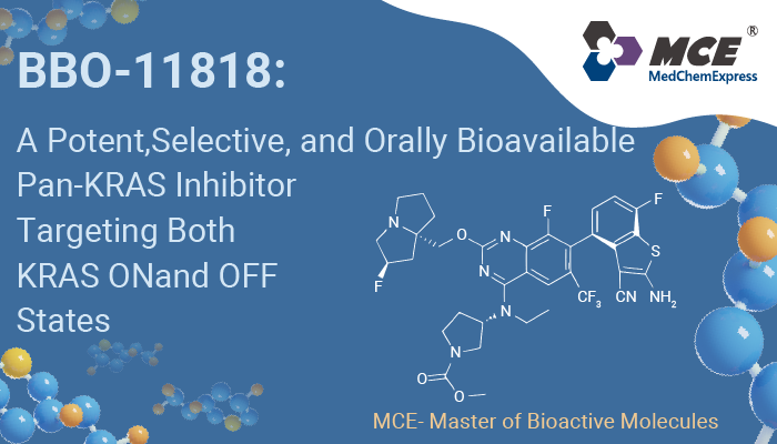
BBO-11818 is a potent, selective, orally bioavailable, and noncovalent pan-KRAS inhibitor targeting multiple clinically relevant KRAS mutants in both ON and OFF states.
Blog
BBO-11818 is a potent, selective, orally bioavailable, and noncovalent pan-KRAS inhibitor targeting multiple clinically relevant KRAS mutants in both ON and OFF states.
Products
All
-
All
-
Inhibitors & Agonists
-
Isotope-Labeled Compounds
-
Fluorescent Dye
-
Peptides
-
Recombinant Proteins
-
Antibodies
-
Screening Libraries
-
Kits
-
Inhibitory Antibodies
-
Reference Standards
-
Induced Disease Models Products
-
Enzyme
-
Biochemical Assay Reagents
-
Natural Products
| Cat. No. | Product Name | Information | Application | Publication |
|---|---|---|---|---|
| HY-10201 | Sorafenib |
Sorafenib (Bay 43-9006) is a potent oral active multikinase inhibitor. Sorafenib blocks autophosphorylation and activity of receptor tyrosine kinases (VEGFR-2, VEGFR-3) and RAF family kinases, thereby suppressing the RAF/MEK/ERK and PI3K/Akt pathways, inhibiting STAT3 phosphorylation, and selectively inhibiting the MAPK pathway in cancer cells. Sorafenib induces cell cycle arrest, autophagy, apoptosis, and PARP cleavage, reduces Bcl-2, Bcl-XL, cyclin D1 levels, and activates Bak and Bax. Sorafenib inhibits tumor growth and metastasis in mouse and rat models. Sorafenib can be used for cancer research, such as colon, breast, non-small-cell lung cancer (NSCLC), ovarian, pancreatic, melanoma, colorectal and hepatocellular carcinoma.
|
Raf
VEGFR
FLT3
Autophagy
Apoptosis
STAT
Akt
MMP
Cadherin
p38 MAPK
ERK
MEK
PI3K
PARP
Bcl-2 Family
Ephrin Receptor
PDGFR
Gastric Cancer
Liver Cancer
Ovarian Cancer
Digestive System Inflammation
Small Cell Lung Cancer
Non-Small Cell Lung Cancer
Metastatic Breast Cancer
Metastatic Colorectal Cancer
Metastatic Prostate Cancer
Multiple Myeloma
Metastatic Pancreatic Cancer
SARS-CoV-2 Infection
Alzheimer's Disease
Parkinson's Disease
Obesity
Lung Fibrosis
|
316
|
| HY-50895 | Gefitinib |
Gefitinib (ZD1839) is a potent, selective and orally active EGFR tyrosine kinase inhibitor with an IC50 of 33 nM. Gefitinib selectively inhibits EGF-stimulated tumor cell growth (IC50 of 54 nM) and that blocks EGF-stimulated EGFR autophosphorylation in tumor cells. Gefitinib also induces autophagy and cell apoptosis, which can be used for cancer related research, such as Lung cancer and breast cancer .
|
Colorectal Cancer
Prostate Cancer
Liver Cancer
Pancreatic Cancer
Ovarian Cancer
Lipid Metabolism
Non-Small Cell Lung Cancer
Lung Adenocarcinoma
Metastatic Breast Cancer
SARS-CoV-2 Infection
|
222
|
| HY-15772 | Osimertinib |
Osimertinib (AZD9291) is a covalent, orally active, irreversible, and mutant-selective EGFR inhibitor with an apparent IC50 of 12 nM against L858R and 1 nM against L858R/T790M, respectively. Osimertinib overcomes T790M-mediated resistance to EGFR inhibitors in lung cancer.
|
Lung Cancer
Colorectal Cancer
Prostate Cancer
Liver Cancer
Pancreatic Cancer
Ovarian Cancer
Glucose Metabolism
SARS-CoV-2 Infection
Hepatitis C Virus Infection
Obesity
|
208
|
| HY-N0565B | Doxycycline hyclate |
Doxycycline hyclate, an antibiotic, is an orally active and broad-spectrum metalloproteinase (MMP) inhibitor. Doxycycline hyclate shows antibacterial activity and anti-cancer cell proliferation activity. Doxycycline hyclate can be used to construct gene expression regulation models.
|
Breast Cancer
Pancreatic Cancer
Viral Infection
Parasitic Infection
Pain
SARS-CoV-2 Infection
Staphylococcus Aureus Infection
Chlamydia Infection
Lung Fibrosis
|
197
|
| HY-N0565 | Doxycycline |
Doxycycline, an antibiotic, is an orally active and broad-spectrum metalloproteinase (MMP) inhibitor. Doxycycline shows antibacterial activity and anti-cancer cell proliferation activity.
|
Breast Cancer
Pancreatic Cancer
Viral Infection
Parasitic Infection
Pain
SARS-CoV-2 Infection
Staphylococcus Aureus Infection
Chlamydia Infection
Lung Fibrosis
|
197
|
| HY-B0496 | PMSF |
PMSF (Phenylmethylsulfonyl fluoride) is a nonspecific and irreversible serine proteases inhibitor. PMSF inhibits some thiol proteases, non-protease enzymes, and acetylcholinesterase. PMSF can be used to prepare protein extracts from tissue and cell lysates.
|
|
163
|
| HY-50896 | Erlotinib |
Erlotinib (CP-358774) is a selective, orally active EGFR tyrosine kinase inhibitor. Erlotinib also acts as a substrate and inhibitor of OATP2B1, with an IC50 of approximately 0.079 μM for inhibiting OATP2B1-mediated uptake of estrone 3-sulfate. Erlotinib blocks EGFR phosphorylation, downstream signal transduction, as well as the growth and proliferation of cancer cells. Erlotinib inhibits MMP-10-mediated renal injury, fibrotic lesions, the ERK1/2, GSK-3β and β-catenin signaling pathways, fibronectin, α-SMA, collagen deposition, and renal injury markers. Erlotinib is metabolized via CYP3A to produce the active metabolite OSI-420. Erlotinib can be used in research related to non-small cell lung cancer, gastric cancer, papillary renal cell carcinoma, pancreatic cancer, renal fibrosis, and other conditions.
|
|
147
|
| HY-10261 | Afatinib |
Afatinib (BIBW 2992) is an orally active, potent and irreversible dual specificity inhibitor of ErbB family (EGFR and HER2), with IC50 values of 0.5 nM, 0.4 nM, 10 nM and 14 nM for EGFRwt, EGFRL858R, EGFRL858R/T790M and HER2, respectively. Afatinib can be used for the research of esophageal squamous cell carcinoma (ESCC), non-small cell lung cancer (NSCLC) and gastric cancer.
|
Lung Cancer
Breast Cancer
Colorectal Cancer
Prostate Cancer
Gastric Cancer
Liver Cancer
Leukemia/Lymphoma/Myeloma
Pancreatic Cancer
Ovarian Cancer
Neurodegenerative Disease
Depression
Pain
Digestive System Inflammation
Hepatitis C Virus Infection
Obesity
Lung Fibrosis
Rheumatoid Arthritis
|
113
|
| HY-12270 | T-5224 |
T-5224 is a transcription factor c-Fos/activator protein (AP)-1 inhibitor with anti-inflammatory effects, which specifically inhibits the DNA binding activity of c-Fos/c-Jun without affecting other transcription factors. T-5224 inhibits the IL-1β-induced up-regulation of Mmp-3, Mmp-13 and Adamts-5 transcription.
|
|
109
|
| HY-50898 | Lapatinib |
Neurological, Eye or Ear Disease
Lung Cancer
Colorectal Cancer
Prostate Cancer
Gastric Cancer
Liver Cancer
Pancreatic Cancer
Ovarian Cancer
HER-2 Positive Breast Cancer
Metastatic Breast Cancer
SARS-CoV-2 Infection
Hepatitis C Virus Infection
Obesity
|
100
|
|
| HY-100229 | Aloxistatin |
Aloxistatin (E64d) is a cell-permeable and irreversible broad-spectrum cysteine protease inhibitor. Aloxistatin (E64d) exhibits entry-blocking effect for MERS-CoV.
|
|
95
|
| HY-14596 | Genistein |
Neurological, Eye or Ear Disease
Lung Cancer
Colorectal Cancer
Prostate Cancer
Liver Cancer
Pancreatic Cancer
Ovarian Cancer
Digestive System Inflammation
SARS-CoV-2 Infection
Hepatitis C Virus Infection
Obesity
|
94
|
|
| HY-L028 | CNS-Penetrant Compound Library |
The blood-brain barrier (BBB) is the complex network of brain microvessels. It protects the brain from the external bloodstream environment and supplies the brain with the required nutrients for normal function. However, blood-brain barrier is also the obstacle to deliver beneficial drugs to treat CNS (central nervous system) diseases or brain tumors, as it has the least permeable capillaries in the entire body due to physical barriers (tight junctions). Therefore, it is crucial to discover drugs which can cross this barrier for the treatment of brain-based diseases, such as Alzheimer’s disease (AD), Parkinson’s disease (PD) and epilepsy.
MCE offers a unique collection of 1,170 compounds with confirmed CNS-Penetrant property. It’s a useful tool for the discovery of drugs used for brain diseases, such as brain tumors, mental disorders, and neurodegenerative diseases.
|
|
91
|
| HY-L031 | Small Molecule Immuno-Oncology Compound Library |
Immuno-Oncology is a type of immunotherapy that has the specific purpose of treating cancer. It works by stimulating our immune system to fight back. Normally, our immune system is able to destroy cancer cells in our body, however sometimes cancer cells can adapt and mutate, effectively hiding from our immune system. This is when tumors can develop and become a threat to our health. Immuno-oncology involves mobilizing lymphocytes to recognize and eliminate cancer cells using the body’s immune system. There are several immuno-oncology treatments available, including Immune cell therapy (CAR-T), monoclonal antibodies (mABs) and checkpoint inhibitors, cytokines and cancer vaccines.
MCE Small Molecule Immuno-Oncology Compound Library offers 831 bioactive tumor immunology compounds that target some important checkpoints such as PD1/PD-L1, CXCR, Sting, IDO, TLR, etc. This library is a useful tool for Immuno-oncology research.
|
|
88
|
| HY-L017 | Stem Cell Signaling Compound Library |
Adult stem cells are important for tissue homeostasis and regeneration due to their ability to self-renew and generate multiple types of differentiated daughters. Self-renewal is reflected by their capacity to undergo multiple/limitless divisions. Several signaling pathways are involved in self-renewal of stem cells, that is, Notch, Wnt, and Hedgehog pathways or Polycomb family proteins. Recent studies mainly focus on cancer stem cell (CSCs), induced pluripotent stem cell (iPSCs), neural stem cell and maintenance of embryonic stem cell pluripotency. Among them, CSCs have been believed to be responsible for tumor initiation, growth, and recurrence that have implications for cancer therapy.
MCE owns a unique collection of 2,931 compounds that can be used for stem cell regulatory and signaling pathway research.
|
|
86
|
| HY-L097 | Animal Disease Model Inducer Library |
Animal disease models are used in a variety of settings in basic research, such as studies on mechanisms of disease progression and evaluation new drugs. Animal models can be broadly classified into five categories: 1) experimental, 2) spontaneous, 3) negative, 4) orphan, 5) genetically engineered. Experimental models, which are induced artificially in the laboratory, are most common. Some small molecular compounds are usually used as inducers for animal models, such as Ceruletide for inflammatory model, Azoxymethane for tumor model. These inducers are useful tool in building animal models.
MCE offers a unique collection of 52 animal model inducers, involving inflammatory model, tumor model, nervous disease model, etc. MCE Animal Disease Model library is a powerful tool for the establishment of animal disease models.
|
|
83
|
| HY-L105S | Peptide Compound Library |
Peptides, composed of amino acids, serve as crucial building blocks for proteins and have gained significant attention in drug development over the past decade. The advancements in production, modification, and analytical technologies have led to a surge in the potential applications of peptides in medicine. Peptides offer a number of advantages over small molecule drugs, including: greater target specificity and efficacy, more predictable metabolic profiles, easier delivery to where they are needed in the body, and fewer side effects. Peptides are increasingly appearing in all branches of medicine as components of innovative drugs, imaging agents, diagnostic agents, and other complex drugs such as peptide-drug conjugates. To date, more than 80 peptide drugs have been approved to treat a variety of diseases, including microbial infections, obesity, anti-diabetes, and cancer, as well as to develop cell targeting platforms and improve cell penetration properties.
MCE designs a unique collection of 867 peptide compounds. HY-L105S is a peptide compound library that can be provided with solution form based on HY-L105, and can be applied to peptides-based drug development.
|
|
83
|
| HY-L135 | Cancer Stem Cells Compound Library |
With the progress of modern cancer therapy, the life of cancer patients has been extended. However, after initial treatment and recovery, the development of secondary tumors often leads to cancer recurrence. Cancer stem cells are a small number of cells that tumor growth and reproduction depend on.
Cancer stem cells have strong self-renewal ability, which is the direct cause of tumor occurrence. In addition, cancer stem cells also have the ability to differentiate into different cell types, playing a crucial role in tumor metastasis and development. Chemotherapy and radiotherapy induced DNA damage and apoptosis are common cancer treatments. However, cancer stem cells can effectively protect cancer cells from apoptosis by activating DNA repair ability. Cancer stem cells are regarded as the key "seed" of tumor occurrence, development, metastasis and recurrence. Since its first discovery in leukemia in 1994, cancer stem cells have been considered a promising therapeutic target for cancer treatment.
MCE supplies a unique collection of 3,540 compounds targeting key proteins in cancer stem cells. MCE Cancer Stem Cells Compound Library is a useful tool for cancer stem cells related research and anti-cancer drug development.
|
|
83
|
| HY-L161 | Cytokine Inhibitors Library |
Cytokines are a kind of low molecular soluble proteins synthesized and secreted by immunogen, mitogen or other factors. They have functions of regulating innate and adaptive immune responses, promoting hematopoiesis, stimulating cell activation, proliferation and differentiation. The process of releasing a large number of cytokines is also called “Cytokine storm”, which can cause damage to many tissues and organs in the body. Cytokine is involved in the pathogenesis of many human diseases, including cancer, diabetes, chronic inflammatory diseases and so on. Cytokine inhibitors are a class of essential compounds that act by directly inhibiting the synthesis and release of cytokine or blocking the binding of cytokine to their receptors. Cytokine inhibitors are important compounds for the study of tumor and autoimmune diseases.
MCE designs a unique collection of 1,416 cytokine inhibitors, mainly targeting the receptor interleukin (IL), colony-stimulating factor (CSF), interferon (IFN), tumor necrosis factor (TNF), growth factor (GF) and chemokine, which is an effective tool for development and research of anti-cancer, anti-chronic inflammatory diseases and anti-autoimmune diseases compounds.
|
|
83
|
| HY-L179 | Radiosensitizer Library |
Radiotherapy is a common treatment for various cancers, and more than 50% of cancer patients require radiotherapy during the disease treatment. With advances in radiation technology and a better understanding of tumor biology, the efficacy of radiation therapy has gradually improved, and more and more patients have benefited from it. However, even with the use of advanced radiotherapy techniques, there are still many malignant tumor cells with low sensitivity to radiation, leading to the radiation effect is not ideal. To solve this problem, radiosensitizers have received more and more attention. Radiosensitizer is a kind of drug that can enhance the radiosensitivity of tumor cells and improve the effect of radiotherapy. Radiation sensitizers act in a variety of ways, such as killing hypoxic cells, enhancing DNA damage, inhibiting DNA damage repair, and blocking cell cycle progression, making tumor cells more susceptible to radiation damage and death than surrounding normal cells.
MCE designs a unique collection of 39 compounds with definite reported radiosensitization. It can be used for drug combination research in anti-cancer treatment.
|
|
83
|
| HY-L204 | Lactic Acid Metabolic Compound Library |
Lactic acid metabolism is one of the key metabolic pathways within living organisms. It plays a crucial role not only in cellular energy conversion but is also closely related to a variety of physiological and pathological processes. The production and clearance of lactic acid are important indicators of cellular metabolic balance, and its abnormal regulation may lead to conditions such as lactic acidosis, muscle fatigue, and hereditary metabolic diseases. Moreover, lactic acid is closely related to the malignancy of tumors and is considered a biomarker for malignant tumors and poor prognosis. Lactic acid can serve as a metabolic substrate to support the metabolic needs of tumor cells under hypoxic conditions, and it can also cause acidification of the tumor microenvironment, suppress immune cell function to promote immune evasion, and induce drug resistance in tumor cells. Currently, targeting lactic acid-lactylation and its related metabolic pathways has become a new research avenue for cancer treatment. In-depth exploration of the molecular mechanisms of lactic acid metabolism can help in screening lead compounds that regulate the lactic acid metabolism.
MCE contains 582 small molecule compounds targeting enzymes involved in lactic acid metabolism. This library is of significant value for researching the role of lactate metabolism in the mechanisms of diseases.
|
|
83
|
| HY-L919 | MCE CNS BBB Lead-like Library |
With the aging population and increasing competitive pressures, neurodegenerative diseases of the central nervous system (CNS) have become a serious medical challenge in modern society, including Parkinson's disease, Alzheimer's disease, brain tumors, and multiple sclerosis. However, the success rate of CNS drug development remains remarkably low, primarily due to the blood-brain barrier (BBB).
The blood-brain barrier (BBB) is a semipermeable barrier structure that surrounds the microvasculature of the CNS. In capillaries, the wedged endothelial cells are tightly packed and wedge-shaped, lining the interior of the vessels to form extensive tight junctions. Along with a range of receptors, transporters, efflux pumps, and other cellular components, this barrier regulates the entry and exit of molecules between the bloodstream and the brain. The intact BBB blocks the passage of most blood-borne substances into the brain, preventing nearly 100% of large-molecule drugs and over 98% of small-molecule drugs from entering.
Compared to non-CNS drugs, physicochemical properties such as hydrogen bonds, lipophilicity, and molecular weight significantly influence a compound's ability to cross the BBB. Using artificial intelligence (AI) algorithms to predict BBB permeability, a predicted value greater than 0.75 indicates that the compound has strong potential to cross the BBB, providing a promising starting point for CNS drug discovery.
|
|
83
|
| HY-L920 | CNS MPO Lead-like Library |
With the aging population and increasing competitive pressures, neurodegenerative diseases of the central nervous system (CNS) have become a serious medical challenge in modern society, including Parkinson's disease, Alzheimer's disease, brain tumors, and multiple sclerosis.
The CNS MPO (Multi-Parameter Optimization) score is a widely recognized algorithm in medicinal chemistry. Developed by Pfizer, this method is based on an analysis of approved CNS drugs and their interior CNS drug candidates, establishing the CNS MPO rules. It incorporates six key physicochemical properties (ClogP, ClogD, MW, TPSA, HBD, and pKa) to prospectively optimize CNS drug attributes—such as high blood-brain barrier (BBB) permeability, low P-gp efflux liability, low metabolic clearance, and high safety—thereby improving the clinical success rate of CNS drug candidates.
The CNS MPO compound library is a collection of compounds with CNS MPO scores greater than 5, specifically designed for CNS drug discovery.
|
|
83
|
| HY-P7058 | TNF-alpha/TNFSF2 Protein, Human |
Tumour Necrosis Factor alpha (TNF alpha) is a potent pro-inflammatory cytokine. TNF alpha binds to its receptors, mainly TNFR1 and TNFR2, and then transmits molecular signals for biological functions such as inflammation and cell death. TNF alpha stimulates NF-κB pathway via TNFR2 promotes cancer growth, invasion, and metastasis. Anti-TNF-α MAb significantly suppresses the tumor development in colitis-associated cancer (CAC) mouse. TNF alpha as a proneurogenic factor activates the SAPK/JNK Pathway and can facilitate neuronal replacement and brain repair in response to brain injury. TNF-alpha/TNFSF2 Protein, Human is a recombinant protein consisting of 157 amino acids (V77-L233) and is produced in E. coli.
Species: Human; Source: E. coli |
|
74
|
| HY-P9907 | Trastuzumab |
Trastuzumab is a humanized IgG1 monoclonal antibody that selectively binds to HER2 with high affinity. Trastuzumab can be used for the research of HER2-positive metastatic breast cancer and gastric cancer. (Note: The product specifications below only indicate the effective content of Trastuzumab. The component ratio of this product is Trastuzumab : excipients = 1:0.6-1:0.9.)
Species: Human |
Non-Small Cell Lung Cancer
Luminal Breast Cancer
HER-2 Positive Breast Cancer
Triple-Negative Breast Cancer
Metastatic Breast Cancer
|
71
|
| HY-18234A | Leupeptin hemisulfate |
Leupeptin hemisulfate is a broad-spectrum protease inhibitor. By inhibiting the activation of the PTEN/PI3K/Akt/NF-κB/ERK1/2/p38 signaling pathway, Leupeptin hemisulfate significantly reduces LPS-induced NO and ROS production, mitochondrial membrane potential hyperpolarization, phagocytic activity, pro-inflammatory cytokine release, and M1 polarization in mouse peritoneal macrophages, and reverses autophagic flux impairment. It also decreases Concanavalin A (HY-P2149)-induced proliferation index of mouse splenic lymphocytes and the Th1/IL-10 and Th2/IL-10 cytokine ratios, thereby modulating innate and adaptive immune responses. Leupeptin hemisulfate inhibits blood coagulation and tumorigenesis in mouse skin. Leupeptin hemisulfate can be used in research on chronic inflammatory diseases and skin tumorigenesis.
Source: Streptomyces roseus MB-26-AI |
Ser/Thr Protease
Cathepsin
Autophagy
SARS-CoV
Atg8/LC3
NO Synthase
COX
p62
Arginase
PTEN
NF-κB
PI3K
Akt
p38 MAPK
ERK
Reactive Oxygen Species (ROS)
|
68
|
| HY-15768 | Ilomastat |
|
67
|
|
| HY-P9905 | Cetuximab |
Non-Small Cell Lung Cancer
Lung Adenocarcinoma
Triple-Negative Breast Cancer
Metastatic Breast Cancer
|
57
|
|
| HY-100350 | CA-074 methyl ester |
CA-074 methyl ester is a specific inhibitor of Cathepsin B, which has potent bioactivities such as neuroprotective, anti-cancer, and anti-inflamatory effects.
|
|
46
|
| HY-D0843 | N-Ethylmaleimide |
N-Ethylmaleimide (NEM) derives from maleic acid, it can alkylates free sulfhydryl. N-Ethylmaleimide is an irreversible cysteine protease inhibitor. N-ethylmaleimide specific inhibits phosphate transport in mitochondria. N-Ethylmaleimide inhibits prolyl endopeptidase with an IC50 value of 6.3 μM. N-Ethylmaleimide can be used to modify cysteine residues in proteins and peptides.
|
|
46
|
| HY-15695 | Puromycin aminonucleoside |
Puromycin aminonucleoside (NSC 3056) is the aminonucleoside portion of the antibiotic puromycin, and used in nephrosis animal models. Puromycin aminonucleoside induces apoptosis. Puromycin aminonucleoside is a reversible inhibitor of dipeptidyl peptidase II and cytosol alanyl aminopeptidase. Puromycin aminonucleoside induces secretion of cell migrasome.
|
|
37
|
| HY-103350 | CA-074 |
CA-074 is a potent inhibitor of cathepsin B with a Ki of 2 to 5 nM.
|
|
32
|
| HY-K2005 | High Sensitivity ECL Kit |
High Sensitivity ECL Kit enables low picogram detection of antigen by oxidizing luminol in the presence of HRP and peroxide. This reaction produces a prolonged chemiluminescence which can be visualized on X-ray film or digital imaging systems. The 100 mL is defined as the base specification. All larger sizes correspond to incremental volumes of this base. |
|
32
|
| HY-100223 | Calpeptin |
Calpeptin is a potent, cell penetrating calpain inhibitor, with an ID50 of 40 nM for Calpain I in human platelets. Calpeptin is also an inhibitor of cathepsin K.
|
|
29
|
| HY-15282 | E-64 |
E-64 (Proteinase inhibitor E 64) is a potent irreversible inhibitor against general cysteine proteases with IC50 of 9 nM for papain.
|
|
25
|
| HY-K6002 | Basement Membrane Matrix |
MCE Basement Membrane Matrix is primarily composed of natural basement membrane matrix extracted from mouse tumors.This product is mainly used for studies of tumor invasion, angiogenesis and organoids culture while avoiding color interference in subsequent experiments. |
|
25
|
| HY-K1047 | AntiFade Mounting Medium (with DAPI) |
Anti-Fade Mounting Medium (with DAPI) is a mounting reagent designed to retard fluorescence photobleaching and preserve signal intensity during long-term imaging. For immediate observation of samples, please select HY-K1048, a non-solidifying anti-fade mounting medium with DAPI. The 5 mL is defined as the base specification. All larger sizes correspond to incremental volumes of this base. |
|
22
|
| HY-E70005A | Collagenase, Type I |
Collagenase, Type I is a microbially derived matrix metalloproteinases (MMPs) and zinc peptidase. Collagenase, Type I breaks down collagens 1, 3, 7, 8, 10, gelatin, proteoglycans, aggrecan.
|
|
21
|
| HY-E70005D | Collagenase IV, Clostridium histolytica |
Collagenase, Type IV (EC 3.4.24.3) is a microbially derived matrix metalloproteinases (MMPs) and zinc peptidase. Collagenase, Type IV degrades type IV collagen and type VII collagen, the main components of the basement membrane, and can also decompose basement matrix and elastin.
|
|
21
|
| HY-K6001 | Basement Membrane Matrix (Phenol Red) |
MCE Basement Membrane Matrix (Phenol Red) is primarily composed of a natural basement membrane matrix derived from mouse tumors. This product is mainly for studies of tumor invasion, angiogenesis, and organoid culture. |
|
21
|
| HY-110251 | DFHBI-1T |
DFHBI-1T is a membrane-permeable RNA aptamers-activated fluorescence probe (ex/em=472 nm/507 nm). DFHBI-1T binds to RNA aptamers (Spinach, Spinach2, iSpinach, and Broccoli) and causes specific fluorescence and lower background fluorescence. DFHBI-1T is used to image RNA in live cells.
|
|
20
|
| HY-B0239 | Chloramphenicol |
Chloramphenicol is an orally active, potent and broad-spectrum antibiotic. Chloramphenicol shows antibacterial activity. Chloramphenicol represses the oxygen-labile transcription factor and hypoxia inducible factor-1 alpha (HIF-1α) in hypoxic A549 and H1299 cells. Chloramphenicol suppresses the mRNA levels of vascular endothelial growth factor (VEGF) and glucose transporter 1, eventually decreasing VEGF release. Chloramphenicol can be used for anaerobic infections and lung cancer research.
Source: Streptomyces venezuelae |
|
19
|
| HY-P1645 | Papain |
Papain is a cysteine protease of the peptidase C1 family. Papain enhances red cell agglutination by anti-D and anti-A, and increases red cell sensitivity to K cell-mediated lysis in ADCC assays. Papain can induce pulmonary emphysema. Papain can be used for the researches of Rh haemolytic disease of the newborn and pulmonary emphysema.
|
|
17
|
| HY-E70005B | Collagenase, Type II |
Collagenase, Type II is a microbially derived matrix metalloproteinases (MMPs) and zinc peptidase. Collagenase, Type II breaksdown collagens1, 3, 5, 7, 8, 10, fibronectin, gelatin, aggrecann.
|
|
16
|
| HY-B0134A | Bestatin hydrochloride |
Bestatin hydrochloride is an inhibitor of CD13 (Aminopeptidase N)/APN and leukotriene A4 hydrolase, used for cancer research.
Source: Actinomycetes |
|
13
|
| HY-B0134 | Bestatin |
Bestatin is a natural, broad-spectrum, and competitive CD13 (Aminopeptidase N)/APN and leukotriene A4 hydrolase inhibitor. Bestatin has anticancer effects.
Source: Streptomyces olivoreticuli |
|
13
|
| HY-138298 | Trastuzumab deruxtecan (solution) |
Trastuzumab deruxtecan (T-DXd; DS-8201a) (solution) is an anti-human epidermal growth factor receptor 2 (HER2) antibody-drug conjugate (ADC). Trastuzumab deruxtecan is composed of a humanized anti-HER2 antibody, an enzymatically cleavable peptide-linker, a topoisomerase I inhibitor (a toxin component of Dxd), and the drug-linker conjugate for ADC is Deruxtecan (HY-13631E). Trastuzumab deruxtecan can be used for the research of HER2-positive breast cancer and gastric cancer.
|
|
12
|
| HY-138298A | Trastuzumab deruxtecan |
Trastuzumab deruxtecan (DS-8201a) is an anti-human epidermal growth factor receptor 2 (HER2) antibody-drug conjugate (ADC). Trastuzumab deruxtecan is composed of a humanized anti-HER2 antibody, an enzymatically cleavable peptide-linker, a topoisomerase I inhibitor (a toxin component of Dxd), and the drug-linker conjugate for ADC is Deruxtecan (HY-13631E). Trastuzumab deruxtecan can be used for the research of HER2-positive breast cancer and gastric cancer.
|
|
12
|
| HY-148905 | p-Aminophenylmercuric acetate |
p-Aminophenylmercuric acetate is an organomercurial activator of matrix metalloproteinases (MMP). P-Aminophenylmercuric acetate participates in the activation and inhibition of MMP-8 by attacking protein sulfhydryl or inducing cysteine switching reaction. p-Aminophenylmercuric acetate promotes the shedding of betacellulin precursor (pro-BTC). p-Aminophenylmercuric acetate influences the binding of agonists and antagonists to the opiate receptor.
|
|
11
|
| HY-P70697 | TNF-alpha/TNFSF2 Protein, Rat |
Tumour Necrosis Factor alpha (TNF alpha) is a potent pro-inflammatory cytokine. TNF alpha binds to its receptors, mainly TNFR1 and TNFR2, and then transmits molecular signals for biological functions such as inflammation and cell death. TNF alpha stimulates NF-κB pathway via TNFR2 promotes cancer growth, invasion, and metastasis. Anti-TNF-α MAb significantly suppresses the tumor development in colitis-associated cancer (CAC) mouse. TNF alpha as a proneurogenic factor activates the SAPK/JNK Pathway and can facilitate neuronal replacement and brain repair in response to brain injury .
Species: Rat; Source: E. coli |
|
10
|
| HY-P70426A | TNF-alpha/TNFSF2 protein, Human (His) |
Tumour Necrosis Factor alpha (TNF alpha) is a potent pro-inflammatory cytokine. TNF alpha binds to its receptors, mainly TNFR1 and TNFR2, and then transmits molecular signals for biological functions such as inflammation and cell death. TNF alpha stimulates NF-κB pathway via TNFR2 promotes cancer growth, invasion, and metastasis. Anti-TNF-α MAb significantly suppresses the tumor development in colitis-associated cancer (CAC) mouse. TNF alpha as a proneurogenic factor activates the SAPK/JNK Pathway and can facilitate neuronal replacement and brain repair in response to brain injury. TNF alpha/TNFSF2 protein, Human (His) is a recombinant protein with a N-Terminal His label, It consists of 157 amino acids (V77-L233) and is produced in CHO cells.
Species: Human; Source: E. coli |
|
8
|
| HY-P80914 | TNF alpha Antibody |
|
8
|
|
| HY-K2006 | Sensitive ECL Kit |
Sensitive ECL Kit enables low picogram detection of antigen by oxidizing luminol in the presence of HRP and peroxide. This reaction produces a prolonged chemiluminescence which can be visualized on X-ray film or digital imaging systems. The 100 mL is defined as the base specification. All larger sizes correspond to incremental volumes of this base. |
|
8
|
| HY-14807 | Tosedostat |
Tosedostat (CHR-2797) is an orally active aminopeptidase inhibitor. CHR-2797 exerts antiproliferative effects against a range of tumor cell lines.
|
|
7
|
| HY-113952 | Actinonin |
Actinonin ((-)-Actinonin) is a naturally occurring antibacterial agent produced by Actinomyces. Actinonin inhibits aminopeptidase M, aminopeptidase N and leucine aminopeptidase. Actinonin is a potent reversible peptide deformylase (PDF) inhibitor with a Ki of 0.28 nM. Actinonin also inhibits MMP-1, MMP-3, MMP-8, MMP-9, and hmeprin α with Ki values of 300 nM, 1,700 nM, 190 nM, 330 nM, and 20 nM, respectively. Actinonin is an apoptosis inducer. Actinonin has antiproliferative and antitumor activities.
Source: actinomycete |
|
7
|
| HY-108467 | GGsTop |
GGsTop (Nahlsgen) is a potent, non-toxic, highly selective and irreversible γ-GGT inhibitor, with a Ki of 170 μM for Human GGT. GGsTop shows a pKa of 9.71, also exhibits Kons of 150 and 51 M-1 s-1 against E.coli GGT and human GGT, respectively. GGsTop protects hepatic ischemia-reperfusion injury in rat model.
|
|
7
|
| HY-D1063 | IR-780 |
IR-780 is a near-infrared fluorescent probe for in vivo imaging of tumor cells. IR-780 is transported into tumor cells via OATPs and ABCB10, with uptake dependent on glycolytic activity and plasma membrane potential. IR-780 preferentially accumulates in tumor cell mitochondria, including those of drug-resistant cancer cells, without chemical conjugation. IR-780 generates reactive oxygen species (ROS), induces hyperthermia and apoptosis, inhibits tumor growth and recurrence, and modulates HSP70 expression upon ultrasound or 808 nm laser exposure. IR-780 acts as a sonosensitizer, photodynamic and photothermal agent, and drug delivery carrier, with low acute imaging-dose toxicity and rapid vital organ clearance. IR-780 can be used for the research of cancer, such as breast cancer, lung cancer, and non-small cell lung cancer (NSCLC).
|
|
7
|
| HY-112641A | AkaLumine hydrochloride |
AkaLumine hydrochloride is a D-luciferin (HY-12591A) analogue with a Km of 2.06 μM for recombinant firefly luciferase (Fluc) protein. AkaLumine hydrochloride emits near-infrared (NIR) light (λmax=677 nm) in reactions with native Fluc. AkaLumine hydrochloride has high tissue-penetration and increases detection sensitivity from deep-tissue targets.
|
|
7
|
| HY-133125 | ERAP1-IN-1 |
ERAP1-IN-1 is an endoplasmic reticulum aminopeptidase 1 (ERAP1) inhibitor. ERAP1-IN-1 competitively inhibits ERAP1 activity towards a nonamer peptide representative of physiological substrates.
|
|
5
|
| HY-139109 | IR-783 |
IR-783 (ADS 780WS) is a heptamethine cyanine dye. IR-783 induces Mitochondrial membrane potential loss, ATP depletion, mitochondrial permeability transition pore opening, Cytochrome c release and Apoptosis in breast cancer cells. IR-783 promotes the translocation of Drp1 from the cytosol to mitochondria. IR-783 increases the expression of mitochondrial fission proteins such as MFF and Fission-1. IR-783 possesses imaging, cancer-targeting and anticancer properties. IR-783 exerts anticancer effects against breast cancer. IR-783 can be used in breast cancer-related research.
|
|
4
|
| HY-111653 | CycLuc1 |
CycLuc1 is a blood-brain barrier permeable luciferase substrate that displays near-infrared (NIR) emission with a peak luminescence wavelength of 599 nm. CycLuc1 can be used for in vivo bioluminescence imaging.
|
|
3
|
| HY-141637 | 2-Deoxy-2-fluoro-D-glucose |
2-Deoxy-2-fluoro-D-glucose (2-Fluoro-2-deoxy-D-glucose) is a glucose analog that can be absorbed by cells. 2-Deoxy-2-fluoro-D-glucose is capable of being labelled with 18F for positron emission tomography (PET) imaging. 18F-2-Deoxy-2-fluoro-D-glucose can be used to detect breast cancer and gastric cancer.
|
|
2
|
| HY-N6843 | Arnicolide D |
Arnicolide D is a sesquiterpene lactone that can be isolated from Centipeda minima. Arnicolide D is cytotoxic to tumor cells and can induce cell cycle arrest, apoptosis, and oncosis in tumor cells. Arnicolide D has anti-tumor activity.
|
Lung Cancer
Breast Cancer
Colorectal Cancer
Prostate Cancer
Gastric Cancer
Liver Cancer
Leukemia/Lymphoma/Myeloma
Pancreatic Cancer
Ovarian Cancer
Depression
Pain
Digestive System Inflammation
Obesity
Lung Fibrosis
Rheumatoid Arthritis
|
2
|
| HY-75954 | 2-Hydroxyhexanoic acid |
2-Hydroxyhexanoic acid is a metabolite. 2-Hydroxyhexanoic acid is elevated in metastatic pancreatic neuroendocrine tumors. 2-Hydroxyhexanoic acid does not promote the proliferation, migration or invasion of pancreatic neuroendocrine tumor cells. 2-Hydroxyhexanoic acid can be used in the research of metastatic pancreatic neuroendocrine tumors.
|
|
2
|
| HY-103226 | SC-57461A |
SC-57461A is a potent, orally active, nonpeptide, and selective inhibitor of Leukotriene A4 (LTA4) hydrolase with IC50s of 2.5 nM, 3 nM, and 23 nM for recombinant human, mouse, and rat LTA4 hydrolase, respectively.
|
|
2
|
| HY-101932 | TNP-470 |
TNP-470 is a methionine aminopeptidase-2 inhibitor and also an angiogenesis inhibitor.
|
|
2
|
| HY-W016586 | Acivicin |
Acivicin (AT-125), a natural product produced by Streptomyces sviceus is a γ-glutamyl transpeptidase (GGT) inhibitor. Acivicin can across the blood-brain barrier and has anti-cancer, anti-parasitic properties.
Source: Streptomyces sviceus |
|
2
|
| HY-139579 | Pafolacianine |
Pafolacianine (OTL 38) is a fluorescent imaging agent made of near-infrared dye used in detecting tumor lesions during surgical procedures. Pafolacianine excites at 774 to 775 nm and emits at 794 to 796 nm.
|
|
2
|
| HY-B0494 | Bufexamac |
Inflammation or Immune System Disease
Breast Cancer
Liver Cancer
Leukemia/Lymphoma/Myeloma
Pancreatic Cancer
|
2
|
|
| HY-W053583 | Tetraxetan |
Tetraxetan (DOTA) is a macrocyclic chelating agent. Tetraxetan can form stable coordination complexes with a variety of metal ions (e.g., 68Ga, 111In, 177Lu). Tetraxetan can be conjugated with targeting molecules (e.g., RGD peptide, folic acid) to enable the complex to target specific biomolecules or cells. Tetraxetan can be used for imaging studies of tumors, including melanoma and folate receptor-positive tumors (e.g., cervical cancer) .
|
|
2
|
| HY-136886 | IR-820 |
IR-820 (New Indocyanine Green) is an infrared blood pool contrast agent. IR-820 also is normally used as a laser and near-infrared dye to detect and quantify diseased tissue in live animals.
|
|
2
|
| HY-P80938 | p53 Antibody |
p53 Antibody is a Rabbit-derived and non-conjugated IgG polyclonal antibody, targeting to p53.
Host: Rabbit; Reactivity: Human, Mouse, Rat, Monkey |
|
2
|
| HY-K6008 | Basement Membrane Matrix HC |
MCE High-Concentration Basement Membrane Matrix (Phenol Red-Free) is primarily composed of natural basement membrane matrix extracted from mouse tumors. It is rich in extracellular matrix components and various naturally occurring growth factors, and is mainly used for the establishment of animal models and 3D tumor models. |
|
2
|
| HY-K6011 | Tumor Tissue Dissociation Solution |
MCE Tumor Tissue Dissociation Solution is able to gently and efficiently dissociate tumor tissue samples into cell suspensions or cell aggregates, which can then be used for the subsequent construction of tumor organoids. |
|
2
|
| HY-P1043 | NGR peptide |
NGR peptide containing the Asn-Gly-Arg (NGR) motif. NGR peptide binds to APN/CD13. NGR peptide is directly conjugated to imaging agents that can be used for tumor imaging.
|
|
1
|
| HY-D1905 | ICG acid |
ICG acid is a fluorescent dye used in medical diagnostics. ICG acid has absorption peaking at 800 nm and can absorb the near IR laser energy and release heat in the dyed tissue. ICG acid has been used in fluorescence-guided surgery to identify critical structures, including intra-abdominal tumors.
|
|
1
|
| HY-D1464A | CH1055 triethylamine |
CH1055 triethylamine is a near-infrared II (NIR-II) fluorescent dye that can be used as a fluorescent probe for in vivo imaging. CH1055 triethylamine can be conjugated with antibodies or ligands for specific imaging (for example the tumor imaging).
|
|
1
|
| HY-P6083 | RTSPSSR |
RTSPSSR is synthesized peptide, which binds specifically to claudin-1 and visulizes the CRC tumor in mice, through near-infrared fluorescence imaging.
|
|
1
|
| HY-D1464 | CH1055 |
CH1055 is a near-infrared II (NIR-II) fluorescent dye that can be used as a fluorescent probe for in vivo imaging. CH1055 can be conjugated with antibodies or ligands for specific imaging (for example the tumor imaging).
|
|
1
|
| HY-128350A | FGTI-2734 mesylate |
FGTI-2734 mesylate is a RAS C-terminal mimetic dual farnesyl transferase (FT) and geranylgeranyl transferase-1 (GGT) inhibitor with IC50s of 250 nM and 520 nM for FT and GGT, respectively. FGTI-2734 mesylate can prevent membrane localization of KRAS, hence solving KRAS resistance problem and thwarting mutant KRAS patient-derived pancreatic tumors.
|
|
1
|
| HY-N0438 | Pimpinellin |
Pimpinellin is a constituent of Cyrtomium fortumei (J.). Pimpinellin inhibits the growth of tumor cells via the induction of tumor cell apoptosis.
|
|
1
|
| HY-Y0395 | 3-Carboxyphenylboronic acid |
3-Carboxyphenylboronic acid is a biochemical reagent with multiple functions including nanoparticle enhancement, perovskite passivation, fluorescence quenching, and adhesion. 3-Carboxyphenylboronic acid improves tumor penetration and pulmonary retention of nanoparticles, and serves as a tumor-targeting ligand for carboxymethyl chitosan nanoparticles. 3-Carboxyphenylboronic acid optimizes film crystallization via coordinate chelation of free lead/iodide ions in perovskites. 3-Carboxyphenylboronic acid acts as an additive for n-i-p perovskite solar cells. Conjugation of 3-Carboxyphenylboronic acid with PPEG8 quenches the near-infrared fluorescence of single-walled carbon nanotubes, enabling aqueous dispersion of carbon nanotubes through binding to the diol structure of saccharides.
|
|
1
|
| HY-P1043A | NGR peptide Trifluoroacetate |
NGR peptide Trifluoroacetatecontaining the Asn-Gly-Arg (NGR) motif. NGR peptide Trifluoroacetate binds to APN/CD13. NGR peptide Trifluoroacetate is directly conjugated to imaging agents that can be used for tumor imaging.
|
|
1
|
| HY-W088089 | ICG-carboxylic acid |
ICG-carboxylic acid is near-infrared (NIR) fluorescent probe. ICG is a fluorescent dye used in medical diagnostics. ICG has absorption peaking at 800 nm and can absorb the near IR laser energy and release heat in the dyed tissue (Ex/Em = 785/810 nm).
|
|
1
|
| HY-D1041 | ICG NHS ester |
ICG NHS ester (ICG-OSu) is a near-infrared fluorescent agent ICG derivative. ICG NHS ester is amine-reactive and has been widely used to design in vivo imaging probes.
|
|
1
|
| HY-P72257 | Cellular tumor antigen P53/TP53, Human (His-SUMO) |
The cellular tumor antigen P53/TP53 is a tumor suppressor that can induce growth arrest or apoptosis in a variety of tumor types. It regulates the cell cycle by negatively controlling genes critical for division. Cellular tumor antigen P53/TP53, Human (His-SUMO) is the recombinant human-derived Cellular tumor antigen P53/TP53, expressed by E. coli , with N-SUMO, N-6*His labeled tag.
Species: Human; Source: E. coli |
|
1
|
| HY-P80257 | p53 Antibody (YA250) |
p53 Antibody (YA250) is a Rabbit-derived and non-conjugated IgG monoclonal antibody, targeting to p53.
Host: Rabbit; Reactivity: Human |
|
1
|
| HY-P9943 | Igovomab |
Igovomab is a monoclonal antibody for imaging. Igovomab can be used for ovarian cancer research.
Species: Human |
|
/
|
| HY-154996 | Gamma-Glutamyl Transferase-IN-2 |
Gamma-Glutamyl Transferase-IN-2 (compound 4dq) is a β-carboline 1-hydrazide inhibitor with antifungal and antibacterial activities, targeting to glutamyltransferase. Gamma-Glutamyl Transferase-IN-2 acts function by resulting the accumulation of reactive oxygen species, destruction of cell membranes, and dysregulation of histone acetylation.
|
|
/
|
| HY-P992449 | REGN2878 |
REGN2878 (PRLR ADC antibody) is a monoclonal antibody targeting the prolactin receptor (PRLR) and can block prolactin‑mediated activation of PRLR. REGN2878 exhibits an equilibrium dissociation constant (KD) of 1.05 nM and an IC50 of 0.344 nM for human PRLR. REGN2878 can be rapidly internalized and degraded in lysosomes by PRLR‑positive tumor cells, showing antigen‑specific binding and targeted enrichment properties. REGN2878 derivatives can be used as an immunoPET agent for antigen‑specific imaging of PRLR‑related tumors, and can also serve as a component of ADCs to exert anti‑tumor activity in breast cancer xenograft models. REGN2878 can be used in the research of breast cancer and prostate cancer. Isotype Comparison HY-P99001.
Species: Human |
|
/
|
| HY-D2281 | FAP Ligand 2 |
|
/
|
|
| HY-P992367 | hIMB1636 |
hIMB1636 is a humanized monoclonal antibody targeting Trop2. By binding to the conformational Trop2 epitope, hIMB1636 regulates related signaling pathways, triggers lysosomal endocytosis, and further induces apoptosis, cell cycle arrest, and antibody-dependent cellular cytotoxicity. hIMB1636 effectively inhibits tumor cell proliferation, migration and in vivo tumor growth, and also exerts bystander killing effect and mediates long-term retention. hIMB1636 can be conjugated with NOTA/DOTA for radiolabeling to enable immuno-PET imaging, or prepared as hIMB1636-LDP-AE to significantly inhibit the growth of breast cancer and lung cancer xenografts.
Species: Human |
|
/
|
| HY-P5520 | GB-6 |
GB-6 is a short linear peptide that targets the gastrin releasing peptide receptor (GRPR). GRPR is overexpressed in pancreatic cancer. Based on the tumor selectivity and tumor-specific accumulation properties of GB-6, GB-6 labeled with near infrared (NIR) fluorescent dyes or radionuclide netium-99m (99mTc) can be used as a high-contrast imaging probe. GB-6 has excellent in vivo stability, with tumor to pancreatic and intestinal fluorescence signal ratios of 5.2 and 6.3, respectively, in SW199 0 subcutaneous xenograft models. GB-6 can rapidly target tumors and accurately delineate tumor boundaries, which has broad application prospects.
|
|
/
|
| HY-162051 | CYP1B1-IN-6 |
CYP1B1-IN-6 (compound 19) is a fluorescence molecular probes which inhibits CYP1B1 activity. CYP1B1-IN-6 can identify tumor sites in fluorescence imaging and photoacoustic imaging modes.
|
|
/
|
| HY-D3284 | CSF1 |
CSF1 is a silicon-rhodamine-based, fluorescence-activatable probe with selectivity for Cu+. CSF1 colocalizes with the endoplasmic reticulum in live cells.
|
|
/
|
| HY-P10788 | CK3 peptide |
CK3 peptide is an NRP-1 targeting peptide with the amino acid sequence CLKADKAKC. The CK3 peptide specifically binds to NRP-1 to target breast cancer cells. Single-photon emission computed tomography (SPECT) and near-infrared fluorescence (NIRF) imaging show significant accumulation of CK3 peptide in xenograft tumors in nude mice. CK3 peptide can be used for molecular imaging studies of breast cancer.
|
|
/
|
| HY-D2997 | 1,2-Dimethylpyridinium iodide-Bodipy |
1,2-Dimethylpyridinium iodide-Bodipy (Probe 1) is an H2S fluorescent probe. Self-assembled 1,2-Dimethylpyridinium iodide-Bodipy exhibits highly efficient responsiveness to H2S. The assembled form of 1,2-Dimethylpyridinium iodide-Bodipy emits near-infrared fluorescence at 718 nm upon H2S activation.
|
|
/
|
| HY-P10648 | Herceptide |
Herceptide (HER2-targeting peptide) is a HER2-targeting peptide, and can be further conjugated to the near-infrared fluorescent dye indocyanine green (HY-D0711) (ICG) for developing theranostic agents.
|
|
/
|
| HY-D3141 | DR probe |
DR probe is a "dual-key-and-lock" fluorescent probe designed based on the Resorufin (HY-123533) scaffold (Ex/Em = 647 nm/663-738 nm). DR probe can be sequentially activated by hydrogen peroxide to form the intermediate SR, which acts as a tyrosinase substrate to trigger a fluorescence turn-on signal. DR probe can distinguish normal melanocytes from melanoma cells. DR probe supports cell imaging and can be applied to mouse melanoma models to achieve melanoma diagnosis with higher accuracy and lower false-positive rates. DR probe is applicable to relevant research on melanoma.
|
|
/
|
| HY-D3140 | MB-m-borate |
MB-m-borate is a double-locked near-infrared fluorescence-activated probe (Ex/Em ≈ 647 nm/684 nm). MB-m-borate undergoes cascade activation by hydrogen peroxide and tyrosinase to release the fluorophore methylene blue, thereby generating a fluorescence activation response. MB-m-borate enables precise detection of melanoma in melanoma cells and mouse models. MB-m-borate can be used for melanoma research.
|
|
/
|
| HY-P992341 | D8P1C1 |
D8P1C1 is a high-affinity ADAM17 inhibitor (with a Kd of 180 pM targeting ADAM17-ECD) that reduces the shedding and phosphorylation of EGFR ligands. D8P1C1 inhibits cancer cell proliferation in vitro and tumor growth in xenograft models. 89Zr-DFO-D8P1C1 radioimmunological PET imaging shows its substantial accumulation in ovarian tumor xenografts, serving as a platform for generating bispecific T-cell engager derivatives. D8P1C1 can be applied to research on related diseases including triple-negative breast cancer, various types of ovarian cancer, lung adenocarcinoma, glioma, and colon cancer.
Species: Human |
|
/
|
| HY-P11595 | TBZ-1 |
TBZ-1 is a fluorescent probe that monitors the activity of immunoproteasome (Proteasome) (iCP) in cells. TBZ-1 undergoes fluorescence activation after being selectively cleaved by β5i, which is used for intracellular activity monitoring. TBZ-1 is applicable to cancer-related research.
|
|
/
|
| HY-D3352 | EMI-137 |
EMI-137 (AGSCYCSGPPRFECWCYETEGT-Cy5) is a fluorescent imaging agent targeting the c-Met receptor. EMI-137 specifically binds to the extracellular domain of the c-Met receptor and is used for fluorescent visualization of c-Met-expressing cells and tissues. EMI-137 can be applied to research related to colorectal cancer.
|
|
/
|
| HY-N17633 | Mulberrofuran L |
Mulberrofuran L is a 2-arylbenzofuran compound. Mulberrofuran L exhibits certain cytotoxicity against tumor cells. Mulberrofuran L can be used in tumor research.
|
|
/
|
| HY-165424 | NIR-BG2 |
NIR-BG2 is a fluorescent indicator targeting senescence-associated β-galactosidase (SA-β-Gal). NIR-BG2 contains a hemicyanine fluorophore caged by SA-β-Gal; cleavage of its β-galactosyl group by SA-β-Gal generates an electrophilic quinone methide, which can form covalent bonds with adjacent proteins to achieve long-term signal retention. NIR-BG2 activates a near-infrared fluorescent signal upon recognition of SA-β-Gal. NIR-BG2 can be used for non-invasive in vivo imaging of cellular senescence in mouse xenograft models. The excitation/emission wavelengths are 675/708 nm.
|
|
/
|
| HY-D3353 | PSMA-SulfoCy7 |
PSMA-SulfoCy7 is a high-affinity imaging agent targeting PSMA/GCPII (with a Ki of 18.1 nM for human PSMA). PSMA-SulfoCy7 regulates PSMA-dependent NAAG hydrolysis. PSMA-SulfoCy7 exhibits excellent in vivo imaging capability, enabling clear visualization of PSMA-expressing tumors in xenograft models, with no obvious toxicity even at a dose of 87.9 mg/kg. PSMA-SulfoCy7 is widely used in prostate cancer-related studies.
|
|
/
|
| HY-P10232 | Hynic-toc |
Hynic-toc (HYNIC-Tyr3-octreotide), a octreotide derivative, can be conjugated with radioactive element for tumor imaging. Hynic-toc can be used for the synthesis/research of Radionuclide-Drug Conjugates (RDCs).
|
|
/
|
| HY-P99500 | Cetuximab sarotalocan |
Cetuximab sarotalocan (RM-1929) is an IRdye700DX (near-infrared photosensitizing dye) conjugate of Cetuximab (anti-EGFR monoclonal antibody). Cetuximab sarotalocan can be used for head and neck cancers research.
|
|
/
|
| HY-141637S | 2-Deoxy-2-fluoro-D-glucose-13C |
2-Deoxy-2-fluoro-D-glucose-13C is the 13C labeled 2-Deoxy-2-fluoro-D-glucose (HY-141637). 2-Deoxy-2-fluoro-D-glucose (2-Fluoro-2-deoxy-D-glucose) is a glucose analog that can be absorbed by cells. 2-Deoxy-2-fluoro-D-glucose is capable of being labelled with 18F for positron emission tomography (PET) imaging. 18F-2-Deoxy-2-fluoro-D-glucose can be used to detect breast cancer and gastric cancer.
|
|
/
|
| HY-P992340 | CTB006 |
CTB006 is a monoclonal antibody targeting a humanized chimeric recombinant anti-DR5. CTB006 specifically binds to and activates DR5, thereby inducing tumor cell apoptosis, inhibiting tumor growth and reducing tumor drug resistance. 177Lu-radiolabeled CTB006 can deliver targeted radiotherapy to tumor cells; while 89Zr- or 177Lu-labeled CTB006 can serve as a PET/CT imaging agent for detecting DR5 expression levels in preclinical tumor models and screening cancers with DR5 overexpression. CTB006 can be applied to research related to gastrointestinal cancer, colorectal cancer and other solid tumors.
Species: Human |
|
/
|
| HY-P992459 | SGM-101 Antibody |
SGM-101 Antibody is a chimeric monoclonal antibody targeting carcinoembryonic antigen (CEA). SGM-101 Antibody can serve as a tumor-specific fluorescent imaging probe after being covalently conjugated with the near-infrared fluorescent dye BM104. SGM-101 Antibody accumulates in CEA-positive tumor tissues via antigen-antibody specific binding, and emits near-infrared fluorescence to enable imaging of tumor lesions. SGM-101 Antibody can be used in research related to gastric cancer, colorectal cancer, pancreatic cancer, non-small cell lung cancer, breast cancer, peritoneal carcinomatosis, and liver metastases.
Species: Human |
|
/
|
| HY-P10052 | CBO-P11 |
CBO-P11 specifically binds to receptor of VEGFR-2 and is used as targeting ligand for tumor angiogenesis. CBO-P11 is modified with a nearinfrared cyanine dye bearing an alkyne function, allowing both “click” coupling on azido-modified nanoparticles and fluorescence labelling.
|
|
/
|
| HY-168943 | PLGA nanoparticles, 100nm |
PLGA nanoparticles, 100nm, are biodegradable tumor-targeting nanocarriers that can serve as drug delivery enhancers and multidrug resistance inhibitors. PLGA nanoparticles, 100nm, passively enriched through the EPR effect and actively targeted via ligand-mediated endocytosis, can encapsulate various antitumor active molecules. PLGA nanoparticles, 100nm can specifically recognize multiple types of tumor cells and improve cellular uptake, cytotoxicity and antiproliferative effects after modification with ligands such as transferrin, folic acid, and RGD. PLGA nanoparticles, 100nm can encapsulate various therapeutic drugs and can be surface modified to achieve targeting, imaging, and prolonged circulation time, making them suitable for cancer-related research.
|
|
/
|
| HY-P992275 | Anti-Nicastrin Antibody (A5226A) |
Anti-Nicastrin Antibody (A5226A) is a monoclonal antibody against Nicastrin and an inhibitor of γ-secretase. Anti-Nicastrin Antibody (A5226A) recognizes the fully glycosylated mature presenilin enhancer in the active γ-secretase complex and inhibits its activity via competition for substrate binding. Anti-Nicastrin Antibody (A5226A) abrogates the growth of cancer cells dependent on γ-secretase activity. Anti-Nicastrin Antibody (A5226A) serves as an imaging tool to visualize the endocytic trafficking of active γ-secretase, and also acts as a detection reagent to evaluate the endocytic efficiency of γ-secretase. Anti-Nicastrin Antibody (A5226A) can be used in studies related to non-small cell lung cancer, T-cell acute lymphoblastic leukemia and Alzheimer's disease.
Species: Human |
|
/
|
| HY-P3196 | Tozuleristide |
Tozuleristide is a tumor-targeting agent covalently conjugated to a derivative of the near-infrared fluorescent dye indocyanine green (ICG). Tozuleristide has the ability to penetrate the blood-brain barrier. Tozuleristide can be used in research related to fluorescence-guided surgery (FGS).
|
|
/
|
| HY-D3186 | HMRef-βGal |
HMRef-βGal is a fluorescent probe and a substrate responsive to β-galactosidase (β-galactosidase) (Ex/Em=498 nm/505-600 nm). After being cleaved by β-galactosidase, HMRef-βGal triggers significant fluorescence enhancement via intramolecular spirocyclic function regulation. HMRef-βGal generates bright fluorescence in cancer cells with elevated β-galactosidase activity, enabling visualization of tiny peritoneal metastases in mouse models. HMRef-βGal exhibits low in vitro cytotoxicity and low acute in vivo toxicity in mice. HMRef-βGal can be used for preclinical fluorescence-guided diagnosis and cytoreductive surgery of peritoneal metastases, including compatibility with real-time naked-eye detection and endoscopic imaging, as well as for studies related to peritoneal metastases of ovarian cancer.
|
|
/
|
| HY-D3151 | RHBCu |
RHBCu is a rhodamine-based azophenol derivative and a fluorescent indicator for Cu2+. RHBCu exhibits high selectivity for Cu2+ ions and enables fluorescent imaging of Cu2+ in cancer cells.
|
|
/
|
| HY-168943A | PLGA nanoparticles, 200nm |
PLGA nanoparticles, 200nm, are biodegradable tumor-targeting nanocarriers that can serve as drug delivery enhancers and multidrug resistance inhibitors. PLGA nanoparticles, 200nm, passively enriched through the EPR effect and actively targeted via ligand-mediated endocytosis, can encapsulate various antitumor active molecules. PLGA nanoparticles, 200nm can specifically recognize multiple types of tumor cells and improve cellular uptake, cytotoxicity and antiproliferative effects after modification with ligands such as transferrin, folic acid, and RGD. PLGA nanoparticles, 200nm can encapsulate various therapeutic drugs and can be surface modified to achieve targeting, imaging, and prolonged circulation time, making them suitable for cancer-related research.
|
|
/
|
| HY-154993 | Gamma-Glutamyl Transferase-IN-1 |
Gamma-Glutamyl Transferase-IN-1 (compound 4de) is a β-carboline 1-hydrazide inhibitor with antifungal and antibacterial activities, targeting to glutamyltransferase. Gamma-Glutamyl Transferase-IN-1 acts function by resulting the accumulation of reactive oxygen species, destruction of cell membranes, and dysregulation of histone acetylation.
|
|
/
|
| HY-W016586R | Acivicin (Standard) |
Articaine (Standard) is the analytical standard of Articaine. This product is intended for research and analytical applications. Articaine (Hoe-045 free base) is an amide agent that can suppress or relieve pain. containing an ester group, reversibly binding to the α-subunit of the voltage-gated sodium channels within the inner cavity of the nerve, can provide effective pain relief. Articaine ameliorates LPS-induced acute kidney injury via inhibition of NF-κB activation and the NLRP3 inflammasome pathway.
|
|
/
|
| HY-D3170 | C-HBrO-GGT |
C-HBrO-GGT is a dual-target fluorescent indicator with specificity for γ-glutamyl transpeptidase (GGT) and hypobromous acid (HBrO). C-HBrO-GGT acts as a substrate for GGT and HBrO, undergoing enzymatic or chemical modification to trigger channel-specific fluorescence, with sequential activation requiring initial GGT hydrolysis. C-HBrO-GGT enables simultaneous in vitro and in vivo fluorescence detection of GGT and HBrO. C-HBrO-GGT identifies mature atherosclerotic plaque positions and provides early warning of plaque formation before visual or classical immunofluorescent detection. C-HBrO-GGT can be used for the research of atherosclerosis (Ex/Em = 370 nm/500 nm).
|
|
/
|
| HY-NP186 | Helix pomatia Agglutinin |
Helix pomatia Agglutinin is a N-acetylgalactosamine (GalNAc) binding lectin, found in the reproductive gland of a Roman snail. Helix pomatia Agglutinin binds with high affinity (kD = 1.9-2.4 μM) steroid hormones: testosterone and progesterone. Helix pomatia Agglutinin interacts with adenine (kD = 5.4 μM) and arylaminonaphthalene sulfonate TNS (kD = 12 μM). Helix pomatia Agglutinin is commonly used for characterizing, imaging, or targeting glycoconjugates, and is also a very useful tool for glycomics analysis . Helix pomatia Agglutinin can be used for cancer prognosis study.
|
|
/
|
| HY-W035399S1 | D-Lysine-d4 dihydrochloride |
D-Lysine-d4 dihydrochloride is the d4-labeled D-Lysine (HY-Y1091). D-Lysine is the D-enantiomer of L-Lysine (HY-N0469). D-Lysine is metabolically inert and not utilized for protein synthesis by mammalian ribosomes. D-Lysine blocks renal uptake of 111In/90Y-Octreotide (HY-P0036)-based probes without inhibiting uptake by tumor/receptor tissues, and thus acts as a renoprotective agent in diagnostic imaging and peptide receptor radionuclide therapy (PRRT). D-Lysine specifically inhibits the early steps of non-enzymatic glycation by competing with glucose via its free amino group, theoretically, it can serve as a glycation competitor that "does not interfere with protein synthesis" under chronic hyperglycemia in diabetes. D-Lysine can be used in research related to cancer and diabetes.
|
|
/
|
| HY-168943B | PLGA nanoparticles, 500nm |
PLGA nanoparticles, 500nm, are biodegradable tumor-targeting nanocarriers that can serve as drug delivery enhancers and multidrug resistance inhibitors. PLGA nanoparticles, 500nm, passively enriched through the EPR effect and actively targeted via ligand-mediated endocytosis, can encapsulate various antitumor active molecules. PLGA nanoparticles, 500nm can specifically recognize multiple types of tumor cells and improve cellular uptake, cytotoxicity and antiproliferative effects after modification with ligands such as transferrin, folic acid, and RGD. PLGA nanoparticles, 500nm can encapsulate various therapeutic drugs and can be surface modified to achieve targeting, imaging, and prolonged circulation time, making them suitable for cancer-related research.
|
|
/
|
| HY-W782087A | p-SCN-Bn-PCTA hydrochloride |
p-SCN-Bn-PCTA hydrochloride is a Bifunctional Chelator used for radiometal labeling. p-SCN-Bn-PCTA hydrochloride can conjugate with peptides or antibodies via its isothiocyanate group. PCTA-RGD, formed by the conjugation of p-SCN-Bn-PCTA with cyclo-RGDyK, retains binding activity towards αvβ3 integrin with an IC50 of 1.8 μM. p-SCN-Bn-PCTA hydrochloride can be used for the construction of PET radiotracers and imaging studies related to pancreatic cancer, colorectal cancer, and tumor angiogenesis.
|
|
/
|
| HY-D2285 | MOR-CES2 |
|
/
|
|
| HY-P992339 | cT84.66 |
cT84.66 is a highly efficient tumor-targeting agent against carcinoembryonic antigen (CEA). cT84.66 engages in bivalent binding with CEA via variable region antigen-binding sites, enabling precise targeting of CEA-producing tumor cells for delivery of therapeutic radiation. cT84.66 exhibits high tumor uptake rate, rapid clearance rate, and excellent tumor-to-blood ratio. With dual functions as an imaging agent and an antibody-directed radiotherapy agent, cT84.66 is widely used in studies of colorectal cancer and metastatic CEA-positive malignancies.
Species: Human |
|
/
|
| HY-146343 | BMI-Glu |
BMI-Glu is a selective γ-glutamyl transpeptidase (GGT) fluorescent probe with a Km of 69.63 µM. BMI-Glu has high sensitivity, good water solubility and biocompatibility.
|
|
/
|
| HY-105129AS | Pimonidazole-d10 |
Pimonidazole-d10 is the deuterium labeled Pimonidazole. Pimonidazole is a novel hypoxia marker for complementary study of tumor hypoxia and cell proliferation in tumor. Pimonidazole accumulates in hypoxic cells via covalent binding with macromolecules or by forming reductive metabolites after reduction of its nitro group, it can be used for qualitative and quantitative assessment of tumor hypoxia .
|
|
/
|
| HY-P11026 | DOTA-TMVP1446 |
DOTA-TMVP1446 is a VEGFR-3 targeting peptide. DOTA-TMVP1446 labeled with 68Ga accurately detectes the status of lymph node metastasis, even in micrometastatic tumors, in the B16-F10 tumor mice model. DOTA-TMVP1446 can be used as a radiotracer for cancer-metastatic sentinel lymph nodes (m-SLN) imaging.
|
|
/
|
| HY-114133 | Depreotide |
Depreotide is a nove tumor tarcer, can be complexed with technetium-99m (99mTc-depreotide) for optimal imaging properties. 99mTc-depreotide somatostain receptor imaging has been playing an important role in medical diagnosis research. Depreotide can be used for the synthesis/research of Radionuclide-Drug Conjugates (RDCs).
|
|
/
|
| HY-P10964 | Tezusomant |
Tezusomant is a growth hormone receptor antagonist. Tezusomant is promising for research on diseases caused by excessive growth hormone secretion, such as acromegaly.
|
|
/
|
| HY-P10674 | QMT-CBT |
QMT-CBT is a near-infrared aggregation-induced luminescent photon. After activation of caspase1 (Cas1), QMT-CBT undergoes a CBT-Cys click reaction to form a cyclic dimer QMT-Dimer (the first aggregate) and assembles into nanoparticles (the second aggregate), turning the AIE signal "on" to enhance imaging of Alzheimer's disease (AD).
|
|
/
|
| HY-P991650 | MDX1201 |
MDX1201 is a humanized monoclonal antibody targeting PSMA1/7. MDX1201 conjugated with a fluorescent dye AlexaTM488 can be used as a probe for near-infrared fluorescence imaging (NIRF) imaging of prostate cancer.
Species: Human |
|
/
|
| HY-P3707 | Tumor targeted pro-apoptotic peptide |
Tumor targeted pro-apoptotic peptide (CNGRC-GG-D(KLAKLAK)2) is an anti-tumor peptide. Tumor targeted pro-apoptotic peptide disrupts mitochondrial membranes and promotes apoptosis, showing anticancer activity in mice.
|
|
/
|
| HY-151544 | PNE-Lyso |
PNE-Lyso is a activatable fluorescent probe. PNE-Lyso can be used to detect intracellular pH and hexosaminidases with two kinds of fluorescence signals. PNE-Lyso can be used to distinguish apoptosis from necrosis through visualizing lysosome morphology. PNE-Lyso is capable of investigating the agent-induced cell death process.
|
|
/
|
| HY-B0926S | Diatrizoic acid-d6 |
Diatrizoic acid-d6 (Diatrizoate-d6; Amidotrizoic acid-d6) is the deuterium labeled Diatrizoic acid (HY-B0926). Diatrizoic acid (Diatrizoate) is an iodinated radiocontrast agent and has the potential for radiographic imaging of the airways. Diatrizoic acid induces mitochondrial turnover and oxidative stress, and activating apoptosis by dysregulating calcium.
|
|
/
|
| HY-Y1217S | 1-Cyclohexanol-d12 |
1-Cyclohexanol12 (Hydroxycyclohexane12; NSC 5471112) is the deuterium labeled 1-Cyclohexanol (HY-Y1217). 1-Cyclohexanol is an orally active Cramer Class I flavor ingredient that can be used in fragrances. 1-Cyclohexanol has a wide margin of safety and is environmentally friendly.
|
|
/
|
| HY-Y1804S | D-Lysine-d4 monohydrochloride |
D-Lysine-d4 monohydrochloride is the d4 D-Lysine monohydrochloride (HY-Y1804). D-Lysine monohydrochloride is the D-enantiomer of L-Lysine (HY-N0469). D-Lysine monohydrochloride is metabolically inert and not utilized for protein synthesis by mammalian ribosomes. D-Lysine monohydrochloride blocks renal uptake of 111In/90Y-Octreotide (HY-P0036)-based probes without inhibiting uptake by tumor/receptor tissues, and thus acts as a renoprotective agent in diagnostic imaging and peptide receptor radionuclide therapy (PRRT). D-Lysine monohydrochloride specifically inhibits the early steps of non-enzymatic glycation by competing with glucose via its free amino group, theoretically, it can serve as a glycation competitor that "does not interfere with protein synthesis" under chronic hyperglycemia in diabetes. D-Lysine monohydrochloride can be used in research related to cancer and diabetes.
|
|
/
|
| HY-D3190 | BODIPY−DOX |
BODIPY-DOX is a conjugate composed of BODIPY and Doxorubicin (HY-15142A), as well as a pH-activated fluorescent probe for M1 macrophages and an apoptosis inducer. BODIPY-DOX undergoes pH-induced hydrazone bond cleavage in acidic M1 macrophage phagosomes, thereby releasing cytotoxic Doxorubicin (Dox) and inhibiting the function of pro-inflammatory M1 macrophages. BODIPY-DOX highly selectively inhibits the production of relevant pro-inflammatory cytokines by mouse and human monocyte-derived M1 macrophages, while exerting minimal effects on M2 or unactivated macrophages. Therefore, BODIPY-DOX enables simultaneous fluorescent tracing, differentiation and elimination of specific macrophage subsets, and exhibits the potential to regulate tissue regeneration in zebrafish models.
|
|
/
|
| HY-W752886 | Estradiol 3-d3 (β-D-Glucuronide) sodium |
Estradiol 3-d3 (β-D-Glucuronide) sodium is the deuterium labeled Estradiol 3-(β-D-Glucuronide) sodium (HY-N7755). Estradiol 3-(β-D-Glucuronide) sodium is the glucuronic acid derivative of estradiol (HY-B0141). Estradiol 3-(β-D-Glucuronide) sodium is radiolabeled for use in tumor imaging and biodistribution studies.
|
|
/
|
| HY-D2334 | AlF-NOTA-c-d-VAP |
AlF-NOTA-c-d-VAP is a peptide positron emission tomography (PET) probe that used for targeted tumor imaging of GRP78. AlF-NOTA-c-d-VAP demonstrates high stability in vitro and in vivo.
|
|
/
|
| HY-141637S1 | 2-Deoxy-2-fluoro-D-glucose-13C6,d7 |
2-Deoxy-2-fluoro-D-glucose-13C6,d7 is the 13C-labeled and deuterium labeled 2-Deoxy-2-fluoro-D-glucose (HY-141637). 2-Deoxy-2-fluoro-D-glucose (2-Fluoro-2-deoxy-D-glucose) is a glucose analog that can be absorbed by cells. 2-Deoxy-2-fluoro-D-glucose is capable of being labelled with 18F for positron emission tomography (PET) imaging. 18F-2-Deoxy-2-fluoro-D-glucose can be used to detect breast cancer and gastric cancer.
|
|
/
|
| HY-B1259R | 6-Acetamidohexanoic acid (Standard) |
6-Acetamidohexanoic acid (Acexamic acid; 6-Acetamidocaproic acid) (Standard) is the analytical standard of 6-Acetamidohexanoic acid. This product is intended for research and analytical applications. 6-Acetamidohexanoic acid (Acexamic acid; 6-Acetamidocaproic acid) is a metabolite of Hexamethylene bisacetamide (HMBA) (HY-124284) with anti-pulmonary fibrosis activity. 6-Acetamidohexanoic acid does not induce differentiation of human promyelocytic leukemia cells, but is taken up by such cells. 6-Acetamidohexanoic acid serves as a carboxylic acid substrate component for constructing carboxylesterase-responsive near-infrared phototheranostic probes. 6-Acetamidohexanoic acid is applicable to research related to pulmonary fibrosis, refractory hypoxemia and cervical cancer.
|
|
/
|
| HY-168943C | PLGA nanoparticles, 1μm |
PLGA nanoparticles, 1μm, are biodegradable tumor-targeting nanocarriers that can serve as drug delivery enhancers and multidrug resistance inhibitors. PLGA nanoparticles, 1μm, passively enriched through the EPR effect and actively targeted via ligand-mediated endocytosis, can encapsulate various antitumor active molecules. PLGA nanoparticles, 1μm can specifically recognize multiple types of tumor cells and improve cellular uptake, cytotoxicity and antiproliferative effects after modification with ligands such as transferrin, folic acid, and RGD. PLGA nanoparticles, 1μm can encapsulate various therapeutic drugs and can be surface modified to achieve targeting, imaging, and prolonged circulation time, making them suitable for cancer-related research.
|
|
/
|
| HY-Y1217R | 1-Cyclohexanol (Standard) |
1-Cyclohexanol (Hydroxycyclohexane; NSC 54711) (Standard) is the analytical standard of 1-Cyclohexanol (HY-Y1217). This product is intended for research and analytical applications. 1-Cyclohexanol is an orally active Cramer Class I flavor ingredient that can be used in fragrances. 1-Cyclohexanol has a wide margin of safety and is environmentally friendly.
|
|
/
|
| HY-W800698 | BP Fluor 430 alkyne |
BP Fluor 430 Alkyne is a green-fluorescent alkyne-activated probe routinely used for imaging of moderate to high abundance azide-containing biomolecules. BP Fluor 430 Alkyne reacts with azides via a copper-catalyzed click reaction (CuAAC) to form a stable triazole linker.
BP Fluor 430 is a bright, photostable, green-fluorescent probe optimally excited near 430 nm. This probe is water-soluble and its fluorescence is pH independent over a wide pH range.
|
|
/
|
| HY-D2729 | Cy3-NO2-Tre |
Cy3-NO2-Tre is a nitroreductase-responsive cyanine-based fluorescent probe that specifically labels Mycobacterium tuberculosis (Mtb). Cy3-NO2-tre generated fluorescence after activation by a specific nitroreductase, Rv3368c, which is conserved in the Mycobacteriaceae. Cy3-NO2-tre effectively imaged mycobacteria within infected host cells, tracked the infection process, and visualized Mycobacterium smegmatis being endocytosed by macrophages (Ex/Em= 540-550/575-625 nm).
|
|
/
|
| HY-Y1804R | D-Lysine monohydrochloride (Standard) |
D-Lysine monohydrochloride (Standard) is the analytical standard of D-Lysine monohydrochloride (HY-Y1804). This product is intended for research and analytical applications. D-Lysine monohydrochloride is the D-enantiomer of L-Lysine (HY-N0469). D-Lysine monohydrochloride is metabolically inert and not utilized for protein synthesis by mammalian ribosomes. D-Lysine monohydrochloride blocks renal uptake of 111In/90Y-Octreotide (HY-P0036)-based probes without inhibiting uptake by tumor/receptor tissues, and thus acts as a renoprotective agent in diagnostic imaging and peptide receptor radionuclide therapy (PRRT). D-Lysine monohydrochloride specifically inhibits the early steps of non-enzymatic glycation by competing with glucose via its free amino group, theoretically, it can serve as a glycation competitor that "does not interfere with protein synthesis" under chronic hyperglycemia in diabetes. D-Lysine monohydrochloride can be used in research related to cancer and diabetes.
|
|
/
|
| HY-122607S | DPA-714-d10 |
DPA-714-d10 is the deuterium labeled DPA-714 (HY-122607). DPA-714 is a high affinity translocator protein (TSPO) ligand (Ki=7 nM), which is designed with a fluorine atom in its structure, allowing labelling with fluorine -18 and in vivo imaging using positron emission tomography. 18FDPA-714 successfully evaluates for the specific imaging of inflammation in various models of neuroinflammation and in a brain tumor model.
|
|
/
|
| HY-D1876 | ZY-2 |
ZY-2 is a specific fluorescent probe for pyruvate kinase M2 (PKM2). ZY-2 can image in PKM2-positive cells in a time- and concentration-dependent manner. ZY-2 can be used for the detection of cancer cells.
|
|
/
|
| HY-P10292 | NOTA-Pep-1L |
NOTA-Pep-1L specifically targets the IL13RA2 receptor for tumor imaging. NOTA-Pep-1L can be used for the synthesis/research of Radionuclide-Drug Conjugates (RDCs).
|
|
/
|
| HY-P2022 | Pentetreotide |
Pentetreotide is a diethylenetriaminopentaacetic (DTPA) conjugate of Octreotide (HY-P0036). Pentetreotide is a somatostatin analog that, when labeled with 111In, can be used for nuclear medicine imaging. Somatostatin receptor scintigraphy with Pentetreotide is effective in visualizing neuroendocrine tumors, and its metastasis.
|
|
/
|
| HY-P10965 | Zovaglutide |
Zovaglutide (ZT002) is a long-acting, selective GLP-1 receptor agonist. Zovaglutide enhances albumin binding capacity via dual fatty acid chain modification. Zovaglutide exerts metabolic effects through central and peripheral GLP-1 pathways, thereby promoting satiety, reducing caloric intake and enhancing glucose-dependent insulin secretion, with no activity against GIP or glucagon receptors. Zovaglutide can be used in research on type 2 diabetes or obesity.
|
|
/
|
| HY-151704 | ICG-alkyne |
ICG-alkyne is a short-wave infrared (SWIR)-emitting cyanine fluorescent probe based on alkyne-derivatized indocyanine green. ICG-alkyne can undergo click chemistry reaction to conjugate with antibody molecules via binding to the azide groups introduced into antibodies. ICG-alkyne enables SWIR fluorescent molecular imaging of breast tumors in living mice. ICG-alkyne can be used in breast cancer-related research.
|
|
/
|
| HY-Y1217S1 | 1-Cyclohexanol-d11 |
1-Cyclohexanol11 (Hydroxycyclohexane11; NSC 5471111) is the deuterium labeled 1-Cyclohexanol (HY-Y1217). 1-Cyclohexanol is an orally active Cramer Class I flavor ingredient that can be used in fragrances. 1-Cyclohexanol has a wide margin of safety and is environmentally friendly.
|
|
/
|
| HY-W250149 | Chlorophosphonazo III |
Chlorophosphonazo III is a cell membrane-permeable contrast agent and spectrophotometric reagent. Chlorophosphonazo III forms stable 1:1 complexes with intracellular calcium, magnesium, uranium, titanium, zirconium, thorium, scandium, equivalent actinides and protactinium. Chlorophosphonazo III generates photoacoustic signals correlated with Ca2+ concentrations, which is applicable for imaging 3D tumor cell cultures and tumor spheroids. Chlorophosphonazo III exhibits no cytotoxicity, can be used for spectrophotometric detection of target ions, and the absorbance of its complexes remains stable for up to one week within the pH range of 2.2 to 7.0.
|
|
/
|
| HY-15936 | L-Glutamic acid γ-(3-carboxy-4-nitroanilide) ammonium salt |
L-Glutamic acid γ-(3-carboxy-4-nitroanilide) ammonium salt is a donor substrate of gamma-glutamyltransferase (GGT) that can be used to measure GGT activity.
|
|
/
|
| HY-W035399S | D-Lysine-d8 dihydrochloride |
D-Lysine-d8 dihydrochloride is the d8-labeled D-Lysine (HY-Y1091). D-Lysine is the D-enantiomer of L-Lysine (HY-N0469). D-Lysine is metabolically inert and not utilized for protein synthesis by mammalian ribosomes. D-Lysine blocks renal uptake of 111In/90Y-Octreotide (HY-P0036)-based probes without inhibiting uptake by tumor/receptor tissues, and thus acts as a renoprotective agent in diagnostic imaging and peptide receptor radionuclide therapy (PRRT). D-Lysine specifically inhibits the early steps of non-enzymatic glycation by competing with glucose via its free amino group, theoretically, it can serve as a glycation competitor that "does not interfere with protein synthesis" under chronic hyperglycemia in diabetes. D-Lysine can be used in research related to cancer and diabetes.
|
|
/
|
| HY-P10896 | NOTA-P2-RM26 |
NOTA-P2-RM26 is an antagonist of Bombesin (HY-P0195) analogs, and the gastrin-releasing peptide receptor (GRPR/BB2) is a molecular target for prostate cancer visualization. NOTA-P2-RM26 is labeled with 111In and 68Ga for prostate cancer imaging studies using PET and SPECT/CT.
|
|
/
|
| HY-101183 | THK5351 |
THK5351 can be radiolabeled and used as a radiotracer for in vivo imaging of tau pathology in the brain.
|
|
/
|
| HY-P991577 | DS-8895 |
DS-8895(DS-8895A) is an anti-EphA2 monoclonal antibody with specific binding to EphA2 receptors and EphA2-expressing cells. DS-8895, when conjugated with 89Zr, 111In, or 125I, supports molecular imaging of EphA2 expression in xenograft models. DS-8895 allows noninvasive measurement of EphA2 expresssion in tumors in vivo..
Species: Human |
|
/
|
| HY-W115607 | PEG4000-bis-amine |
PEG4000-bis-amine (Poly(ethylene glycol)-bis-amine 4000) is a diamine-terminated polyethylene glycol that acts as a linker for conjugate synthesis. PEG4000-bis-amine facilitates the development of MUC1 aptamer-based near-infrared fluorescent probes, and also conjugates folic acid with the near-infrared dye ICG-Der-01 to form a tumor-targeting probe. PEG4000-bis-amine supports the synthesis of folic acid-PEG-DSPE conjugates for the preparation of folic acid-targeted biliposomes. PEG4000-bis-amine is widely used in studies related to breast cancer, non-small cell lung cancer, hepatocellular carcinoma, rheumatoid arthritis, and tumors with high folate receptor expression.
|
|
/
|
| HY-P10943 | APO-15 |
APO-15 is a phosphatidylserine-binding fluorescent probe and apoptosis imaging reagent. APO-15 exhibits high chemical stability under proteolytic and oxidative conditions, enables quantification and imaging of drug-induced apoptosis in preclinical mouse models, and is applicable to fixed tissue samples and multiple in vivo administration routes (Ex = 488 nm; Em = 525 nm). APO-15 can be used in studies related to acute lung injury and breast cancer.
|
|
/
|
| HY-110251A | DFHBI-2T |
DFHBI-2T is a membrane-permeable, RNA aptamer-activated fluorescent probe (Ex/Em=500 nm/523 nm), with a Kd value of 1300 nM for the RNA aptamer Spinach2. DFHBI-2T binds to Spinach2, converting the fluorophore to a fluorescent state, stabilizing its planar structure to facilitate the radiative fluorescence decay pathway, and forming a complex with red-shifted excitation and emission peaks. DFHBI-2T can be used for RNA imaging in live cells. It is also applicable to research related to fragile X-associated tremor/ataxia syndrome.
|
|
/
|
| HY-W130177 | Iodocholine iodide |
Iodocholine iodide is a non-toxic, metabolizable "green" catalyst that can catalyze the free radical polymerization of functional polymers. Iodocholine iodide is also the non-radioactive iodide of Carbon-11 choline. C-11 Choline can be used in PET imaging and non-informative bone scintigraphy, CT or MRI to monitor various types of cancer.
|
|
/
|
| HY-P11051 | GE11, Cys Conjugated |
GE11, Cys Conjugated is a peptide conjugate. GE11, Cys Conjugated consists of GE11 (HY-P10128) conjugated to a GGGGC sequence. GE11, Cys Conjugated can be used to synthesize stimulus-responsive peptide nanomaterials (SRPNs) for photoacoustic imaging and cancer research, such as studies on triple-negative breast cancer (TNBC).
|
|
/
|
| HY-Y1804 | D-Lysine monohydrochloride |
D-Lysine monohydrochloride is the D-enantiomer of L-Lysine (HY-N0469). D-Lysine monohydrochloride is metabolically inert and not utilized for protein synthesis by mammalian ribosomes. D-Lysine monohydrochloride blocks renal uptake of 111In/90Y-Octreotide (HY-P0036)-based probes without inhibiting uptake by tumor/receptor tissues, and thus acts as a renoprotective agent in diagnostic imaging and peptide receptor radionuclide therapy (PRRT). D-Lysine monohydrochloride specifically inhibits the early steps of non-enzymatic glycation by competing with glucose via its free amino group, theoretically, it can serve as a glycation competitor that "does not interfere with protein synthesis" under chronic hyperglycemia in diabetes. D-Lysine monohydrochloride can be used in research related to cancer and diabetes.
|
|
/
|
| HY-126710 | OU749 |
OU749 is a potent γ-glutamyl transpeptidase (GGT) inhibitor with a Ki value of 17.6 µM. OU749 shows cytotoxicity.
|
|
/
|
| HY-P10956 | Colulintide |
Colulintide (LY3541105) is an amylin-calcitonin dual receptor agonist with Amylin as its scaffold. Colulintide is applicable to research related to type 2 diabetes and obesity.
|
|
/
|
| HY-N9459 | 2-Amino-2-deoxyglucose hydrochloride |
2-Amino-2-deoxyglucose hydrochloride (D-Glucosamine Hydrochloride) is a glucose analog that is specifically recognized and transported by the cell membrane GLUT1. 2-Amino-2-deoxyglucose hydrochloride acts as a tumor-targeting ligand and a guiding molecule for the synthesis of prodrug conjugates, thus delivering drugs precisely to tumor cells. 2-Amino-2-deoxyglucose hydrochloride is applicable to diagnostic imaging and therapeutic efficacy monitoring of solid tumors and various cancers (e.g., breast cancer, glioblastoma). 2-Amino-2-deoxyglucose hydrochloride also helps bacteria resist lysozyme digestion by integrating into the non-N-acetylated residues of Streptococcus pneumoniae peptidoglycan. 2-Amino-2-deoxyglucose hydrochloride is used in studies on tumor metabolism and the exploration of bacterial drug resistance mechanisms.
Source: Shrimp, crab and other crustaceans |
|
/
|
| HY-P991319 | TAB-004 |
TAB-004 is a murine IgG1 monoclonal antibody that specifically binds to the tumor-associated hypoglycosylated mucin 1 (tMUC1), with high selectivity for human tMUC1. TAB-004 can be conjugated with Indocyanine green (ICG) (HY-D0711) for in vivo targeted imaging. TAB-004 can be used for the research of early detection, tumor progression monitoring and cancer stem cell targeting in breast cancer and pancreatic cancer.
Species: Human |
|
/
|
| HY-D1456 | TEPC466 |
TEPC466 is a novel TEPP-46-based aggregation-induced emission (AIE) probe. TEPC466 shows a high degree of selectivity and sensitivity for the detection of PKM2 protein via the AIE effect. EPC466 can be used for the detection of PKM2. TEPC466 is successfully applied in imaging the PKM2 protein in colorectal cancer cells with low toxicity. TEPC466 is a useful tool for cancer diagnosis and research.
|
|
/
|
| HY-P5290 | HYNIC-PSMA |
HYNIC-PSMA is a ligand for molecular imaging of tumors. Hynic-psma consists of two components: HYNIC (6-hydrazinonicotinamide) and PSMA (Prostate-Specific Membrane Antigen). HYNIC is a compound used to attach radioactive isotopes to targeted molecules, such as 188Re-HYNIC-PSMA. PSMA is a membrane antigen that is specifically expressed on the surface of prostate cancer cells. HYNIC-PSMA can be used in prostate cancer research. HYNIC-PSMA can be used for the synthesis/research of Radionuclide-Drug Conjugates (RDCs).
|
|
/
|
| HY-DY1047 | DFHBI-1T (solution) |
DFHBI-1T (solution) is a membrane-permeable RNA aptamers-activated fluorescence probe (ex/em=472 nm/507 nm). DFHBI-1T binds to RNA aptamers (Spinach, Spinach2, iSpinach, and Broccoli) and causes specific fluorescence and lower background fluorescence. DFHBI-1T is used to image RNA in live cells.
Solvent and concentration: DMSO: 20 mM |
|
/
|
| HY-D1873 | 800CW acid |
800CW acid is an orally active near-infrared (NIR) dye (excitation/emission 774/789 nm). 800CW acid can be used for intestinal permeability testing. 800CW acid can be used for in vivo imaging studies.
|
|
/
|
| HY-D2363 | DOTA.SA.FAPi TFA |
DOTA.SA.FAPi TFA is a selective FAP inhibitor with an IC50 value of 0.9 nM against FAP. DOTA.SA.FAPi TFA forms stable complexes with 68Ga and serves as a PET radiotracer for preclinical imaging of FAP-expressing tumors. DOTA.SA.FAPi TFA can be used in the research of colorectal adenocarcinoma and various other cancers.
|
|
/
|
| HY-P5287 | cRGDfK-thioacetyl ester |
cRGDfK-thioacetyl ester is a bioactive polypeptide molecule. cRGDfK peptide has a selective affinity for integrins. cRGDfK peptide can modify NIR fluorescent probes for cancer targeting imaging.
|
|
/
|
| HY-P10959A | Macupatide acetate |
Macupatide acetate is a glucose-dependent insulinotropic polypeptide (GIP) receptor agonist. Macupatide acetate improves insulin secretion responses and insulin sensitivity.Macupatide acetate can be used for the research of type 2 diabetes.
|
|
/
|
| HY-19997 | Mannose triflate |
Mannose triflate is a glucose analog. Mannose triflate is a precursor for 18F-FDG synthesis for PET applications. Mannose triflate binds to 18F via SN2 nucleophilic substitution reaction. Mannose triflate can be used for an imaging technique in detection of cancer.
|
|
/
|
| HY-100133A | DOTA-ADIBO TFA |
DOTA-ADIBO TFA is a DOTA-derived bifunctional chelator (BFC) that allows drug conjugation via an uncatalyzed, copper-free cycloaddition reaction. DOTA-ADIBO TFA enables the construction of fusion chelator systems that can be further used to synthesize radiotracers after Cu[64] modification. Positron emission tomography imaging of tumors expressing integrin αvβ6.
|
|
/
|
| HY-B0594S | Iohexol-d5 |
Iohexol-d5 is deuterium labeled Iohexol. Iohexol is a radiographic contrast agent and can be applied for myelography, computerized tomography?(cisternography, ventriculography) and MicroCT imaging in vivo.
|
|
/
|
| HY-Y1091 | D-Lysine |
D-Lysine is the D-enantiomer of L-Lysine (HY-N0469). D-Lysine is metabolically inert and not utilized for protein synthesis by mammalian ribosomes. D-Lysine blocks renal uptake of 111In/90Y-Octreotide (HY-P0036)-based probes without inhibiting uptake by tumor/receptor tissues, and thus acts as a renoprotective agent in diagnostic imaging and peptide receptor radionuclide therapy (PRRT). D-Lysine specifically inhibits the early steps of non-enzymatic glycation by competing with glucose via its free amino group, theoretically, it can serve as a glycation competitor that "does not interfere with protein synthesis" under chronic hyperglycemia in diabetes. D-Lysine can be used in research related to cancer and diabetes.
|
|
/
|
| HY-D1275 | CAY10731 |
CAY10731 (compound 3) is a highly selective fluorescent probe for detection of hydrogen sulfide (H2S). CAY10731 is used to monitor exo- and endogenous H2S in both cancer and normal cells. CAY10731 is applied for imaging of H2S in living tissues at variable depths and in nematodes.
|
|
/
|
| HY-Y1217 | 1-Cyclohexanol |
1-Cyclohexanol (Hydroxycyclohexane; NSC 5471) is an orally active Cramer Class I flavor ingredient that can be used in fragrances. 1-Cyclohexanol has a wide margin of safety and is environmentally friendly.
|
|
/
|
| HY-P10137 | Satoreotide |
Satoreotide (JR11) is a somatostatin receptor subtype 2 (sstr2)-selective antagonist. Satoreotide can be used as a PET imaging agent when labeled with 68Ga. Satoreotide can be used for cancer research.
|
|
/
|
| HY-W250574 | NOTA-bis(tBu)ester |
NOTA-bis (tBu) ester is a derivative of NOTA (HY-134418). NOTA-bis (tBu) ester is radiolabeled with 68Ga to form ZD2-(68Ga-NOTA) after conjugation with ZD2. ZD2-(68Ga-NOTA) can be used for PET imaging studies of pancreatic cancer.
|
|
/
|
| HY-P10131 | 3BP-3940 |
3BP-3940 is a highly potent and selective peptide inhibitor of FAP that targets cancer-associated fibroblasts (CAFs) in the tumor microenvironment. 3BP-3940 can be labeled with radionuclides (such as Ga-68) for precise tumor imaging or Lu-177 for the development of targeted anticancer technologies. 3BP-3940 accumulates in tumor lesions and can be used to diagnose and inhibit various solid cancers and CAFs-related diseases.
|
|
/
|
| HY-W066890 | 4-Nitrocatechol |
4-Nitrocatechol is a Lipoxygenase inhibitor. 4-Nitrocatechol can serve as a ligand to stabilize the formation of metal complexes. As a (+) MALDI matrix, 4-Nitrocatechol significantly enhances the MALDI-MS detection and imaging of low-molecular-weight compounds in animal and plant tissue sections.
|
|
/
|
| HY-P1997 | Ferrichrome Iron-free |
Ferrichrome Iron-free (Desferrichrome; DFC; N-Desferriferrichrome) is the ligand form of iron-loaded Ferrichrome when it is not bound to Fe3+. Ferrichrome Iron-free induces apoptosis in gastric cancer cells by activating the JNK-DDIT3 apoptotic axis. As an iron chelator, Ferrichrome Iron-free enables dual-modal imaging of HER2 via conjugation with SiR and Trastuzumab (HY-P9907). Ferrichrome Iron-free can be used in the research of gastric cancer and HER2-overexpressing cancers.
|
|
/
|
| HY-137298 | LYS006 |
LTA4H-IN-1 is a potent inhibitor of leukotriene A4 hydrolase (LTA4H) extracted from patent WO2015092740A1, example 29, has an IC50 of 2 nM. LTA4H-IN-1 can be used for the research of inflammatory and autoimmune disorders.
|
|
/
|
| HY-W012634 | Benzothiazole |
Benzothiazole is a natural occurring heterocyclic nuclei. Benzothiazole nucleus possesses a number of biological activities such as anticancer, antimicrobial, antidiabetic, anti-inflammatory, antileishmanial, and antiviral. Furthermore, Benzothiazole nucleus can function as an efficacious β-amyloid imaging agent.
|
|
/
|
| HY-D1555 | Cyanine7 DBCO |
Cyanine7 DBCO (Cy7 DBCO) is a near-infrared fluorescent dye and an efficient bio-orthogonal quencher. Cyanine7 DBCO is formed by covalent connection of the near-infrared fluorescent dye Cy7 and the dibenzocyclooctyne (DBCO) functional group. After reacting with N3-Cy5-COOH, the fluorescence of Cy5 decreases by 90% within 90 minutes, and rapid signal attenuation can be observed within 2-5 minutes. Cyanine7 DBCO can be used for the study of deep tissue imaging and receptor-targeted therapeutic strategies.
|
|
/
|
| HY-145746 | Sulfo-Cy5 azide |
Sulfo-Cy5 azide is a near-infrared fluorescent probe with favorable click chemistry reactivity. Sulfo-Cy5 azide enables fluorescence imaging, tissue and cellular visualization of PD-L1 in tumors, and site-specific modification of anti-PD-L1 antibodies. Sulfo-Cy5 azide has been employed for RNA labeling and imaging. Sulfo-Cy5 azide can be conjugated to targeting agents for fluorescence imaging in atherosclerosis and breast cancer models (Ex/Em = 645/670 nm).
|
|
/
|
| HY-P10959 | Macupatide |
Macupatide is a glucose-dependent insulinotropic polypeptide (GIP) receptor agonist. Macupatide improves insulin secretion responses and insulin sensitivity.Macupatide can be used for the research of type 2 diabetes.
|
|
/
|
| HY-20167A | H-Glu(OtBu)-OtBu hydrochloride |
H-Glu(OtBu)-OtBu hydrochloride is a key intermediate that can be used to synthesize prostate-specific membrane antigen (PSMA) targeting probes. H-Glu(OtBu)-OtBu hydrochloride can reduce nonspecific background binding through negatively charged linkers, improve tumor/background contrast, and can be used in prostate cancer PET/SPECT imaging studies.
|
|
/
|
| HY-D1872 | 800CW NHS ester |
800CW NHS ester is a near-infrared fluorescent dye active ester that can conjugate with amine-containing small molecules, antibodies, peptides, and other substances to form fluorescent conjugates. 800CW NHS ester exhibits high fluorescence quantum yield and low non-specific binding properties. 800CW NHS ester can be used in biomedical research such as microbial infection imaging, tumor imaging, and biochemical detection.
|
|
/
|
| HY-D1711 | MHI-148 |
MHI-148 is a near-infrared heptamethine cyanine dye with tumor-targeting properties for cancer detection, diagnosis and research. MHI-148 is immediately taken up and accumulated by lysosomes and mitochondria of tumor cells, but not in lysosomes and mitochondria of normal cells.
|
|
/
|
| HY-P99289 | Epratuzumab |
Epratuzumab (Antibody hLL 2) is an anti-CD22 IgG1 monoclonal antibody. Epratuzumab is also a tumor-imaging agent and an immunomodulatory agent. Epratuzumab can induce CD22 phosphorylation. Epratuzumab has been used to research non-Hodgkin’s lymphomas (NHL) and certain autoimmune diseases.
Species: Human |
|
/
|
| HY-N6664 | Gum arabic |
Gum Arabic is an orally active complex branched polysaccharide. Gum Arabic can be isolated from the Acacia senegal tree. Gum Arabic upregulates the expression of maturation markers (CD86, CD40, and CD54), promotes ERK1/2 phosphorylation, and inhibits Apoptosis. Gum Arabic exhibits antimalarial effects against Plasmodium berghei ANKA. Gum Arabic exhibits hepatoprotective, renal, and cardiovascular protective activities. Gum Arabic improves obesity. Gum Arabic is commonly used as a stabilizer and thickener. Gum Arabic can be used in the research of brain tumor imaging.
|
Cancer
Neurological, Eye or Ear Disease
Blood or Cardio-cerebrovascular Disease
Metabolic or Endocrine Disease
Bacterial Infection
|
/
|
| HY-NP163A | Wheat germ agglutinin-AF488 |
Wheat germ agglutinin-AF488 (WGA-AF488) is a cell membrane-specific staining agent prepared by conjugating wheat germ agglutinin with the Alexa Fluor 488 (HY-D1304) fluorescent dye, and it binds to cell surface glycoproteins with high affinity. Wheat germ agglutinin-AF488 is applied in fluorescence microscopy and confocal imaging techniques, and it can clearly label the membrane structures of various cells including breast cancer cells, enabling high-resolution visual observation. Wheat germ agglutinin-AF488 is used in studies of breast cancer and triple-negative breast cancer to observe cell morphology and membrane dynamic changes.
|
|
/
|
| HY-P76124 | VHL Protein, Human (His) |
VHL protein is an important component of the von Hippel-Lindau ubiquitination complex and plays a key role in ubiquitination and proteasomal degradation. As a target recruitment subunit in the E3 ubiquitin ligase complex, VHL specifically recruits hydroxylated hypoxia-inducible factor (HIF) under normoxic conditions and regulates cellular responses to oxygen levels. VHL Protein, Human (His) is the recombinant human-derived VHL protein, expressed by E. coli , with N-6*His labeled tag.
Species: Human; Source: E. coli |
|
/
|
| HY-P73443 | TNF-alpha/TNFSF2 Protein, Canine |
Tumour Necrosis Factor alpha (TNF alpha) is a potent pro-inflammatory cytokine. TNF alpha binds to its receptors, mainly TNFR1 and TNFR2, and then transmits molecular signals for biological functions such as inflammation and cell death. TNF alpha stimulates NF-κB pathway via TNFR2 promotes cancer growth, invasion, and metastasis. Anti-TNF-α MAb significantly suppresses the tumor development in colitis-associated cancer (CAC) mouse. TNF alpha as a proneurogenic factor activates the SAPK/JNK Pathway and can facilitate neuronal replacement and brain repair in response to brain injury. TNF-alpha/TNFSF2 Protein, Canine is a recombinant protein produced in E. coli.
Species: Canine; Source: E. coli |
|
/
|
| HY-P71703 | p53 Protein, Rat (His) |
The p53 protein acts as a tumor suppressor, regulates the cell cycle and induces growth arrest or apoptosis.It activates genes that inhibit cell division and trigger apoptosis by controlling the expression of various proteins.p53 Protein, Rat (His) is the recombinant rat-derived p53 protein, expressed by E.coli , with N-6*His labeled tag.
Species: Rat; Source: E. coli |
|
/
|
| HY-P705518 | Cellular tumor antigen P53/TP53 Protein, Human (His) |
The cellular tumor antigen P53/TP53 is a tumor suppressor that can induce growth arrest or apoptosis in a variety of tumor types. It regulates the cell cycle by negatively controlling genes critical for division. Cellular tumor antigen P53/TP53, Human (His) is the recombinant human-derived Cellular tumor antigen P53/TP53 protein, expressed by E. coli, with N-6*His labeled tag.
Species: Human; Source: E. coli |
|
/
|
| HY-P72261 | Wilms tumor protein/WT1 Protein, Human (His-SUMO) |
Wilms tumor protein (WT1) is a key transcription factor that critically regulates cell development and survival by binding to 5'-GCG(T/G)GGGCG-3'. It oversees target genes such as erythropoietin (EPO) and is critical for the development of the genitourinary system. Wilms tumor protein/WT1 Protein, Human (His-SUMO) is the recombinant human-derived Wilms tumor protein/WT1 protein, expressed by E. coli , with N-SUMO, N-6*His labeled tag.
Species: Human; Source: E. coli |
|
/
|
| HY-P74657 | p53 Protein, Rhesus Macaque |
p53 protein, a tumor suppressor, induces growth arrest or apoptosis in various tumors, regulating cell division through gene control, including cyclin-dependent kinase inhibitors. It stimulates BAX and FAS antigen expression or represses Bcl-2 to induce apoptosis. p53's multifaceted role underscores its importance in orchestrating cellular responses, maintaining genomic integrity, and countering aberrant cell proliferation. p53 Protein, Rhesus Macaque is the recombinant Rhesus Macaque-derived p53 protein, expressed by E. coli , with tag free.
Species: Rhesus Macaque; Source: E. coli |
|
/
|
| HY-P85931 | Wilms Tumor Protein Antibody (YA5623) |
Wilms Tumor Protein Antibody (YA5623) is a Mouse-derived and non-conjugated IgG2b monoclonal antibody, targeting to Wilms Tumor Protein.
Host: Mouse; Reactivity: Human, Mouse, Rat, Pig |
|
/
|
| HY-P82532 | Wilms Tumor Protein Antibody (YA2277) |
Wilms Tumor Protein Antibody (YA2277) is a Rabbit-derived and non-conjugated IgG monoclonal antibody, targeting to Wilms Tumor Protein.
Host: Rabbit; Reactivity: Human, Mouse |
|
/
|
| HY-P82532A | Wilms Tumor Protein Antibody (YA2277)(PBS only) |
Wilms Tumor Protein Antibody (YA2277) is a Rabbit-derived and non-conjugated IgG monoclonal antibody, targeting to Wilms Tumor Protein.
Host: Rabbit; Reactivity: Human, Mouse |
|
/
|
| HY-P86440 | Wilms Tumor Protein Antibody (YA6132) |
Wilms Tumor Protein Antibody (YA6132) is a Rabbit-derived and non-conjugated IgG monoclonal antibody, targeting to Wilms Tumor Protein.
Host: Rabbit; Reactivity: Human, Mouse, Rat |
|
/
|
| HY-P86923 | Wilms Tumor Protein Antibody(YA6616) |
Wilms Tumor Protein Antibody(YA6616) is a Rabbit-derived and non-conjugated IgG monoclonal antibody, targeting to Wilms Tumor Protein.
Host: Rabbit; Reactivity: Mouse |
|
/
|
| HY-P811181 | TPD52L2 Antibody |
TPD52L2 Antibody is a Rabbit-derived and non-conjugated IgG Polyclonal antibody, targeting to TPD52L2.
Host: Rabbit; Reactivity: Human, Mouse, Rat |
|
/
|
| HY-P83948 | p53 Antibody (YA3645) |
p53 Antibody (YA3645) is a Mouse-derived and non-conjugated IgG1 monoclonal antibody, targeting to p53.
Host: Mouse; Reactivity: Human |
|
/
|
| HY-K0608 | Masson Staining Kit |
MCE Masson Staining Kit can simultaneously stain various tissue components, such as cell nuclei, collagen fibers, and muscle fibers. It features low toxicity, environmental friendliness, simple operation, and stable performance. The staining results show clear coloration and high contrast. The stained sections can be stored for long periods with minimal fading, facilitating long-term preservation and image analysis. This kit is widely used in studies of connective tissue, muscle tissue, and collagen fibers, and is suitable for histological observation and related pathological analyses. |
|
/
|
| HY-K0609 | Modified Masson Staining Kit |
MCE Modified Masson Staining Kit employs Celestine Blue hematoxylin for light nuclear staining, offering shorter differentiation time compared to conventional methods. It features low toxicity, environmental friendliness, simple operation, and stable performance. The staining results show clear coloration and high contrast. The stained sections can be stored for long periods with minimal fading, facilitating long-term preservation and image analysis. This kit is widely used in studies of connective tissue, muscle tissue, and collagen fibers, and is suitable for histological observation and related pathological analyses. |
|
/
|
| HY-K2035 | Fluc mRNA (N1-Me-pUTP) |
MCE Fluc mRNA (N1-Me-pUTP) is widely used for evaluating lipid nanoparticle (LNP) delivery efficiency, validating mRNA transfection reagents, assessing in vitro and in vivo mRNA expression, IVIS bioluminescence imaging, and screening novel nucleic acid delivery systems. It is commonly used as a positive control or reporter mRNA. |
|
/
|
| HY-KD1109 | NIR Red Fluorescent Rapid Labeling Kit (CY7) |
Near-infrared Fluorescent Rapid Labelling Kit (CY7) enables red fluorescent labelling of proteins. Maximum excitation/ emission wavelengths: 750/773 nm. Based on NHS ester chemistry, the NHS ester-activated fluorescent dye reacts with primary amines in the antibody/protein to be labelled at pH 7-9, forming stable amide bonds to achieve conjugation. Typically, one IgG molecule can bind 2–8 molecules of CY7. The entire experiment can be completed within 2 hours.
|
|
/
|
| HY-KD1110 | NIR Red Fluorescent Rapid Labeling Kit (CY7.5) |
The Near-Infrared Fluorescence Rapid Labelling Kit (CY7.5) enables red fluorescent labelling of proteins. Maximum excitation/ emission wavelengths: 788/814 nm. Based on NHS ester chemistry, the NHS ester-activated fluorescent dye reacts with primary amines in the antibody/protein to be labelled at pH 7-9, forming stable amide bonds to achieve conjugation. Typically, one IgG molecule can bind 2-8 molecules of CY7.5. The entire procedure can be completed within two hours.
|
|
/
|
| HY-KD1111 | NIR Red Fluorescent Rapid Labeling Kit (AF750) |
Near-infrared Fluorescent Rapid Labelling Kit (AF750) enables red fluorescent labelling of proteins. Maximum excitation/ Emission Wavelength: 750/775 nm. Based on NHS ester chemistry, the NHS ester-activated fluorescent dye reacts with primary amines in pH 7-9 solutions to form stable amide bonds, thereby achieving conjugation with antibodies/proteins. Typically, one IgG molecule can bind 2-8 molecules of AF750. The entire procedure can be completed within two hours.
|
|
/
|
Targets/Pathways
