




 LinkedIn
 Researchgate
 Instagram
 X
X Facebook
 WhatsApp
 Reddit
 Mail
MailSARS-CoV-2 contains four main structural proteins: spike (S), membrane (M), envelope (E), and nucleocapsid (N) proteins. All the proteins and subcellular structures of CoVs are promising targets for SARS-CoV-2 research.
Articles
All
-
All
-
Scientific Reviews
-
EDM
-
Blog
Most Recent
-
Most Recent
-
Oldest
-
 Scientific Reviews
-
 Scientific Reviews
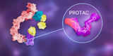
PROTAC, which exploit the ubiquitin-proteasome pathway to specifically degrade target proteins. PROTACs not only solve the problem of undruggability but they also have other advantages compared to traditional drug targeting strategies.
Scientific Reviews
PROTAC, which exploit the ubiquitin-proteasome pathway to specifically degrade target proteins. PROTACs not only solve the problem of undruggability but they also have other advantages compared to traditional drug targeting strategies. -
 Scientific Reviews
PROTAC — Target Selection and Design
Scientific Reviews
PROTAC — Target Selection and Design2022-07-08
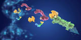
A PROTAC molecule consists of three components: a target protein binding ligand, an E3 ligase ligand, and a linker connecting these two moieties. Here, we will discuss the conventional approaches for the rational design of PROTAC molecules. -
 Scientific Reviews
Organoids represent an important bridge between 2D cultures and in vivo mouse/human models. The organoid technology exerts enormous potential in evaluation of efficacy and toxicity of drugs, regenerative medicine, and precision medicine.In this article, we will briefly introduce organoid technology.
Scientific Reviews
Organoids represent an important bridge between 2D cultures and in vivo mouse/human models. The organoid technology exerts enormous potential in evaluation of efficacy and toxicity of drugs, regenerative medicine, and precision medicine.In this article, we will briefly introduce organoid technology. -
 Scientific Reviews
Development of Intestinal Organoid
Scientific Reviews
Development of Intestinal Organoid2022-07-28
3D organoid is one of the revolutionary developments in biomedical field during the past 10 years. The establishment of intestinal organoids is an important milestone in the development of organoid technology. -
 Scientific Reviews
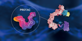
BacPROTACs is composed of a POI ligand, a chemical linker and a ClpCNTD anchor. BacPROTACs can induce in vitro and in vivo degradation of non-eukaryotic proteins in bacteria without the ubiquitin proteasome system.
Scientific Reviews
BacPROTACs is composed of a POI ligand, a chemical linker and a ClpCNTD anchor. BacPROTACs can induce in vitro and in vivo degradation of non-eukaryotic proteins in bacteria without the ubiquitin proteasome system. -
 Scientific Reviews
RNA therapeutics have changed the landscape of drug development, which possess broader spectrum of drug targets, simplicity and efficiency in development and manufacturing.In this article, we will discuss the underlying mechanisms of RNA-based drugs on the market or in clinical stages.
Scientific Reviews
RNA therapeutics have changed the landscape of drug development, which possess broader spectrum of drug targets, simplicity and efficiency in development and manufacturing.In this article, we will discuss the underlying mechanisms of RNA-based drugs on the market or in clinical stages. -
 Scientific Reviews
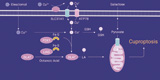
Tsvetkov et al. published in Science and demonstrated a copper-induced programmed cell death — Cuproptosis. As a novel programmed cell death, excess copper triggers abnormal aggregation of lipoylated proteins in TCA cycle and clearance of Fe-S cluster proteins, ultimately leading to cell death.
Scientific Reviews
Tsvetkov et al. published in Science and demonstrated a copper-induced programmed cell death — Cuproptosis. As a novel programmed cell death, excess copper triggers abnormal aggregation of lipoylated proteins in TCA cycle and clearance of Fe-S cluster proteins, ultimately leading to cell death. -
 Scientific Reviews
A comprehensive explanation of ferroptosis
Scientific Reviews
A comprehensive explanation of ferroptosis2022-09-15
Ferroptosis is a new type of RCD that depends on iron and characterized by the accumulation of lipid peroxides. In this article, we will pay our attention on ferroptosis and briefly discusses its mechanism. -
 Scientific Reviews
It's has been proved that p53, as a tumor suppressor gene and immune guardian, may become a destroyer through its own mutation. Moreover, the mechanism of p53 was found to be related to ferroptosis. This article mainly explores the mechanism between p53 and ferroptosis in detail.
Scientific Reviews
It's has been proved that p53, as a tumor suppressor gene and immune guardian, may become a destroyer through its own mutation. Moreover, the mechanism of p53 was found to be related to ferroptosis. This article mainly explores the mechanism between p53 and ferroptosis in detail. -
Scientific Reviews
Organoids have great potential in research of organ development, disease modeling, drug screening and, precision and regenerative medicin. In this article, we will expalin the origin of the organoids. Adult stem cells (ASCs) or pluripotent stem cells (PSCs) , which is the better choice.
-
 Scientific Reviews
Autophagy, derived from the Greek meaning "eating of self", plays an indispensable role in maintaining homeostasis. p27 is an inhibitor of cyclin CDKs, but how p27 regulates autophagy remains unknown. This article will cover the mechanism of autophagy and p27-related cell cycle regulation.
Scientific Reviews
Autophagy, derived from the Greek meaning "eating of self", plays an indispensable role in maintaining homeostasis. p27 is an inhibitor of cyclin CDKs, but how p27 regulates autophagy remains unknown. This article will cover the mechanism of autophagy and p27-related cell cycle regulation. -
 Scientific Reviews
AlphaFold2 can predict disease-related protein structures at low cost, and then find potential drugs for these diseases through drug repositioning, virtual screening, and other methods. ZINC is a public database summarizing information about billions of compounds. AlphaFold2 + ZINC20 speeds up the virtual screening process and improves the computing speed.
Scientific Reviews
AlphaFold2 can predict disease-related protein structures at low cost, and then find potential drugs for these diseases through drug repositioning, virtual screening, and other methods. ZINC is a public database summarizing information about billions of compounds. AlphaFold2 + ZINC20 speeds up the virtual screening process and improves the computing speed. -
 Scientific Reviews
CMA (chaperone-mediated autophagy) plays an essential role in maintaining neuronal protein stability and preventing neurodegeneration. In this article, we will comprehensively clarify the role of CMA in the occurrence and development of neurodegenerative diseases.
Scientific Reviews
CMA (chaperone-mediated autophagy) plays an essential role in maintaining neuronal protein stability and preventing neurodegeneration. In this article, we will comprehensively clarify the role of CMA in the occurrence and development of neurodegenerative diseases. -
 Scientific Reviews
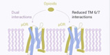
As powerful pain relievers, the opioids morphine and fentanyl have been "checked" by their side effects (listed as controlled substances). How to reduce its side effects? What is its mechanism? This research will explore its mechanism.
Scientific Reviews
As powerful pain relievers, the opioids morphine and fentanyl have been "checked" by their side effects (listed as controlled substances). How to reduce its side effects? What is its mechanism? This research will explore its mechanism. -
 Scientific Reviews
Efferocytosis is the process in which phagocytes remove programmed dead cells. It prevents secondary necrosis of dying cells from releasing harmful cell contents (such as oxides and proteases) that may cause inflammation. Here, we will introduce three stages of efferocytosis: Find, Eat, Digest.
Scientific Reviews
Efferocytosis is the process in which phagocytes remove programmed dead cells. It prevents secondary necrosis of dying cells from releasing harmful cell contents (such as oxides and proteases) that may cause inflammation. Here, we will introduce three stages of efferocytosis: Find, Eat, Digest. -
 Scientific Reviews
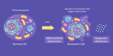
Understanding the mechanism of aging not only has guiding significance for prolonging human life but also has important clinical significance for the prevention and treatment of diseases in the elderly population , thus, improving their life quality and well-being.
Scientific Reviews
Understanding the mechanism of aging not only has guiding significance for prolonging human life but also has important clinical significance for the prevention and treatment of diseases in the elderly population , thus, improving their life quality and well-being. -
 Scientific Reviews
Mammalian cells can also photosynthesize like plants! Photosynthesis can improve cell anabolism and exhibit good clinical effect in degenerative diseases (osteoarthritis).
Scientific Reviews
Mammalian cells can also photosynthesize like plants! Photosynthesis can improve cell anabolism and exhibit good clinical effect in degenerative diseases (osteoarthritis). -
 Scientific Reviews
The latest study of Cell magazine "Neural mechanism underlying depressive-like state associated with social status loss" considers social factors as a breakthrough point. It has been found that the downward transition of social status induces depression-like behavior in mice whereas improves the depressive state by restoring their social environment.
Scientific Reviews
The latest study of Cell magazine "Neural mechanism underlying depressive-like state associated with social status loss" considers social factors as a breakthrough point. It has been found that the downward transition of social status induces depression-like behavior in mice whereas improves the depressive state by restoring their social environment. -
 Scientific Reviews
PROTAC - Design Strategy for Targeting
Scientific Reviews
PROTAC - Design Strategy for Targeting2023-04-23
Protein degradation targeting chimera (PROTAC) is a technology that uses the ubiquitin proteasome pathway to silent target protein. However, PROTAC still has problems such as solubility, membrane permeability, and selectivity. In this article, we have summarized three strategies for optimization: light-controlled linker, PAC molecule, and specific E3 ligase. -
 Scientific Reviews
Necroptosis, also known as necroptosis, is a form of regulated necrotizing cell death mediated by RIP1 and RIP3 kinases. Necroptosis is a process that prevents the self-destruction of activated cells that are blocked by apoptosis. Necroptosis plays a tumor suppressor role in most cases. It may provide benefits in the researches of a variety of human diseases involving immune inflammation and cell death.
Scientific Reviews
Necroptosis, also known as necroptosis, is a form of regulated necrotizing cell death mediated by RIP1 and RIP3 kinases. Necroptosis is a process that prevents the self-destruction of activated cells that are blocked by apoptosis. Necroptosis plays a tumor suppressor role in most cases. It may provide benefits in the researches of a variety of human diseases involving immune inflammation and cell death. -
 Scientific Reviews
Stem cell classification and its application
Scientific Reviews
Stem cell classification and its application2023-05-18
Stem cells (SCs) have the unique ability to self-renew and differentiate into different cell types. SCs can differentiate into various types of tissue cells under specific conditions. Additionally, they can be further cultured to form different tissues and organs in the human body. Stem cells have numerous applications in various fields, including cell therapy, organ transplantation, neurodegenerative disease modeling, and drug screening. -
 Scientific Reviews
Mitophagy:Mechanisms and Detection
Scientific Reviews
Mitophagy:Mechanisms and Detection2023-05-25
About 60 years ago, Christian de Duve first used the term "autophagy" to describe his observation of the degradation of mitochondria and other intracellular structures in lysosomes of rat liver. Over the years, autophagy has remained a beloved topic of research by the National Natural Science Foundation of China (NSFC).Today, let's talk about mitochondrial autophagy. -
 Scientific Reviews
Structure of Lipid Droplets
Scientific Reviews
Structure of Lipid Droplets2023-06-15
Huh? Lipid droplets? Organelles? In the past, biological data usually only show the traditional organelles, such as mitochondria, Golgi apparatus, endoplasmic reticulum, etc., lipid droplets are often not mentioned by people. Today, we will make a systematic explanation of lipid droplets, so that everyone has a clear understanding! -
 Scientific Reviews
How to Perform Western Blot?
Scientific Reviews
How to Perform Western Blot?2023-07-13
Western blot is one of the most frequently performed experiments in molecular biology, biochemistry and immunology. This article describes in detail how to do WB. -
 Scientific Reviews
Organoid Culture: Questions & Answers
Scientific Reviews
Organoid Culture: Questions & Answers2023-07-26
In the last issue, we conducted a live lecture on the theme of organoid culture. Today, we have a special topic to solve your doubts in the last live class! -
 Scientific Reviews
A suitable model is crucial in drug screening experiments. Organs can mimic the three-dimensional functional structure of internal organs, have similar spatial organization to corresponding organs, maintain some key characteristics, and reproduce some physiological functions. They are widely used for modeling and personalized drug screening of diseases such as cancer, infectious diseases, and rare diseases.
Scientific Reviews
A suitable model is crucial in drug screening experiments. Organs can mimic the three-dimensional functional structure of internal organs, have similar spatial organization to corresponding organs, maintain some key characteristics, and reproduce some physiological functions. They are widely used for modeling and personalized drug screening of diseases such as cancer, infectious diseases, and rare diseases. -
 Scientific Reviews
FDA Annual Review | Record-breaking number of new drug approvals in 2023!
Scientific Reviews
FDA Annual Review | Record-breaking number of new drug approvals in 2023! -
 Scientific Reviews
Are frozen cells always damaged? Is the survival rate of revived cells low? When is it appropriate to freeze cells? What should be considered when reviving them? Today, we're sharing a guide to avoid pitfalls in cell freezing and revival!
Scientific Reviews
Are frozen cells always damaged? Is the survival rate of revived cells low? When is it appropriate to freeze cells? What should be considered when reviving them? Today, we're sharing a guide to avoid pitfalls in cell freezing and revival! -
 Scientific Reviews
KRAS, a gene we've heard so much about, has quickly risen to fame after shedding its "undruggable" label. After reading numerous articles, it's easy to feel overwhelmed and wonder: What exactly should we know about this often-discussed but previously "undruggable" target KRAS?
Scientific Reviews
KRAS, a gene we've heard so much about, has quickly risen to fame after shedding its "undruggable" label. After reading numerous articles, it's easy to feel overwhelmed and wonder: What exactly should we know about this often-discussed but previously "undruggable" target KRAS? -
 Scientific Reviews
Compound Screening Guide!
Scientific Reviews
Compound Screening Guide!2024-03-15
How to use compound library? How to design an experiment if you buy a compound library? Want a specific experimental protocol? This article will introduce popular experimental techniques and provide new ideas for publishing high level literature. -
 Scientific Reviews
Wnt/β-catenin and tumor EMT
Scientific Reviews
Wnt/β-catenin and tumor EMT2024-03-18
Epithelial-Mesenchymal Transition (EMT) is closely related to the plasticity of tumor cells and is a necessary process for tumor metastasis. Wnt/β-catenin is one of the main actors involved in the EMT process. Today, we’re here to popularize the tumor EMT and Wnt/β-catenin pathway~ -
 Scientific Reviews
The 2024 AACR meeting concluded successfully in California, USA. Which antitumor drugs stole the show at this conference?
Scientific Reviews
The 2024 AACR meeting concluded successfully in California, USA. Which antitumor drugs stole the show at this conference? -
 Scientific Reviews
SPR, which stands for Surface Plasmon Resonance, essentially works by detecting the interaction between ligands and analytes on a biosensor chip. This in turn allows us to probe the properties and structure of substances. With this technology, we can analyze molecules, proteins, DNA, and various organic and inorganic substances in samples in real-time with precision.
Scientific Reviews
SPR, which stands for Surface Plasmon Resonance, essentially works by detecting the interaction between ligands and analytes on a biosensor chip. This in turn allows us to probe the properties and structure of substances. With this technology, we can analyze molecules, proteins, DNA, and various organic and inorganic substances in samples in real-time with precision. -
 Scientific Reviews
Science | A "new" mechanism for non-ubiquitinated Midnolin-proteasomal degradation pathway
Scientific Reviews
Science | A "new" mechanism for non-ubiquitinated Midnolin-proteasomal degradation pathway2024-04-26
“ubiquitin-mediated protein degradation” won the Nobel Prize in Chemistry in 2004! In fact, proteasomes degrade not only ubiquitinated proteins but also non-ubiquitinated ones. The mechanism remains shrouded in mystery. After reading this piece today, you might have a lightbulb moment! -
 Scientific Reviews
Common Questions and Solutions for WB
Scientific Reviews
Common Questions and Solutions for WB2024-05-09
Come to understand the common problems and solutions of WB, and better complete the experiment! -
 Scientific Reviews
Still struggling to find the preparation methods for various solutions? Save your time! Here comes a nanny-level tutorial!
Scientific Reviews
Still struggling to find the preparation methods for various solutions? Save your time! Here comes a nanny-level tutorial! -
 Scientific Reviews
STZ Induced Diabetes Models
Scientific Reviews
STZ Induced Diabetes Models2024-06-13
Spotlight: How can STZ Help Diabetes Research? -
 Scientific Reviews
How important is the compound library? It connects to drug screening on one end and leads to lead compound modifications on the other, serving as one of the sources of new drug development.
Scientific Reviews
How important is the compound library? It connects to drug screening on one end and leads to lead compound modifications on the other, serving as one of the sources of new drug development. -
 Scientific Reviews
Degrade target proteins through the autophagy-lysosome pathway including LYTAC, AUTAC, and ATTEC have gained increasing attention in recent years due to their significant research potential!
Scientific Reviews
Degrade target proteins through the autophagy-lysosome pathway including LYTAC, AUTAC, and ATTEC have gained increasing attention in recent years due to their significant research potential! -
 Scientific Reviews
Streptavidin-Biotin System
Scientific Reviews
Streptavidin-Biotin System2024-08-03
Streptavidin strongly binding small molecule biotin is one of the most popular non-covalent coupling methods. Streptavidin can be coupled to various carriers such as magnetic beads and agarose matrix, and become a highly specific affinity medium to capture various biotin-labeled ligands. -
 Scientific Reviews
Science’s 2023 Breakthrough: GLP-1R Agonists
Scientific Reviews
Science’s 2023 Breakthrough: GLP-1R Agonists2024-08-13
GLP-1RAs, which achieved great success in 2023 and draws people’s attention back to obesity treatments and GLP-1 therapies worldwide, was chosen as the breakthrough of year 2023 by Science [2]. -
 Scientific Reviews
What Are Popular Anti-tumor Drug Targets?
Scientific Reviews
What Are Popular Anti-tumor Drug Targets?2024-08-20
The rapid development of targeted anti-cancer drugs has spurred diverse research across various modalities. These include small molecules, monoclonal antibodies (mAbs), cell immunotherapies, antibody-drug conjugates (ADCs), and PROTACs (proteolysis targeting chimeras). -
 Scientific Reviews
Cuproptosis, How much do you know?
Scientific Reviews
Cuproptosis, How much do you know?2024-08-22
Cells die in a variety of ways, including apoptosis, pyroptosis, necrosis, and ferroptosis......And, of course, cuproptosis. So, how much do you know about cuproptosis? -
 Scientific Reviews
Virtual Screening and New Uses for Old Drugs
Scientific Reviews
Virtual Screening and New Uses for Old Drugs2024-09-17
With the advancement of medical science, drug screening against various disease targets has become the fundamental strategy for drug development. Currently, computer-based virtual screening techniques are emerging in the field of new drug research due to their efficiency and low cost. Let's explore it today! -
 Scientific Reviews
Recently, Mol Cell reported the discovery of the first ferroptosis marker, Hyperoxidized PRDX3! Let’s take a look together~
Scientific Reviews
Recently, Mol Cell reported the discovery of the first ferroptosis marker, Hyperoxidized PRDX3! Let’s take a look together~ -
 Scientific Reviews
Cuproptosis's Knowledge Points!
Scientific Reviews
Cuproptosis's Knowledge Points!2024-09-25
With the establishment of the cuproptosis mechanism related research has attracted more and more attention from major journals. Expect to use the sharp sword of cuproptosis to stab the tumor cells. -
 Scientific Reviews
2024 Nobel Prize Announcements! Curious about the details? Click to dive into the exciting developments!
Scientific Reviews
2024 Nobel Prize Announcements! Curious about the details? Click to dive into the exciting developments! -
 Scientific Reviews
How they used PKH 26 for cellular studies?
Scientific Reviews
How they used PKH 26 for cellular studies?2024-10-21
We are thrilled to highlight our client study using PKH 26 (MedChemExpress) , a red fluorescent dye that has proven invaluable for in vitro cell labeling and tracing. This innovative research, published in the Journal of Nanobiotechnology. -
Scientific Reviews
Delve into the intricate networks that govern mitochondrial quality control. By examining key mechanisms such as biogenesis, mitochondrial dynamics (fission and fusion), proteolysis, and mitophagy. -
 Scientific Reviews
A New Form of Cell Death: PANoptosis!
Scientific Reviews
A New Form of Cell Death: PANoptosis! -
 Scientific Reviews
Unlocking the Power of Intermittent Fasting: The 16+8 Method
Scientific Reviews
Unlocking the Power of Intermittent Fasting: The 16+8 Method -
 Scientific Reviews
We are thrilled to share the latest advancements in AI technology as highlighted in this insightful article. From groundbreaking innovations to transformative applications, the future of AI is brighter than ever!
Scientific Reviews
We are thrilled to share the latest advancements in AI technology as highlighted in this insightful article. From groundbreaking innovations to transformative applications, the future of AI is brighter than ever! -
 Scientific Reviews
Advancing Chronic Kidney Disease Research with GJ103 and Lamin B1 Antibody!
Scientific Reviews
Advancing Chronic Kidney Disease Research with GJ103 and Lamin B1 Antibody! -
 Scientific Reviews
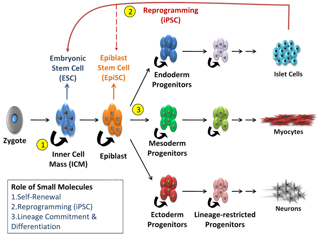
In early January 2025, MIT Tech Review unveiled its annual list of top 10 breakthrough technologies poised to redefine the future. Among these, stem cell therapy stood out for its potential to treat diverse diseases—including neurodegenerative disorders, diabetes, cancer, and heart failure. This groundbreaking approach uses stem cells to replace damaged cells, offering hope for millions worldwide.
Scientific Reviews
In early January 2025, MIT Tech Review unveiled its annual list of top 10 breakthrough technologies poised to redefine the future. Among these, stem cell therapy stood out for its potential to treat diverse diseases—including neurodegenerative disorders, diabetes, cancer, and heart failure. This groundbreaking approach uses stem cells to replace damaged cells, offering hope for millions worldwide. -
 Scientific Reviews
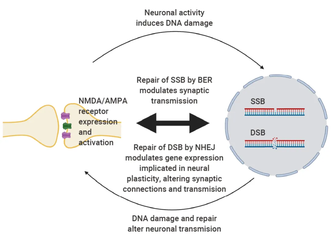
When you hear "inflammation" and "DNA damage," you might immediately think of disease or injury. However, in brains, these two processes are key steps in forming long-term memories, particularly related to specialized cells in our brain called hippocampal neurons.
Scientific Reviews
When you hear "inflammation" and "DNA damage," you might immediately think of disease or injury. However, in brains, these two processes are key steps in forming long-term memories, particularly related to specialized cells in our brain called hippocampal neurons. -
 Scientific Reviews
Autophagy is a fundamental process that degrades various components within the cell.
Scientific Reviews
Autophagy is a fundamental process that degrades various components within the cell. -
 Scientific Reviews
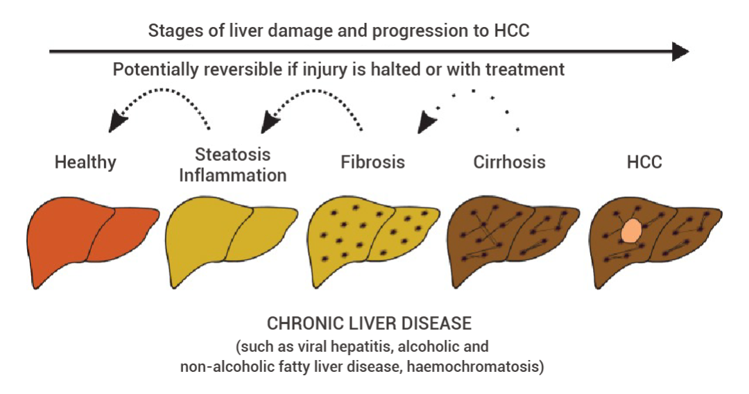
This article will tell you about the common methods of modeling liver disease in animal models.
Scientific Reviews
This article will tell you about the common methods of modeling liver disease in animal models. -
.jpg) Scientific Reviews
nside cells, the homeostasis and degradation of proteins is a precisely regulated process. If proteins cannot be degraded in time, it may lead to the occurrence of various diseases such as neurodegenerative diseases and cancer. This article will tell you the process of how proteins are recognized, labeled and then degraded!
Scientific Reviews
nside cells, the homeostasis and degradation of proteins is a precisely regulated process. If proteins cannot be degraded in time, it may lead to the occurrence of various diseases such as neurodegenerative diseases and cancer. This article will tell you the process of how proteins are recognized, labeled and then degraded! -
 Scientific Reviews
This article introduces some common cardiovascular disease models, inducers, modeling protocols and successful modeling cases in the research.
Scientific Reviews
This article introduces some common cardiovascular disease models, inducers, modeling protocols and successful modeling cases in the research. -
 Scientific Reviews
Do you feel confused when you start culturing cells? This article will show you the basic methods and steps of cell culture!
Scientific Reviews
Do you feel confused when you start culturing cells? This article will show you the basic methods and steps of cell culture! -
 Scientific Reviews
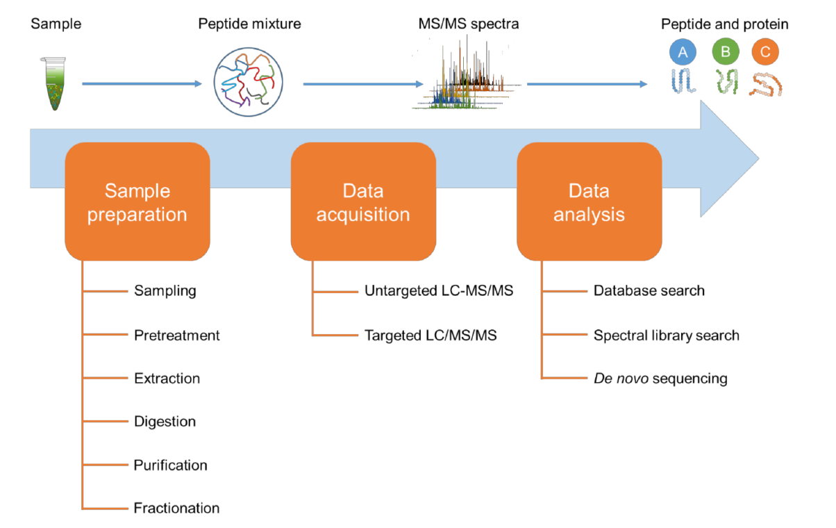
As a pivotal branch in the post - genomic era, proteomics is committed to comprehensively elucidating the types, abundances, structures, functions, and interactions of all proteins within living organisms. This article will tell you the key steps of proteomics sample preparation based on mass spectrometry. This article will tell you the key steps of proteomics sample preparation based on mass spectrometry.
Scientific Reviews
As a pivotal branch in the post - genomic era, proteomics is committed to comprehensively elucidating the types, abundances, structures, functions, and interactions of all proteins within living organisms. This article will tell you the key steps of proteomics sample preparation based on mass spectrometry. This article will tell you the key steps of proteomics sample preparation based on mass spectrometry. -
 Scientific Reviews
Cell Migration vs. Invasion: Differences Revealed by Scratch Assays and Transwell Experiments
Scientific Reviews
Cell Migration vs. Invasion: Differences Revealed by Scratch Assays and Transwell Experiments2025-05-30
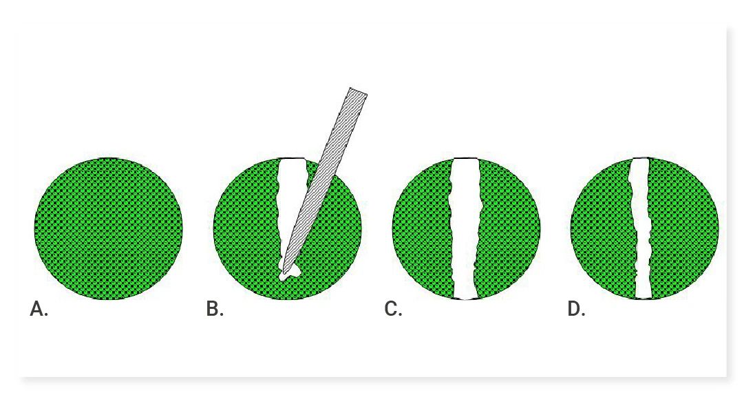
In scientific research, cell migration and invasion are crucial for understanding many important biological processes. This article delves into commonly used detection methods: the scratch assay and Transwell migration/invasion assay. -
 Scientific Reviews
Western blotting is a crucial and fundamental technique in life science research. It plays a significant role in exploring protein expression and function. The following article will comprehensively and thoroughly elaborate on the specific procedures and detailed key points of this experiment.
Scientific Reviews
Western blotting is a crucial and fundamental technique in life science research. It plays a significant role in exploring protein expression and function. The following article will comprehensively and thoroughly elaborate on the specific procedures and detailed key points of this experiment. -
 Scientific Reviews
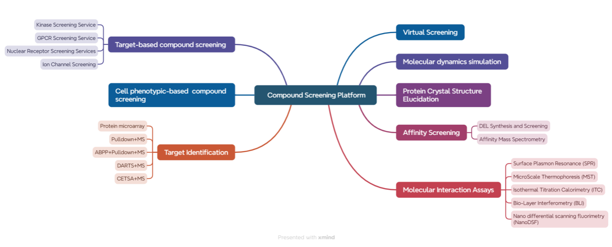
How should drug screening experiments be conducted? How can we ensure the accuracy of the lead compounds identified? This article will take you through how MCE's clients conduct drug screening experiments.
Scientific Reviews
How should drug screening experiments be conducted? How can we ensure the accuracy of the lead compounds identified? This article will take you through how MCE's clients conduct drug screening experiments. -
 Scientific Reviews
In this issue, we will conduct an in-depth interpretation from the dimensions of nanoparticle design, mechanism of action, in vivo and in vitro efficacy, and immune regulation, revealing how this research brings new hope for the treatment of invasive tumors through interdisciplinary innovation!
Scientific Reviews
In this issue, we will conduct an in-depth interpretation from the dimensions of nanoparticle design, mechanism of action, in vivo and in vitro efficacy, and immune regulation, revealing how this research brings new hope for the treatment of invasive tumors through interdisciplinary innovation! -
 Scientific Reviews
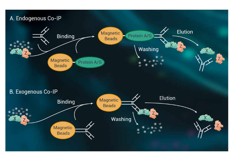
In protein biology, Co-IP is a powerful tool to uncover protein "social networks." But poorly performed, it easily becomes an awkward lab "meet-and-greet." Today, we discuss making Co-IP experiments elegant and efficient—so you pull down target proteins with confidence and precision.
Scientific Reviews
In protein biology, Co-IP is a powerful tool to uncover protein "social networks." But poorly performed, it easily becomes an awkward lab "meet-and-greet." Today, we discuss making Co-IP experiments elegant and efficient—so you pull down target proteins with confidence and precision. -
 Scientific Reviews
IHC, ICC, IF Techniques: A Practical Guide
Scientific Reviews
IHC, ICC, IF Techniques: A Practical Guide2025-07-25
Confused About IHC/ICC/IF? Why Does Immunostaining Seem So Complicated? Read This Now! Master Immunostaining with Confidence! -
 Scientific Reviews
Chromatin Immunoprecipitation (ChIP) Demystified: A Complete Guide to Epigenetic Analysis
Scientific Reviews
Chromatin Immunoprecipitation (ChIP) Demystified: A Complete Guide to Epigenetic Analysis2025-08-05
In this issue, we introduce a powerful technique for detecting interactions between epigenetic regulatory factors and DNA—Chromatin Immunoprecipitation (ChIP)! -
 Scientific Reviews
HTS Breakthroughs Powered by MCE Libraries
Scientific Reviews
HTS Breakthroughs Powered by MCE Libraries2025-08-13
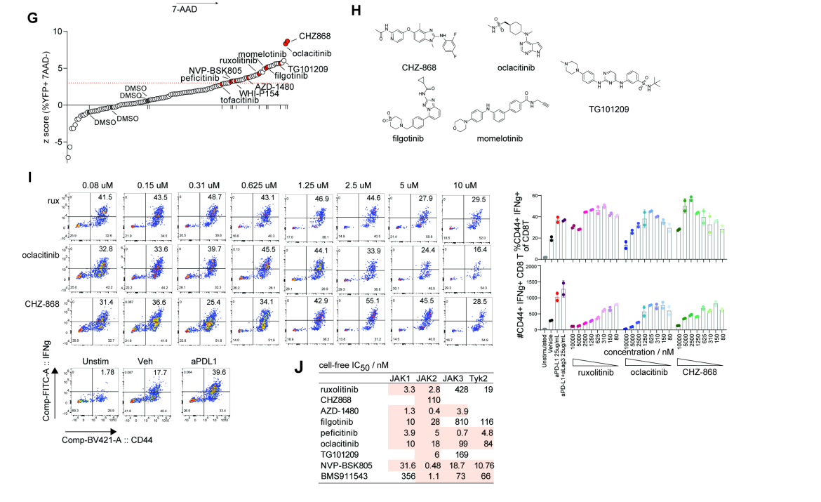
Key High-Throughput Screening Breakthroughs of 2024 Featuring MCE -
 Scientific Reviews
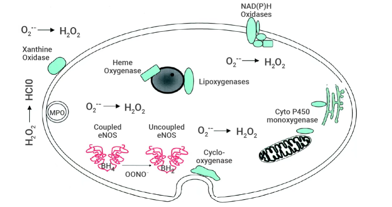
The article introduces the mechanisms of ROS generation and the methods for their detection.
Scientific Reviews
The article introduces the mechanisms of ROS generation and the methods for their detection. -
 Scientific Reviews
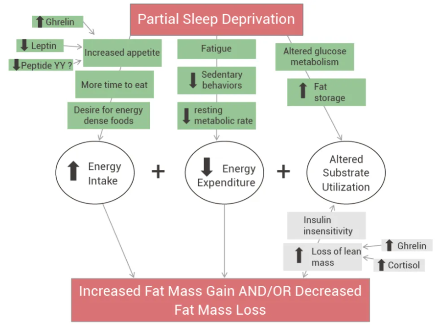
Have you ever noticed that after staying up late, your appetite—especially for high-calorie foods—gets out of control? If this sounds familiar, today’s article might offer some good news.
Scientific Reviews
Have you ever noticed that after staying up late, your appetite—especially for high-calorie foods—gets out of control? If this sounds familiar, today’s article might offer some good news. -
 Scientific Reviews
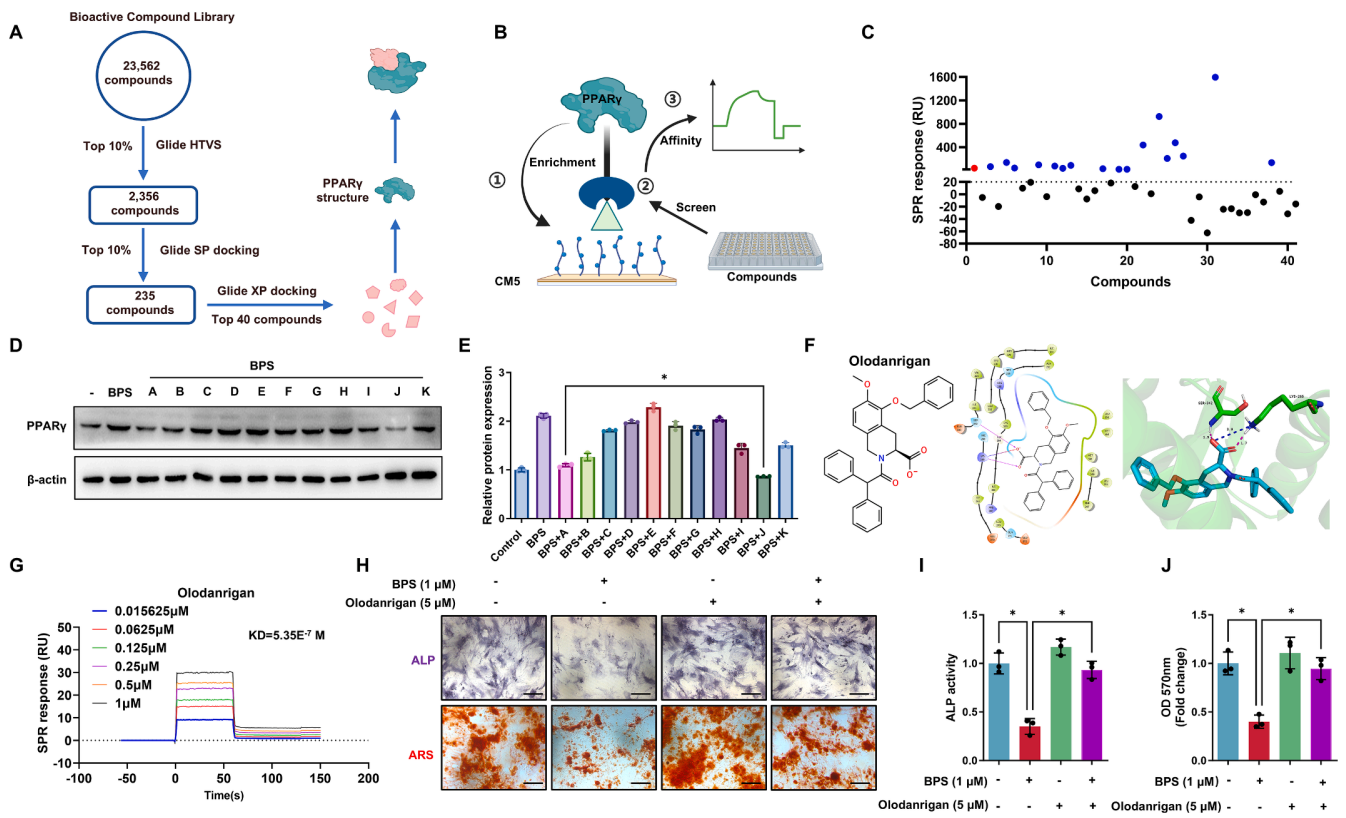
Encountering challenges with the high costs and long timelines of drug screening? Have a defined target but remain uncertain how to efficiently identify active molecules? Unsure how to validate hits generated from virtual screening? The ‘Winning Combination’ of drug screening offers a powerful solution to address these critical obstacles.
Scientific Reviews
Encountering challenges with the high costs and long timelines of drug screening? Have a defined target but remain uncertain how to efficiently identify active molecules? Unsure how to validate hits generated from virtual screening? The ‘Winning Combination’ of drug screening offers a powerful solution to address these critical obstacles. -
 Scientific Reviews
Generating Stable Cell Lines with Lentivirus
Scientific Reviews
Generating Stable Cell Lines with Lentivirus2025-09-16
A step-by-step protocol of establishing stable cell lines using lentivirus -
 Scientific Reviews
A groundbreaking study in Nature Communications reveals the key mechanism behind Idiopathic Pulmonary Fibrosis and identifies an existing drug with the potential to counter it.
Scientific Reviews
A groundbreaking study in Nature Communications reveals the key mechanism behind Idiopathic Pulmonary Fibrosis and identifies an existing drug with the potential to counter it. -
 Scientific Reviews
Finding adipogenic induction media too expensive and tricky to prepare? This comprehensive guide to 3T3-L1 adipogenic differentiation simplifies the process, helping you achieve great results!
Scientific Reviews
Finding adipogenic induction media too expensive and tricky to prepare? This comprehensive guide to 3T3-L1 adipogenic differentiation simplifies the process, helping you achieve great results! -
Scientific Reviews
For all the protein research folks out there, techniques like IP and Co-IP are no stranger, right? And of course, there's a faster and more convenient go-to tool—Protein A/G magnetic beads! In this article, let's chat about how these beads work their magic in classic experiments like IP and Co-IP. -
Scientific Reviews
Microglia– the only immune cells within the brain parenchyma. With advancements in imaging technologies, people’s understanding of microglia has shifted from being viewed as 'resting' cells to 'highly active' cells, particularly due to their dynamic processes that seem to be probing surrounding tissues and monitoring neuronal activity. This has made microglia a focal point of research in the field of neuroscience. -
Scientific Reviews
Cracking the PROTAC Permeability Barrier: CD36-Mediated Endocytosis as a Potential Breakthrough2025-12-03
This article provides an in-depth analysis of cutting-edge literature revealing CD36 as a key mediator of cellular uptake for PROTACs and bRO5 compounds. By structurally optimizing PROTAC molecules to enhance their affinity for CD36, membrane permeability can be markedly improved, leading to significantly enhanced antitumor efficacy. -
 Scientific Reviews
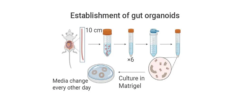
Efficient Generation of Mouse Small Intestinal Organoids: A Complete Experimental Protocol Guide
Scientific Reviews
Efficient Generation of Mouse Small Intestinal Organoids: A Complete Experimental Protocol Guide2025-12-10
How to successfully create mouse small intestine organoids? A detailed, hands-on guide to the entire process, all in one article! -
 Scientific Reviews
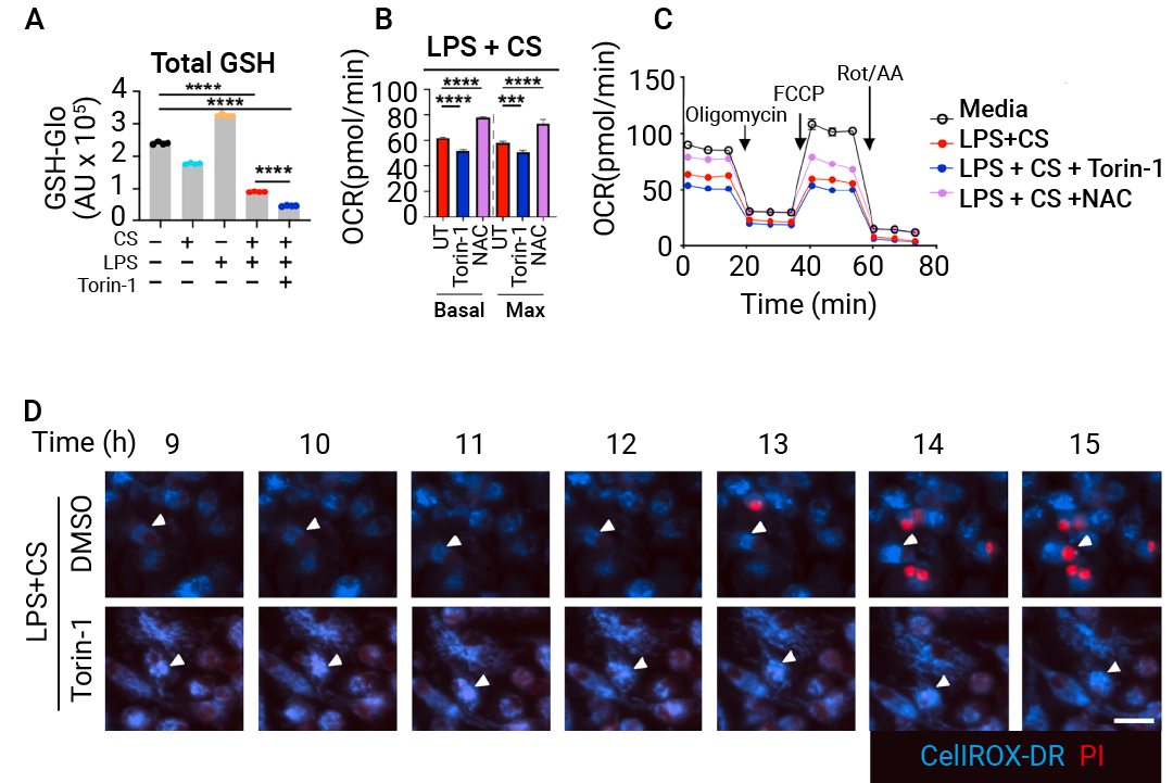
A November 2025 Cell study discovers Mitoxyperilysis, a new mTOR-regulated, caspase-independent cell death pathway driven by innate immune and metabolic dysregulation via mitochondrial-plasma membrane contact and oxidative damage, and verifies its potential to induce tumor necrosis for cancer therapy with key regulators including BAX, BAK1 and BID.
Scientific Reviews
A November 2025 Cell study discovers Mitoxyperilysis, a new mTOR-regulated, caspase-independent cell death pathway driven by innate immune and metabolic dysregulation via mitochondrial-plasma membrane contact and oxidative damage, and verifies its potential to induce tumor necrosis for cancer therapy with key regulators including BAX, BAK1 and BID. -
 Scientific Reviews
In the hunt for the next ‘GLP-1,’ amylin therapeutics, which have shown strong weight-loss results in clinical trials, have become a major focus for both multinational pharma and the scientific community.
Scientific Reviews
In the hunt for the next ‘GLP-1,’ amylin therapeutics, which have shown strong weight-loss results in clinical trials, have become a major focus for both multinational pharma and the scientific community. -
 Scientific Reviews
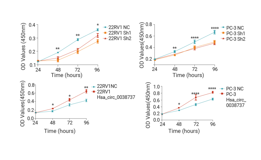
Essential for High-Impact Papers: Present Your CCK-8 Experimental Results in a More Outstanding Way!
Scientific Reviews
Essential for High-Impact Papers: Present Your CCK-8 Experimental Results in a More Outstanding Way!2026-03-18
CCK-8 is a widely used WST‑8‑based reagent for cell proliferation and cytotoxicity assays. It features high sensitivity, reliable results and easy operation, and is applicable to cell viability analysis, drug screening and growth inhibition testing. -
 Scientific Reviews
FISH is a molecular technique using fluorescent probes to detect specific nucleic acids in cells, with high sensitivity and diverse biomedical applications.
Scientific Reviews
FISH is a molecular technique using fluorescent probes to detect specific nucleic acids in cells, with high sensitivity and diverse biomedical applications. -
 Scientific Reviews
This article walks you through the experimental design and workflow of flow cytometry, delivering a clear, dynamic, and professional overview to elevate your research.
Scientific Reviews
This article walks you through the experimental design and workflow of flow cytometry, delivering a clear, dynamic, and professional overview to elevate your research. -
 Scientific Reviews
Molecular glue degraders have evolved from a serendipitous observation to one of the most dynamic and transformative fields in biomedical research.
Scientific Reviews
Molecular glue degraders have evolved from a serendipitous observation to one of the most dynamic and transformative fields in biomedical research. -
 Scientific Reviews
GLP-1 and Obesity Research
Scientific Reviews
GLP-1 and Obesity Research2026-08-14
Obesity substantially increases the risk of chronic diseases such as T2D and cardiovascular disease. The breakout success of GLP-1 therapies has spotlighted GLP-1R and a wave of emerging obesity targets. -
 EDM
This review introduces the major forms of programmed cell death, with a focus on the molecular mechanisms of ferroptosis and the methods used for its detection.
EDM
This review introduces the major forms of programmed cell death, with a focus on the molecular mechanisms of ferroptosis and the methods used for its detection. -
 EDM
This review explores the molecular mechanisms of the DNA damage response, reviews therapeutic strategies targeting DDR pathways in cancer, and examines their roles in cancer drug resistance.
EDM
This review explores the molecular mechanisms of the DNA damage response, reviews therapeutic strategies targeting DDR pathways in cancer, and examines their roles in cancer drug resistance. -
 EDM
This review provides a practical guide to the fundamental mechanisms, regulatory pathways, and detection methods of pyroptosis.
EDM
This review provides a practical guide to the fundamental mechanisms, regulatory pathways, and detection methods of pyroptosis. -
 EDM
Explore lysosomal nutrient sensing, quality control, cellular adaptation, disease mechanisms, and emerging therapeutic approaches.
EDM
Explore lysosomal nutrient sensing, quality control, cellular adaptation, disease mechanisms, and emerging therapeutic approaches. -
 EDM
Explore how mitochondrial quality control, inflammatory signaling, and metabolic–epigenetic reprogramming regulate cellular senescence and the SASP, along with strategies to restore mitochondrial homeostasis.
EDM
Explore how mitochondrial quality control, inflammatory signaling, and metabolic–epigenetic reprogramming regulate cellular senescence and the SASP, along with strategies to restore mitochondrial homeostasis. -
 Blog
EC0489, a SMDC for Cancer Therapy
Blog
EC0489, a SMDC for Cancer Therapy2019-03-25
EC0489, a conjugate of folic acid and desacetyl vinblastine hydrazide, is a SMDC under development for the treatment of solid tumours. -
 Blog
Role of PRMT7 Probe SGC3027 in Cancer
Blog
Role of PRMT7 Probe SGC3027 in Cancer2019-03-27
SGC3027 is the first potent, selective and cell active chemical probe for PRMT7. SGC3027 is also a pro-drug, which converts to the active compound SGC8158 -
 Blog
AZD5991, a macrocyclic molecule with high selectivity for Mcl-1, reduces Mcl-1 protein in AZD5991-sensitive but not in AZD5991-resistant MM cell lines.
Blog
AZD5991, a macrocyclic molecule with high selectivity for Mcl-1, reduces Mcl-1 protein in AZD5991-sensitive but not in AZD5991-resistant MM cell lines. -
 Blog
A Novel and Efficacious RAF Inhibitor RAF709
Blog
A Novel and Efficacious RAF Inhibitor RAF7092019-03-30
RAF709, a novel and efficacious RAF inhibitor, activates the MAPK pathway and shows antitumor activity in tumor cells harboring BRAF or RAS mutations. -
 Blog
CF53 is a highly potent, selective and orally active inhibitor of BET protein, with anti-tumor activity in acute leukemia and breast cancer cell lines.
Blog
CF53 is a highly potent, selective and orally active inhibitor of BET protein, with anti-tumor activity in acute leukemia and breast cancer cell lines. -
 Blog
HJB97 is a BET PROTAC inhibitor with good anti-tumor activity, and effectively blocks the degradation of BRD2, BRD3, and BRD4 proteins induced by BETd-260.
Blog
HJB97 is a BET PROTAC inhibitor with good anti-tumor activity, and effectively blocks the degradation of BRD2, BRD3, and BRD4 proteins induced by BETd-260. -
 Blog
CCB02 is an tubulin binder and has potential in the therapy of cancer cells with extra centrosomes. CCB02 also activates spindle assembly checkpoint.
Blog
CCB02 is an tubulin binder and has potential in the therapy of cancer cells with extra centrosomes. CCB02 also activates spindle assembly checkpoint. -
 Blog
H3B-5942 is a selective and irreversible estrogen receptor covalent antagonist, inactivates both ERαWT and ERα mutation.
Blog
H3B-5942 is a selective and irreversible estrogen receptor covalent antagonist, inactivates both ERαWT and ERα mutation. -
 Blog
AZD3229 is a Potent Pan-KIT Mutant Inhibitor
Blog
AZD3229 is a Potent Pan-KIT Mutant Inhibitor2019-04-04
AZD3229 is a potent, pan-KIT mutant inhibitor with potent single digit nM growth inhibition against a diverse panel of mutant KIT driven Ba/F3 cell lines. -
 Blog
A Lead PROTAC BRD9 Chemical Degrader
Blog
A Lead PROTAC BRD9 Chemical Degrader2019-04-06
PROTAC BRD9 Degrader-1 is a lead PROTAC BRD9 chemical degrader and a selective probe useful for the study of BAF complex biology. -
 Blog
SGC-GAK-1 is a potent, selective, and cell-active GAK inhibitor and shows potent anti-proliferative activity in LNCaP and 22Rv1 cells.
Blog
SGC-GAK-1 is a potent, selective, and cell-active GAK inhibitor and shows potent anti-proliferative activity in LNCaP and 22Rv1 cells. -
 Blog
COH000 is an allosteric, covalent and irreversible inhibitor of SUMO-activating enzyme, with an IC50 of 0.2 μM for SUMOylation in vitro.
Blog
COH000 is an allosteric, covalent and irreversible inhibitor of SUMO-activating enzyme, with an IC50 of 0.2 μM for SUMOylation in vitro. -
 Blog
HS-1371 is a Small Molecule RIP3 Inhibitor
Blog
HS-1371 is a Small Molecule RIP3 Inhibitor2019-04-09
HS-1371 is a novel kinase inhibitor of RIP3-mediated necroptosis, showing an inhibitory effect on S227 auto-phosphorylation of RIP3 at the basal level. -
 Blog
Nevanimibe is a selective and potent ACAT1 inhibitor. An excellent drug candidate in the treatment of adrenocortical cancer.
Blog
Nevanimibe is a selective and potent ACAT1 inhibitor. An excellent drug candidate in the treatment of adrenocortical cancer. -
 Blog
PhiKan 083 is a carbazole derivative, which binds to the surface cavity and stabilizes Y220C (a p53 mutant), with a Kd of 167 μM.
Blog
PhiKan 083 is a carbazole derivative, which binds to the surface cavity and stabilizes Y220C (a p53 mutant), with a Kd of 167 μM. -
 Blog
AZ304, a Dual BRAF Inhibitor Against Cancer
Blog
AZ304, a Dual BRAF Inhibitor Against Cancer2019-04-12
AZ304 is a potent BRAF inhibitor, blocks both wild type BRAF and V600E mutant BRAF activity, with IC50s in the nanomolar range. -
 Blog
Novel Greatwall Kinase Inhibitor GKI-1
Blog
Novel Greatwall Kinase Inhibitor GKI-12019-04-13
GKI-1, a GWL inhibitor, robustly inhibited ROCK1 with an IC50 of ~11 μM, but only weakly affected PKA and had no observable inhibition towards CDK2. -
 Blog
IITZ-01 is a potent lysosomotropic autophagy inhibitor with single-agent antitumor activity, with an IC50 of 2.62 μM for PI3Kγ.
Blog
IITZ-01 is a potent lysosomotropic autophagy inhibitor with single-agent antitumor activity, with an IC50 of 2.62 μM for PI3Kγ. -
 Blog
SSE15206 is a microtubule polymerization inhibitor, with a GI50 of 197 nM in HCT116 cells. Causes aberrant mitosis resulting in G2/M arrest.
Blog
SSE15206 is a microtubule polymerization inhibitor, with a GI50 of 197 nM in HCT116 cells. Causes aberrant mitosis resulting in G2/M arrest. -
 Blog
Erteberel is a Selective ERβ Agonist
Blog
Erteberel is a Selective ERβ Agonist2019-04-16
Erteberel (LY500307) is a synthetic, nonsteroidal estrogen which acts as a selective ERβ agonist and under development for the treatment of schizophrenia. -
 Blog
MBQ-167 is a dual Rac/Cdc42 inhibitor in in metastatic cancer, with IC50s of 103 nM for Rac 1/2/3 and 78 nM for Cdc42 in MDA-MB-231 cells, respectively.
Blog
MBQ-167 is a dual Rac/Cdc42 inhibitor in in metastatic cancer, with IC50s of 103 nM for Rac 1/2/3 and 78 nM for Cdc42 in MDA-MB-231 cells, respectively. -
 Blog
PRN1008 is a Reversible Covalent and Oral Active Inhibitor of Bruton’s Tyrosine Kinase (BTK)
Blog
PRN1008 is a Reversible Covalent and Oral Active Inhibitor of Bruton’s Tyrosine Kinase (BTK)2019-04-18
PRN1008 is a selective, reversible covalent and oral active inhibitor of Bruton’s Tyrosine Kinase (BTK), with an IC50 of 1.3 nM. -
 Blog
Y06036 is a potent and selective BET inhibitor for potential treatment of castration-resistant prostate cancer. With nanomolar inhibition.
Blog
Y06036 is a potent and selective BET inhibitor for potential treatment of castration-resistant prostate cancer. With nanomolar inhibition. -
 Blog
JNJ-64619178 is a selective and pseudo-irreversible PRMT5 inhibitor with an IC50 of 0.14 nM. Has potent Activity In Lung Cancer.
Blog
JNJ-64619178 is a selective and pseudo-irreversible PRMT5 inhibitor with an IC50 of 0.14 nM. Has potent Activity In Lung Cancer. -
 Blog
(E)-AG 99 is an EGFR inhibitor and shows a growth-inhibition on not only serum-starved cells but also normally grown cells. Treatment for Bladder cancer.
Blog
(E)-AG 99 is an EGFR inhibitor and shows a growth-inhibition on not only serum-starved cells but also normally grown cells. Treatment for Bladder cancer. -
 Blog
Cevipabulin is a microtubule-active compound and inhibits the binding of [3H] vinblastine to tubulin, with an IC50 of 18-40 nM for in human tumor cell line.
Blog
Cevipabulin is a microtubule-active compound and inhibits the binding of [3H] vinblastine to tubulin, with an IC50 of 18-40 nM for in human tumor cell line. -
 Blog
BTR-1 potently inhibits cell growth. It induces S phase arrest, affects DNA replication. Dose-dependently induces cytotoxicity in leukemic cell lines.
Blog
BTR-1 potently inhibits cell growth. It induces S phase arrest, affects DNA replication. Dose-dependently induces cytotoxicity in leukemic cell lines. -
 Blog
Y06137 is a potent and selective BET inhibitor, which binds to the BRD4(1) bromodomain with a Kd of 81 nM. Antitumor activity.
Blog
Y06137 is a potent and selective BET inhibitor, which binds to the BRD4(1) bromodomain with a Kd of 81 nM. Antitumor activity. -
 Blog
Borussertib is a covalent-allosteric and first-in-class inhibitor of protein kinase Akt, with an IC50 of 0.8 nM and a Ki of 2.2 nM for Akt-wt.
Blog
Borussertib is a covalent-allosteric and first-in-class inhibitor of protein kinase Akt, with an IC50 of 0.8 nM and a Ki of 2.2 nM for Akt-wt. -
 Blog
TD-428 is a Highly Specific BRD4 Degrader
Blog
TD-428 is a Highly Specific BRD4 Degrader2019-04-29
TD-428, a immunomodulatory drug analog, is a highly specific BRD4 degrader with a DC50 of 0.32 nM. TD-428 reduces c-Myc levels more efficiently than JQ1. -
 Blog
SLLN-15 is an oral activ enhancer of autophagy that activates cytostatic macroautophagy/autophagy in triple-negative breast cancer (TNBC).
Blog
SLLN-15 is an oral activ enhancer of autophagy that activates cytostatic macroautophagy/autophagy in triple-negative breast cancer (TNBC). -
 Blog
CB-6644 is a selective non-ATP-competitive inhibitor of the RUVBL1/2 complex. CB-6644 significantly reduces tumor growth without obvious toxicity.
Blog
CB-6644 is a selective non-ATP-competitive inhibitor of the RUVBL1/2 complex. CB-6644 significantly reduces tumor growth without obvious toxicity. -
 Blog
A1874 is a nutlin-based and BRD4-degrading PROTAC with a DC50 of 32 nM. Effective in inhibiting many cancer cell lines proliferation
Blog
A1874 is a nutlin-based and BRD4-degrading PROTAC with a DC50 of 32 nM. Effective in inhibiting many cancer cell lines proliferation -
 Blog
NSC 228155 is a activator of EGFR and a potent inhibitor of KIX-KID interaction. NSC 228155 shows excellent anti-tumor activity.
Blog
NSC 228155 is a activator of EGFR and a potent inhibitor of KIX-KID interaction. NSC 228155 shows excellent anti-tumor activity. -
 Blog
BSJ-03-123, a Degrader with Proteome-wide Selectivity for CDK6 (PROTAC). Induces a G1 cell-cycle arrest without a measurable increase in apoptosis.
Blog
BSJ-03-123, a Degrader with Proteome-wide Selectivity for CDK6 (PROTAC). Induces a G1 cell-cycle arrest without a measurable increase in apoptosis. -
 Blog
FT671 is a potent, non-covalent and selective USP7 inhibitor with an IC50 of 52 nM and binds to the USP7 catalytic domain with a Kd of 65 nM.
Blog
FT671 is a potent, non-covalent and selective USP7 inhibitor with an IC50 of 52 nM and binds to the USP7 catalytic domain with a Kd of 65 nM. -
 Blog
BSc5371 is a potent and irreversible FLT3 inhibitor. BSc5371 is cytotoxic to FLT3-dependent cell line. BSc5371 has potential to treat Acute Leukemia.
Blog
BSc5371 is a potent and irreversible FLT3 inhibitor. BSc5371 is cytotoxic to FLT3-dependent cell line. BSc5371 has potential to treat Acute Leukemia. -
 Blog
Blog
-
 Blog
MRTX-1257 is a selective, irreversible, covalent and oral active KRAS G12C inhibitor, with an IC50 of 900 pM for KRAS dependent ERK phosphorylation.
Blog
MRTX-1257 is a selective, irreversible, covalent and oral active KRAS G12C inhibitor, with an IC50 of 900 pM for KRAS dependent ERK phosphorylation. -
 Blog
SJ572403 (SJ403) is an inhibitor of disordered protein p27 (Kip1). p27 (Kip1) is a regulator of the CDKs that control eukaryotic cell division.
Blog
SJ572403 (SJ403) is an inhibitor of disordered protein p27 (Kip1). p27 (Kip1) is a regulator of the CDKs that control eukaryotic cell division. -
 Blog
ZM223 is a non-sulfamide NEDD8 activating enzyme (NAE) inhibitor, with IC50 value of 100 nM in cells. ZM223 has potential to treat colon cancer.
Blog
ZM223 is a non-sulfamide NEDD8 activating enzyme (NAE) inhibitor, with IC50 value of 100 nM in cells. ZM223 has potential to treat colon cancer. -
 Blog
JR-AB2-011 inhibits mTORC2 activity by blocking Rictor-mTOR association. JR-AB2-011 has anti-glioblastoma multiforme (GBM) properties.
Blog
JR-AB2-011 inhibits mTORC2 activity by blocking Rictor-mTOR association. JR-AB2-011 has anti-glioblastoma multiforme (GBM) properties. -
 Blog
CHMFL-ABL-039 is a Type II native and drug-resistant mutant BCR-ABL inhibitor for chronic myeloid leukemia. The IC50s are 7.9 nM and 27.9 nM, respectively.
Blog
CHMFL-ABL-039 is a Type II native and drug-resistant mutant BCR-ABL inhibitor for chronic myeloid leukemia. The IC50s are 7.9 nM and 27.9 nM, respectively. -
 Blog
MM-589 is an inhibitor of WDR5 and MLL protein-protein interaction. Binds to WDR5 (IC50=0.90 nM) and inhibits the MLL H3K4 methyltransferase activity.
Blog
MM-589 is an inhibitor of WDR5 and MLL protein-protein interaction. Binds to WDR5 (IC50=0.90 nM) and inhibits the MLL H3K4 methyltransferase activity. -
 Blog
GNE-955 is a potent and orally active pan Pim kinase inhibitor with Kis of 0.018, 0.11, 0.08 nM for Pim1, Pim2, Pim3, respectively.
Blog
GNE-955 is a potent and orally active pan Pim kinase inhibitor with Kis of 0.018, 0.11, 0.08 nM for Pim1, Pim2, Pim3, respectively. -
 Blog
STL127705 is a Ku 70/80 heterodimer protein inhibitor, inhibits Ku70/80-DNA interaction (IC50 of 3.5 μM) and Ku-dependent activation of DNA-PKCS kinase.
Blog
STL127705 is a Ku 70/80 heterodimer protein inhibitor, inhibits Ku70/80-DNA interaction (IC50 of 3.5 μM) and Ku-dependent activation of DNA-PKCS kinase. -
 Blog
SRT 1460, a Sirtuin-1 Activator, Negatively Regulate Pancreatic Cancer Cell Growth and Viability
Blog
SRT 1460, a Sirtuin-1 Activator, Negatively Regulate Pancreatic Cancer Cell Growth and Viability2019-05-18
SRT 1460, a selective SIRT1 activator with an EC1.5 value of 2.9 μM, is more potent than Resveratrol and the closest sirtuin homologues. -
 Blog
MS4077 is an anaplastic lymphoma kinase (ALK) PROTAC (degrader) with a Kd of 37 nM for binding affinity to ALK. Efficacy for breast cancer and lung cancer.
Blog
MS4077 is an anaplastic lymphoma kinase (ALK) PROTAC (degrader) with a Kd of 37 nM for binding affinity to ALK. Efficacy for breast cancer and lung cancer. -
 Blog
A Potent and Specific Tankyrase Inhibitor RK-287107, Blocks Colorectal Cancer Cell Growth
Blog
A Potent and Specific Tankyrase Inhibitor RK-287107, Blocks Colorectal Cancer Cell Growth2019-05-20
RK-287107 is a specific tankyrase inhibitor with IC50s of 14.3 and 10.6 nM for tankyrase-1 and tankyrase-2, respectively. -
 Blog
JI051 is a stabilizer for the Hes1-PHB2 interaction, induces cell-cycle arrest by inhibiting the Notch downstream effector gene Hes1.
Blog
JI051 is a stabilizer for the Hes1-PHB2 interaction, induces cell-cycle arrest by inhibiting the Notch downstream effector gene Hes1. -
 Blog
ORIC-101 is a highly potent and selective glucocorticoid receptor antagonist, with an EC50 of 5.6 nM. ORIC-101 has anti-cancer activity.
Blog
ORIC-101 is a highly potent and selective glucocorticoid receptor antagonist, with an EC50 of 5.6 nM. ORIC-101 has anti-cancer activity. -
 Blog
BGT226 is a Dual PI3K/mTOR Inhibitor
Blog
BGT226 is a Dual PI3K/mTOR Inhibitor2019-05-25
BGT226 (NVP-BGT226) is a PI3K (with IC50s of 4 nM, 63 nM and 38 nM for PI3Kα, PI3Kβ and PI3Kγ)/mTOR dual inhibitor. -
 Blog
SNIPER(TACC3)-1 targets the TACC3 protein for degradation via the ubiquitin-proteasome pathway. SNIPER(TACC3)-1 induces cancer cell death.
Blog
SNIPER(TACC3)-1 targets the TACC3 protein for degradation via the ubiquitin-proteasome pathway. SNIPER(TACC3)-1 induces cancer cell death. -
 Blog
MDL-800 is an allosteric and selective SIRT6 activator. MDL-800 increases SIRT6 deacetylation activity with an EC50 of 10.3 µM
Blog
MDL-800 is an allosteric and selective SIRT6 activator. MDL-800 increases SIRT6 deacetylation activity with an EC50 of 10.3 µM -
 Blog
TAS-114 is a dual dUTPase/dihydropyrimidine dehydrogenase (DPD) inhibitor, can improving the therapeutic efficacy of fluoropyrimidine.
Blog
TAS-114 is a dual dUTPase/dihydropyrimidine dehydrogenase (DPD) inhibitor, can improving the therapeutic efficacy of fluoropyrimidine. -
 Blog
CFI-402257 is a highly selective and oral active TTK/Mps1 inhibitor with an IC50 of 1.7 nM for TTK. CFI-402257 has potential to treat ovary cancer and TNBC.
Blog
CFI-402257 is a highly selective and oral active TTK/Mps1 inhibitor with an IC50 of 1.7 nM for TTK. CFI-402257 has potential to treat ovary cancer and TNBC. -
 Blog
RIPGBM is a selective inducer of apoptosis in glioblastoma multiforme (GBM) cancer stem cells (CSCs) with an EC50 less than 500 nM.
Blog
RIPGBM is a selective inducer of apoptosis in glioblastoma multiforme (GBM) cancer stem cells (CSCs) with an EC50 less than 500 nM. -
 Blog
GGTI-2418 is a selective GGTase I inhibitor. GGTI-2418 inhibits GGTase I and FTase activities with IC50s of 9.5 nM and 53 μM, respectively.
Blog
GGTI-2418 is a selective GGTase I inhibitor. GGTI-2418 inhibits GGTase I and FTase activities with IC50s of 9.5 nM and 53 μM, respectively. -
 Blog
TX1-85-1, a ATP-competitive ligand of Her3, covalent modification of Her3 to inhibit Her3 signaling
Blog
TX1-85-1, a ATP-competitive ligand of Her3, covalent modification of Her3 to inhibit Her3 signaling2019-06-01
TX1-85-1 is a Her3 (ErbB3) inhibitor with an IC50 of 23 nM. TX1-85-1 induces partial degradation of Her3 protein and attenuates Her3-dependent signaling. -
 Blog
Blog
-
 Blog
PF-06821497 (compound 23a) is an orally active EZH2 inhibitor, with a Ki value <0.1 nM against mutant Y641N EZH2. Exhibits robust tumor growth inhibition.
Blog
PF-06821497 (compound 23a) is an orally active EZH2 inhibitor, with a Ki value <0.1 nM against mutant Y641N EZH2. Exhibits robust tumor growth inhibition. -
 Blog
OTS186935 is a methyltransferase SUV39H2 inhibitor with an IC50 of 6.49 nM. OTS186935 reveales significant inhibition of tumor growth in animal models.
Blog
OTS186935 is a methyltransferase SUV39H2 inhibitor with an IC50 of 6.49 nM. OTS186935 reveales significant inhibition of tumor growth in animal models. -
 Blog
GSK3145095 is a RIP1 kinase inhibitor with an IC50 of 6.3 nM. Potently blocks the TNF response and RIP1-dependent inflammatory cytokine MIP-1β production.
Blog
GSK3145095 is a RIP1 kinase inhibitor with an IC50 of 6.3 nM. Potently blocks the TNF response and RIP1-dependent inflammatory cytokine MIP-1β production. -
 Blog
IZCZ-3 is a potent c-MYC transcription inhibitor with antitumor activity. IZCZ-3 induces an apparent accumulation of cells in the G0/G1 phase.
Blog
IZCZ-3 is a potent c-MYC transcription inhibitor with antitumor activity. IZCZ-3 induces an apparent accumulation of cells in the G0/G1 phase. -
 Blog
PF-06465469 is a Covalent Inhibitor of ITK
Blog
PF-06465469 is a Covalent Inhibitor of ITK2019-06-08
PF-06465469 is a potent and covalent inhibitor of ITK with an IC50 of 2 nM. PF-06465469 inhibits MEK1/2 or AKT phosphorylation. -
 Blog
Riviciclib is a Potent CDKs Inhibitor
Blog
Riviciclib is a Potent CDKs Inhibitor2019-06-09
Riviciclib P276-00 free base) is a potent CDK inhibitor, which inhibits CDK9-cyclinT1, CDK4-cyclin D1, and CDK1-cyclinB with IC50s of 20 nM, 63 nM, and 79 nM, respectively -
 Blog
Olutasidenib is a highly potent, selective inhibitor of mutant IDH1 that could be used in the treatment of AML or myelodysplastic syndrome (MDS).
Blog
Olutasidenib is a highly potent, selective inhibitor of mutant IDH1 that could be used in the treatment of AML or myelodysplastic syndrome (MDS). -
 Blog
SJFδ, a 10-atom Linker PROTAC, Degrades p38δ
Blog
SJFδ, a 10-atom Linker PROTAC, Degrades p38δ2019-06-11
SJFδ, a 10-atom Linker PROTAC, Degrades p38δ, degrades p38δwith strong capacity. SJFδ degrades p38δ with a DC50 of 46.17±9.85 nM and a Dmax of 99.41±3.31%. -
 Blog
Gboxin is an oxidative phosphorylation inhibitor that targets glioblastoma. Gboxin inhibits the activity of F0F1 ATP synthase and shows antitumour activity.
Blog
Gboxin is an oxidative phosphorylation inhibitor that targets glioblastoma. Gboxin inhibits the activity of F0F1 ATP synthase and shows antitumour activity. -
 Blog
CBS9106, a Reversible Oral CRM1 Inhibitor, Causes Arrest of the Cell Cycle and Induces Apoptosis
Blog
CBS9106, a Reversible Oral CRM1 Inhibitor, Causes Arrest of the Cell Cycle and Induces Apoptosis2019-06-14
CBS9106 (SL-801) is a reversible oral CRM1 inhibitor antitumor activities. CBS9106 causes arrest of the cell cycle and induces apoptosis. -
 Blog
Ralimetinib (LY2228820) is a selective and ATP-competitive inhibitor of p38 MAPK α/β, with IC50s of 5.3 and 3.2 nM, respectively. Anti-tumor activity.
Blog
Ralimetinib (LY2228820) is a selective and ATP-competitive inhibitor of p38 MAPK α/β, with IC50s of 5.3 and 3.2 nM, respectively. Anti-tumor activity. -
 Blog
BAY-11-7082 inhibits the proliferation and induces the apoptosis of U266 cells through inhibiting NF-κB pathway. BAY 11-7082 ameliorates experimental diabetic neuropathy by modulating neuroinflammation and improving antioxidant defence.
Blog
BAY-11-7082 inhibits the proliferation and induces the apoptosis of U266 cells through inhibiting NF-κB pathway. BAY 11-7082 ameliorates experimental diabetic neuropathy by modulating neuroinflammation and improving antioxidant defence. -
 Blog
NU6140 is a selective CDK2-cyclin A inhibitor (IC50, 0.41 μM). NU6140 also potently inhibits Aurora A and Aurora B, with IC50s of 67 and 35 nM, respectively
Blog
NU6140 is a selective CDK2-cyclin A inhibitor (IC50, 0.41 μM). NU6140 also potently inhibits Aurora A and Aurora B, with IC50s of 67 and 35 nM, respectively -
 Blog
ASP5878 is an oral active inhibitor of FGFR 1, 2, 3, and 4, with IC50 values of 0.47 nM, 0.6 nM, 0.74 nM and 3.5 nM for FGFR 1, 2, 3, and 4 kinase activity.
Blog
ASP5878 is an oral active inhibitor of FGFR 1, 2, 3, and 4, with IC50 values of 0.47 nM, 0.6 nM, 0.74 nM and 3.5 nM for FGFR 1, 2, 3, and 4 kinase activity. -
 Blog
SPP-86 is a selective inhibitor of RET tyrosine kinase, with an IC50 of 8 nM. SPP-86 inhibits RET-induced PI3K/Akt and MAPK signaling.
Blog
SPP-86 is a selective inhibitor of RET tyrosine kinase, with an IC50 of 8 nM. SPP-86 inhibits RET-induced PI3K/Akt and MAPK signaling. -
 Blog
S130, Targeting ATG4B, Inhibits Autophagy and Activates Apoptosis in Colorectal Colon Cancer
Blog
S130, Targeting ATG4B, Inhibits Autophagy and Activates Apoptosis in Colorectal Colon Cancer2019-06-19
S130 is a high affinity, selective inhibitor of ATG4B (a major cysteine protease) with an IC50 of 3.24 µM. S130 suppresses autophagy flux. -
 Blog
BI8622 is a specific inhibitor of the ubiquitin ligase HUWE1 with an IC50 of 3.1 μM. BI8622 suppresses colony formation of Ls174T cells.
Blog
BI8622 is a specific inhibitor of the ubiquitin ligase HUWE1 with an IC50 of 3.1 μM. BI8622 suppresses colony formation of Ls174T cells. -
 Blog
RO-5963 is a dual p53-MDM2 and p53-MDMX inhibitor with IC50s of ~17 nM and ~24 nM, respectively. Potently shows apoptotic activity.
Blog
RO-5963 is a dual p53-MDM2 and p53-MDMX inhibitor with IC50s of ~17 nM and ~24 nM, respectively. Potently shows apoptotic activity. -
 Blog
TH34, an HDAC6/8/10 inhibitor with IC50s of 4.6 μM, 1.9 μM, and 7.7 μM respectively, shows high selectivity over HDAC1/2/3.
Blog
TH34, an HDAC6/8/10 inhibitor with IC50s of 4.6 μM, 1.9 μM, and 7.7 μM respectively, shows high selectivity over HDAC1/2/3. -
 Blog
Alisertib (MLN 8237) is an oral active and selective Aurora A kinase inhibitor with an IC50 of 1.2 nM. To treat hematologic malignancies and solid tumors.
Blog
Alisertib (MLN 8237) is an oral active and selective Aurora A kinase inhibitor with an IC50 of 1.2 nM. To treat hematologic malignancies and solid tumors. -
 Blog
MA242 is a Dual Inhibitor of MDM2 and NFAT1
Blog
MA242 is a Dual Inhibitor of MDM2 and NFAT12019-06-24
MA242 is a dual inhibitor of murine double minute 2 (MDM2) and nuclear factor of activated T cells 1 (NFAT1) for Pancreatic Cancer Therapy. -
 Blog
PTC299 is a dual and orally active DHODH and VEGF inhibitor, has broad and potent activity against hematological cancer cells.
Blog
PTC299 is a dual and orally active DHODH and VEGF inhibitor, has broad and potent activity against hematological cancer cells. -
 Blog
NG25, a Dual Inhibitor of TAK1 and MAP4K2, Enhances Doxorubicin-mediated Apoptosis in Breast Cancer
Blog
NG25, a Dual Inhibitor of TAK1 and MAP4K2, Enhances Doxorubicin-mediated Apoptosis in Breast Cancer2019-06-26
NG25 is a potent dual TAK1 and MAP4K2 inhibitor, with IC50s of 149 nM and 21.7 nM, respectively. NG25 enhances Dox-mediated apoptosis in breast cancer. -
 Blog
GW7604 is an antiestrogen. GW7604 is the metabolite of GW5638, which is a high affinity estrogen receptor (ER) antagonist
Blog
GW7604 is an antiestrogen. GW7604 is the metabolite of GW5638, which is a high affinity estrogen receptor (ER) antagonist -
 Blog
TAS4464 is a highly potent and selective inhibitor of NEDD8 activating enzyme (NAE), with an IC50 of 0.955 nM. TAS4464 shows antitumor activity.
Blog
TAS4464 is a highly potent and selective inhibitor of NEDD8 activating enzyme (NAE), with an IC50 of 0.955 nM. TAS4464 shows antitumor activity. -
 Blog
DK419 is an orally active Wnt/β-catenin inhibitor, with an IC50 of 0.19 μM. DK419 reduces Axin2, β-catenin, c-Myc, Cyclin D1 and Survivin expression.
Blog
DK419 is an orally active Wnt/β-catenin inhibitor, with an IC50 of 0.19 μM. DK419 reduces Axin2, β-catenin, c-Myc, Cyclin D1 and Survivin expression. -
 Blog
AZ82 is a selective kinesin-like protein KIFC1 (HSET/KIFC1) inhibitor, with a Ki of 43 nM and an IC50 of 300 nM for KIFC1.
Blog
AZ82 is a selective kinesin-like protein KIFC1 (HSET/KIFC1) inhibitor, with a Ki of 43 nM and an IC50 of 300 nM for KIFC1. -
 Blog
CH-223191 is a Specific Antagonist of Aryl Hydrocarbon Receptor (AhR) with Anti-Tumor Activity
Blog
CH-223191 is a Specific Antagonist of Aryl Hydrocarbon Receptor (AhR) with Anti-Tumor Activity2019-07-02
CH-223191 is a potent and specific antagonist of aryl hydrocarbon receptor (AhR). CH-223191 blocks the binding of TCDD to AhR with an IC50 of 0.03 µM. -
 Blog
MRT67307, a dual IKKε/TBK1 inhibitor, inhibits ULK1 and ULK2 with IC50s of 45 and 38 nM, respectively. MRT67307 blocks mTOR-dependent autophagy.
Blog
MRT67307, a dual IKKε/TBK1 inhibitor, inhibits ULK1 and ULK2 with IC50s of 45 and 38 nM, respectively. MRT67307 blocks mTOR-dependent autophagy. -
 Blog
NSC 95397 inhibits MKP-1 and suppresses proliferation and induces apoptosis in colon cancer cells through MKP-1 and ERK1/2 pathway.
Blog
NSC 95397 inhibits MKP-1 and suppresses proliferation and induces apoptosis in colon cancer cells through MKP-1 and ERK1/2 pathway. -
 Blog
PTC-028 is an orally bioavailable inhibitor of stem cell factor BMI-1 in ovarian cancer. Depletion of BMI-1 by PTC-028 induces caspase-mediated apoptosis
Blog
PTC-028 is an orally bioavailable inhibitor of stem cell factor BMI-1 in ovarian cancer. Depletion of BMI-1 by PTC-028 induces caspase-mediated apoptosis -
 Blog
DS21360717 is a potent and orally active FER tyrosine kinase inhibitor, with an IC50 of 0.49 nM. DS21360717 has anti-cancer activity.
Blog
DS21360717 is a potent and orally active FER tyrosine kinase inhibitor, with an IC50 of 0.49 nM. DS21360717 has anti-cancer activity. -
 Blog
LY3295668 is a potent, orally active and highly specific Aurora-A kinase inhibitor, with Ki values of 0.8 nM and 1038 nM for AurA and AurB, respectively.
Blog
LY3295668 is a potent, orally active and highly specific Aurora-A kinase inhibitor, with Ki values of 0.8 nM and 1038 nM for AurA and AurB, respectively. -
 Blog
BAY1238097 is a selective inhibitor of BET binding to histones and has strong anti-proliferative activity through down-regulation of c-Myc levels.
Blog
BAY1238097 is a selective inhibitor of BET binding to histones and has strong anti-proliferative activity through down-regulation of c-Myc levels. -
 Blog
CCT020312, the G1/S checkpoint activator, is a selective EIF2AK3/PERK activator. CCT020312 elicits EIF2A phosphorylation in cells.
Blog
CCT020312, the G1/S checkpoint activator, is a selective EIF2AK3/PERK activator. CCT020312 elicits EIF2A phosphorylation in cells. -
 Blog
MRT-83, a Potent Smoothened Antagonist, has Potential to Treat Hh-pathway Related Diseases
Blog
MRT-83, a Potent Smoothened Antagonist, has Potential to Treat Hh-pathway Related Diseases2019-07-10
MRT-83 is a potent antagonist of Smo, with an IC50 in the nanomolar range. MRT-83 antagonizes the up-regulation of Ptc transcription. -
 Blog
MS023 is a selective inhibitor of human type I PRMTs inhibitor, with IC50s of 30, 119, 83, 4 and 5 nM for PRMT1, 3, 4, 6, and 8, respectively.
Blog
MS023 is a selective inhibitor of human type I PRMTs inhibitor, with IC50s of 30, 119, 83, 4 and 5 nM for PRMT1, 3, 4, 6, and 8, respectively. -
 Blog
S55746 (BLC201) is an orally active and selective BCL-2 inhibitor, with a Ki of 1.3 nM.. S55746 (BLC201) has antitumor activity with low toxicity.
Blog
S55746 (BLC201) is an orally active and selective BCL-2 inhibitor, with a Ki of 1.3 nM.. S55746 (BLC201) has antitumor activity with low toxicity. -
 Blog
WYC-209 is a retinoic acid receptor (RAR) agonist. WYC-209 induces apoptosis primarily via the caspase 3 pathway (IC50 = 0.19 μM for mTRCs).
Blog
WYC-209 is a retinoic acid receptor (RAR) agonist. WYC-209 induces apoptosis primarily via the caspase 3 pathway (IC50 = 0.19 μM for mTRCs). -
 Blog
SBI-0206965 is a selective and cell permeable autophagy kinase ULK1 inhibitor with IC50s of 108 nM for ULK1 and 711 nM for the highly related kinase ULK2 .
Blog
SBI-0206965 is a selective and cell permeable autophagy kinase ULK1 inhibitor with IC50s of 108 nM for ULK1 and 711 nM for the highly related kinase ULK2 . -
 Blog
TPCA-1, a Direct Dual Inhibitor of STAT3 and NF-κB, Regresses Mutant EGFR-Associated NSCLC
Blog
TPCA-1, a Direct Dual Inhibitor of STAT3 and NF-κB, Regresses Mutant EGFR-Associated NSCLC2019-07-15
TPCA-1 is a potent and selective inhibitor of IKK-2 with IC50 of 17.9 nM. TPCA-1 is an effective inhibitor of STAT3 phosphorylation, DNA binding. -
 Blog
NMS-P515 is a potent, orally active and stereospecific PARP-1 inhibitor, with a Kd of 16 nM and an IC50 of 27 nM (in Hela cells). Anti-tumor activity.
Blog
NMS-P515 is a potent, orally active and stereospecific PARP-1 inhibitor, with a Kd of 16 nM and an IC50 of 27 nM (in Hela cells). Anti-tumor activity. -
 Blog
ERD-308 is a highly potent PROTAC degrader of ER for ER+ breast cancer treatment. ERD-308 induces >95% of ER degradation at concentrations as low as 5 nM.
Blog
ERD-308 is a highly potent PROTAC degrader of ER for ER+ breast cancer treatment. ERD-308 induces >95% of ER degradation at concentrations as low as 5 nM. -
 Blog
JG-98, an Hsp70 inhibitor, binds tightly to a conserved site on Hsp70 and disrupts the Hsp70-Bag3 interaction. JG-98 shows anti-cancer activities.
Blog
JG-98, an Hsp70 inhibitor, binds tightly to a conserved site on Hsp70 and disrupts the Hsp70-Bag3 interaction. JG-98 shows anti-cancer activities. -
 Blog
ARS-853 is a Selective KRAS (G12C) Inhibitor
Blog
ARS-853 is a Selective KRAS (G12C) Inhibitor2019-07-19
ARS-853 is a selective, covalent KRAS (G12C) inhibitor, with an IC50 of 2.5 μM. ARS-853 treatment also induces apoptosis in four KRASG12C mutant cell lines. -
 Blog
ARS-1620 is an atropisomeric selective KRAS G12C inhibitor for KRAS-mutant cancer with desirable pharmacokinetics. A promising clinical candidate.
Blog
ARS-1620 is an atropisomeric selective KRAS G12C inhibitor for KRAS-mutant cancer with desirable pharmacokinetics. A promising clinical candidate. -
 Blog
MZP-55 is a selective PROTAC degrader of BRD3/4, shows no obvious effect on BRD2. MZP-55 exhibits excellent activity in cancer research.
Blog
MZP-55 is a selective PROTAC degrader of BRD3/4, shows no obvious effect on BRD2. MZP-55 exhibits excellent activity in cancer research. -
 Blog
dBET6 is a potent PROTAC degrader of BET, shows high affinity to BRD4(1), and possess good efficacy in T cell acute lymphoblastic leukemia activity.
Blog
dBET6 is a potent PROTAC degrader of BET, shows high affinity to BRD4(1), and possess good efficacy in T cell acute lymphoblastic leukemia activity. -
 Blog
SAR-260301 is a selective PI3Kβ inhibitor with an IC50 of 23 nM. SAR-260301 is a potent and highly selective PI3Kβ inhibitor for melanoma.
Blog
SAR-260301 is a selective PI3Kβ inhibitor with an IC50 of 23 nM. SAR-260301 is a potent and highly selective PI3Kβ inhibitor for melanoma. -
 Blog
FGTI-2734 is a dual farnesyl and geranylgeranyl transferase-1 inhibitor. FGTI-2734 prevents membrane localization of KRAS and mutant KRAS pancreatic tumors.
Blog
FGTI-2734 is a dual farnesyl and geranylgeranyl transferase-1 inhibitor. FGTI-2734 prevents membrane localization of KRAS and mutant KRAS pancreatic tumors. -
 Blog
GSK2643943A is a Novel DUB Inhibitor
Blog
GSK2643943A is a Novel DUB Inhibitor2019-07-25
GSK2643943A is a novel deubiquitylating enzyme (DUB) inhibitor. GSK2643943A targets USP20/Ub-Rho and shows an IC50 of 160 nM. -
 Blog
M-89 is a specific menin inhibitor, with a Kd of 1.4 nM. M-89 inhibits the Menin-MLL protein-protein interaction and has potential to treat MLL leukemia.
Blog
M-89 is a specific menin inhibitor, with a Kd of 1.4 nM. M-89 inhibits the Menin-MLL protein-protein interaction and has potential to treat MLL leukemia. -
 Blog
APG-115 is an orally active MDM2 protein inhibitor binding to MDM2 protein. APG-115 blocks the interaction of MDM2 and p53 and induces apoptosis.
Blog
APG-115 is an orally active MDM2 protein inhibitor binding to MDM2 protein. APG-115 blocks the interaction of MDM2 and p53 and induces apoptosis. -
 Blog
MBM-55 is a potent, selective Nek2 inhibitor with an IC50 of 1 nM. MBM-55 shows antitumor activities and induces cell cycle arrest and apoptosis.
Blog
MBM-55 is a potent, selective Nek2 inhibitor with an IC50 of 1 nM. MBM-55 shows antitumor activities and induces cell cycle arrest and apoptosis. -
 Blog
GSK3368715 is an orally active and SAM uncompetitive type I PRMT inhibitor that produces a shift in arginine methylation states.
Blog
GSK3368715 is an orally active and SAM uncompetitive type I PRMT inhibitor that produces a shift in arginine methylation states. -
 Blog
MT-802 is a potent BTK degrader based on PROTAC technology, with a DC50 of 1 nM. MT-802 has potential to treat C481S mutant chronic lymphocytic leukemia.
Blog
MT-802 is a potent BTK degrader based on PROTAC technology, with a DC50 of 1 nM. MT-802 has potential to treat C481S mutant chronic lymphocytic leukemia. -
 Blog
Alobresib is an Orally Active BET Bromodomain Inhibitor for Uterine Serous Carcinoma Treatment
Blog
Alobresib is an Orally Active BET Bromodomain Inhibitor for Uterine Serous Carcinoma Treatment2019-07-31
Alobresib is a novel and potent BET bromodomain inhibitor for recurrent/chemotherapy resistant uterine serous carcinoma overexpressing c-Myc. -
 Blog
GNE-207 is an orally bioavailable inhibitor of the bromodomain of CBP, with an IC50 of 1 nM, exhibits a selectively index of >2500-fold against BRD4 (1).
Blog
GNE-207 is an orally bioavailable inhibitor of the bromodomain of CBP, with an IC50 of 1 nM, exhibits a selectively index of >2500-fold against BRD4 (1). -
 Blog
BI-0252 is an orally active, selective MDM2-p53 inhibitor with an IC50 of 4 nM and can induce tumor regressions in all animals of a mouse SJSA-1 xenograft.
Blog
BI-0252 is an orally active, selective MDM2-p53 inhibitor with an IC50 of 4 nM and can induce tumor regressions in all animals of a mouse SJSA-1 xenograft. -
 Blog
ML367 is an ATAD5 Stabilization Inhibitor
Blog
ML367 is an ATAD5 Stabilization Inhibitor2019-08-03
ML367 is a potent inhibitor of ATAD5 stabilization. It blocks DNA repair pathways, and suppresses phosphorylation of RPA32 and CHK1. -
 Blog
DW14800 is a potent PRMT5 inhibitor, exhibits anti-cancer activity. DW14800 reduces H4R3me2s and H3R8me2s, and reduces symmetric dimethylarginine.
Blog
DW14800 is a potent PRMT5 inhibitor, exhibits anti-cancer activity. DW14800 reduces H4R3me2s and H3R8me2s, and reduces symmetric dimethylarginine. -
 Blog
LSN 3213128 is a selective, nonclassical, orally bioavailable antifolate with anti-cancer activity. LSN 3213128 potently and specifically inhibits AICARFT.
Blog
LSN 3213128 is a selective, nonclassical, orally bioavailable antifolate with anti-cancer activity. LSN 3213128 potently and specifically inhibits AICARFT. -
 Blog
CA-5f is a potent late-stage macroautophagy (autophagy) inhibitor via inhibiting autophagosome-lysosome fusion. CA-5f increases LC3B-II and SQSTM1 protein.
Blog
CA-5f is a potent late-stage macroautophagy (autophagy) inhibitor via inhibiting autophagosome-lysosome fusion. CA-5f increases LC3B-II and SQSTM1 protein. -
 Blog
ACBI1 is a PROTAC Degrader of BAF Complex
Blog
ACBI1 is a PROTAC Degrader of BAF Complex2019-08-07
ACBI1 is a potent PROTAC degrader of BAF ATPase subunits SMARCA2, SMARCA4 and PBRM1, with DC50s of 6 nM, 11 nM and 32 nM in MV-4-11 cells, respectively. -
 Blog
TAK-659 is a highly potent, selective, reversible and orally available inhibitor of spleen tyrosine kinase (SYK) and fms related tyrosine kinase 3 (FLT3).
Blog
TAK-659 is a highly potent, selective, reversible and orally available inhibitor of spleen tyrosine kinase (SYK) and fms related tyrosine kinase 3 (FLT3). -
 Blog
dMCL1-2 is a potent and selective degrader of myeloid cell leukemia 1 (MCL1) based on PROTAC, which binds to MCL1 with a KD of 30 nM.
Blog
dMCL1-2 is a potent and selective degrader of myeloid cell leukemia 1 (MCL1) based on PROTAC, which binds to MCL1 with a KD of 30 nM. -
 Blog
VERU-111 (ABI-231) is a potent and orally bioavailable α and β tubulin inhibitor, which displays strong antiproliferative activity.
Blog
VERU-111 (ABI-231) is a potent and orally bioavailable α and β tubulin inhibitor, which displays strong antiproliferative activity. -
 Blog
JH-RE-06, a potent REV1-REV7 interface inhibitor (IC50=0.78 μM; Kd=0.42 μM), targets REV1 that interacts with the REV7 subunit of POLζ.
Blog
JH-RE-06, a potent REV1-REV7 interface inhibitor (IC50=0.78 μM; Kd=0.42 μM), targets REV1 that interacts with the REV7 subunit of POLζ. -
 Blog
BI-882370 is a potent RAF kinase inhibitor with IC50s of 0.4, 0.8, and 0.6 nM for oncogenic BRAFV600E-mutant, the WT BRAF and CRAF kinases , respectively.
Blog
BI-882370 is a potent RAF kinase inhibitor with IC50s of 0.4, 0.8, and 0.6 nM for oncogenic BRAFV600E-mutant, the WT BRAF and CRAF kinases , respectively. -
 Blog
BDP9066 is a potent and selective myotonic dystrophy-related Cdc42-binding kinase MRCK inhibitor with an IC50 of 64 nM for MRCKβ in SCC12 cells.
Blog
BDP9066 is a potent and selective myotonic dystrophy-related Cdc42-binding kinase MRCK inhibitor with an IC50 of 64 nM for MRCKβ in SCC12 cells. -
 Blog
TTT-28 Antagonizes Multidrug Resistance by Selectively Inhibiting the Efflux Activity of ABCB1
Blog
TTT-28 Antagonizes Multidrug Resistance by Selectively Inhibiting the Efflux Activity of ABCB12019-08-15
TTT-28 is a novel selective inhibitor of ABCB1 (P-gp/MDR1) with high efficacy and low toxicity, which selectively blocks the efflux function of ABCB1. -
 Blog
MPT0B392 Induces Apoptosis via Inhibiting Tubulin Polymerization and Inducing c-JNK Activation
Blog
MPT0B392 Induces Apoptosis via Inhibiting Tubulin Polymerization and Inducing c-JNK Activation2019-08-16
MPT0B392 is a novel microtubule depolymerizing agent that triggers induction of the mitotic arrest and induces JNK activation, leading to apoptosis. -
 Blog
ZT-12-037-01 is a ATP-competitive and specific STK19 inhibitor and inhibits oncogenic NRAS-driven melanocyte malignant transformation.
Blog
ZT-12-037-01 is a ATP-competitive and specific STK19 inhibitor and inhibits oncogenic NRAS-driven melanocyte malignant transformation. -
 Blog
NXT629 is a potent, selective, and competitive PPAR-α antagonist and shows high selectivity over other nuclear hormone receptor.
Blog
NXT629 is a potent, selective, and competitive PPAR-α antagonist and shows high selectivity over other nuclear hormone receptor. -
 Blog
GNA002 is a highly potent, specific and covalent EZH2 inhibitor, which efficiently reduces EZH2-mediated H3K27 trimethylation.
Blog
GNA002 is a highly potent, specific and covalent EZH2 inhibitor, which efficiently reduces EZH2-mediated H3K27 trimethylation. -
 Blog
BJE6-106 is a selective PKCδ inhibitor with an IC50 of 0.05 μM and targets selectivity over PKCα. BJE6-106 induces caspase-dependent apoptosis.
Blog
BJE6-106 is a selective PKCδ inhibitor with an IC50 of 0.05 μM and targets selectivity over PKCα. BJE6-106 induces caspase-dependent apoptosis. -
 Blog
PT2977 is an orally active and selective HIF-2α inhibitor with an IC50 of 9 nM. PT2977 is a potential treatment for ccRCC and VHL disease.
Blog
PT2977 is an orally active and selective HIF-2α inhibitor with an IC50 of 9 nM. PT2977 is a potential treatment for ccRCC and VHL disease. -
 Blog
BI-2852 is a potent KRAS inhibitor with nanomolar affinity and reduces pERK and pAKT levels in a dose-dependent manner in a KRAS mutant cell line NCI-H358.
Blog
BI-2852 is a potent KRAS inhibitor with nanomolar affinity and reduces pERK and pAKT levels in a dose-dependent manner in a KRAS mutant cell line NCI-H358. -
 Blog
BI-4924 is a Selective PHGDH Inhibitor
Blog
BI-4924 is a Selective PHGDH Inhibitor2019-08-23
BI-4924 is a lipophilic and highly plasma protein bound selective phosphoglycerate dehydrogenase (PHGDH) inhibitor (IC50=3 nM) with excellent microsomal. -
 Blog
MS31 is a highly selective spindlin 1 inhibitor, which inhibits the interactions between SPIN1 and H3K4me3. MS31 is not toxic to nontumorigenic cells.
Blog
MS31 is a highly selective spindlin 1 inhibitor, which inhibits the interactions between SPIN1 and H3K4me3. MS31 is not toxic to nontumorigenic cells. -
 Blog
CP-10 is a Specific PROTAC Degrader of CDK6
Blog
CP-10 is a Specific PROTAC Degrader of CDK62019-08-25
CP-10 is a PROTAC with highly selective, specific, and remarkable CDK6 degradation (DC50=2.1 nM), which has anti-cancer activity. -
 Blog
AAPK-25 is a potent and selective Aurora kinases and Polo-like Kinases (PLK) dual inhibitor, which shows anti-tumor activity.
Blog
AAPK-25 is a potent and selective Aurora kinases and Polo-like Kinases (PLK) dual inhibitor, which shows anti-tumor activity. -
 Blog
AMG 232 is a potent, selective and orally available inhibitor of p53-MDM2 interaction, with an IC50 of 0.6 nM. AMG 232 binds to MDM2 with a Kd of 0.045 nM.
Blog
AMG 232 is a potent, selective and orally available inhibitor of p53-MDM2 interaction, with an IC50 of 0.6 nM. AMG 232 binds to MDM2 with a Kd of 0.045 nM. -
 Blog
RG7112 is a potent, selective, first clinical and orally active MDM2-p53 inhibitor, with a KD of 11 nM for binding to MDM2.
Blog
RG7112 is a potent, selective, first clinical and orally active MDM2-p53 inhibitor, with a KD of 11 nM for binding to MDM2. -
 Blog
ARV-825 is a PROTAC, and acts as a potent BRD4 degrader, with Kds of 90 and 28 nM for BRD4 BD1 and BRD4 BD2, respectively.
Blog
ARV-825 is a PROTAC, and acts as a potent BRD4 degrader, with Kds of 90 and 28 nM for BRD4 BD1 and BRD4 BD2, respectively. -
 Blog
GMB-475 is a PROTAC BCR-ABL1 degrader, overcomes BCR-ABL1-dependent drug resistance, targets BCR-ABL1 protein and recruits the E3 ligase Von Hippel Lindau.
Blog
GMB-475 is a PROTAC BCR-ABL1 degrader, overcomes BCR-ABL1-dependent drug resistance, targets BCR-ABL1 protein and recruits the E3 ligase Von Hippel Lindau. -
 Blog
BAY-293 is a potent inhibitor of Son of Sevenless 1 (SOS1) and blocks RAS activation via disruption of the KRAS-SOS1 interaction with an IC50 of 21 nM.
Blog
BAY-293 is a potent inhibitor of Son of Sevenless 1 (SOS1) and blocks RAS activation via disruption of the KRAS-SOS1 interaction with an IC50 of 21 nM. -
 Blog
MI-773 is a potent MDM2 inhibitor (Ki, 0.88 nM), blocks p53-MDM2 interaction, and and leads to p53 accumulation, with anti-cancer activity.
Blog
MI-773 is a potent MDM2 inhibitor (Ki, 0.88 nM), blocks p53-MDM2 interaction, and and leads to p53 accumulation, with anti-cancer activity. -
 Blog
CITCO is a selective agonist of constitutive androstane receptor, with an EC50 of 49 nM, with potent activity against brain tumor stem cells.
Blog
CITCO is a selective agonist of constitutive androstane receptor, with an EC50 of 49 nM, with potent activity against brain tumor stem cells. -
 Blog
RRx-001 is a hypoxia-selective epigenetic agent, triggers apoptosis and overcomes drug resistance in multi myeloma cells.
Blog
RRx-001 is a hypoxia-selective epigenetic agent, triggers apoptosis and overcomes drug resistance in multi myeloma cells. -
 Blog
MI-1061 is an Orally Active MDM2 Inhibitor
Blog
MI-1061 is an Orally Active MDM2 Inhibitor2019-09-04
MI-1061 is an orally bioavailable MDM2 inhibitor (IC50=4.4 nM; Ki=0.16 nM). MI-1061 potently activates p53, induces apoptosis, and has anti-tumor activity -
 Blog
dBET57 is a potent and selective degrader of BRD4BD1 based on the PROTAC technology and mediates recruitment to the CRL4CRBN E3 ubiquitin ligase.
Blog
dBET57 is a potent and selective degrader of BRD4BD1 based on the PROTAC technology and mediates recruitment to the CRL4CRBN E3 ubiquitin ligase. -
 Blog
FT113 is a potent and orally active fatty acid synthase inhibitor, with an IC50 of 213 nM for full-length recombinant human FAS, with anti-tumor activity.
Blog
FT113 is a potent and orally active fatty acid synthase inhibitor, with an IC50 of 213 nM for full-length recombinant human FAS, with anti-tumor activity. -
 Blog
SGC-iMLLT is a potent, selective MLLT1/3-histone interactions inhibitor and shows high binding activity towards MLLT1 YEATS domain and MLLT3 YD.
Blog
SGC-iMLLT is a potent, selective MLLT1/3-histone interactions inhibitor and shows high binding activity towards MLLT1 YEATS domain and MLLT3 YD. -
 Blog
YUKA1 is a potent, selective and cell permeable KDM5A inhibitor, with an IC50 of 2.66 μM. YUKA1 exhibits anti-cancer activity.
Blog
YUKA1 is a potent, selective and cell permeable KDM5A inhibitor, with an IC50 of 2.66 μM. YUKA1 exhibits anti-cancer activity. -
 Blog
MD-224 is a human MDM2 degrader based on the PROTAC concept. MD-224 induces rapid degradation of MDM2 at concentrations <1 nM in human leukemia cells.
Blog
MD-224 is a human MDM2 degrader based on the PROTAC concept. MD-224 induces rapid degradation of MDM2 at concentrations <1 nM in human leukemia cells. -
 Blog
BAY 61-3606 is an orally available, ATP-competitive, reversible and highly selective Syk inhibitor and sensitizes apoptosis by in breast cancer.
Blog
BAY 61-3606 is an orally available, ATP-competitive, reversible and highly selective Syk inhibitor and sensitizes apoptosis by in breast cancer. -
 Blog
E3330, an APE1 Redox Inhibitor, Modulates Cell Migration and Invasion in Metastatic Cancer
Blog
E3330, an APE1 Redox Inhibitor, Modulates Cell Migration and Invasion in Metastatic Cancer2019-09-13
E3330 is a direct, orally active AP endonuclease 1 (APE1) inhibitor, which suppresses NF-κB DNA-binding activity. E3330 shows good anticancer properties. -
 Blog
AOH1160, an Orally Active PCNA Inhibitor, Exhibits Efferctive Anti-cancer Activity with Low Toxicity
Blog
AOH1160, an Orally Active PCNA Inhibitor, Exhibits Efferctive Anti-cancer Activity with Low Toxicity2019-09-14
AOH1160 is a potent, first-in-class, orally available PCNA inhibitor and exhibits broad-spectrum anti-cancer activity without causing unacceptable toxicity. -
 Blog
WJ460 is a potent myoferlin (MYOF) inhibitor, which exerts anti-metastatic activity in the nanomolar range in breast cancer cells.
Blog
WJ460 is a potent myoferlin (MYOF) inhibitor, which exerts anti-metastatic activity in the nanomolar range in breast cancer cells. -
 Blog
UPGL00004 is a allosteric glutaminase C inhibitor which strongly inhibits the proliferation of highly aggressive triple-negative breast cancer cell lines.
Blog
UPGL00004 is a allosteric glutaminase C inhibitor which strongly inhibits the proliferation of highly aggressive triple-negative breast cancer cell lines. -
 Blog
TL02-59 is a selective Src-family kinase Fgr inhibitor with an IC50 of 0.03 nM. TL02-59 also inhibits Lyn and Hck. TL02-59 suppresses AML cell growth.
Blog
TL02-59 is a selective Src-family kinase Fgr inhibitor with an IC50 of 0.03 nM. TL02-59 also inhibits Lyn and Hck. TL02-59 suppresses AML cell growth. -
 Blog
IDH889 is an orally available, brain penetrant, allosteric and mutant specific inhibitor of isocitrate dehydrogenase 1 (IDH1) R132 mutations.
Blog
IDH889 is an orally available, brain penetrant, allosteric and mutant specific inhibitor of isocitrate dehydrogenase 1 (IDH1) R132 mutations. -
 Blog
DVD-445 is a potent peptidomimetic covalent TrxR1 inhibitor with an IC50 of 0.60 μM for rat TrxR1. DVD-445 has good anticancer application.
Blog
DVD-445 is a potent peptidomimetic covalent TrxR1 inhibitor with an IC50 of 0.60 μM for rat TrxR1. DVD-445 has good anticancer application. -
 Blog
MPT0E028 is an orally active and selective histone deacetylase (HDAC) inhibitor with IC50s of 53.0 nM, 106.2 nM, 29.5 nM for HDAC1, HDAC2 and HDAC6.
Blog
MPT0E028 is an orally active and selective histone deacetylase (HDAC) inhibitor with IC50s of 53.0 nM, 106.2 nM, 29.5 nM for HDAC1, HDAC2 and HDAC6. -
 Blog
VU0155069 is a selective phospholipase D1 inhibitor with an IC50 of 46 nM, which strongly inhibits the invasive migration of several cancer cell lines.
Blog
VU0155069 is a selective phospholipase D1 inhibitor with an IC50 of 46 nM, which strongly inhibits the invasive migration of several cancer cell lines. -
 Blog
KPT-6566 is a Selective PIN1 Inhibitor
Blog
KPT-6566 is a Selective PIN1 Inhibitor2019-09-23
KPT-6566 is a potent prolyl isomerase PIN1 inhibitor, covalently binds to the catalytic site of PIN1, selectively inhibits and degrades PIN1. -
 Blog
BY27 is a potent, selective BET BD2 inhibitor, shows high BD1/BD2 selectivity for BRD2, BRD3, BRD4, and BRDT. BY27 has anti-cancer activity.
Blog
BY27 is a potent, selective BET BD2 inhibitor, shows high BD1/BD2 selectivity for BRD2, BRD3, BRD4, and BRDT. BY27 has anti-cancer activity. -
 Blog
AZD0424 is a selective Src/Abl kinase inhibitor with potential antineoplastic activity. AZD0424 induces apoptosis and cell cycle arrest in lymphoma cells
Blog
AZD0424 is a selective Src/Abl kinase inhibitor with potential antineoplastic activity. AZD0424 induces apoptosis and cell cycle arrest in lymphoma cells -
 Blog
SS-208 is a selective HDAC6 inhibitor, with an IC50 of 12 nM. SS-208 (25 mg/kg, ip) significantly reduces the tumor growth in melanoma murine model.
Blog
SS-208 is a selective HDAC6 inhibitor, with an IC50 of 12 nM. SS-208 (25 mg/kg, ip) significantly reduces the tumor growth in melanoma murine model. -
 Blog
PTC596, an orally active and selective BMI-1 inhibitor, induces p53-independent mitochondrial apoptosis in acute myeloid leukemia progenitor cells.
Blog
PTC596, an orally active and selective BMI-1 inhibitor, induces p53-independent mitochondrial apoptosis in acute myeloid leukemia progenitor cells. -
 Blog
CSRM617 is a selective inhibitor of ONECUT2 (OC2). CSRM617 induces apoptosis. Well tolerated in the prostate cancer mouse model.
Blog
CSRM617 is a selective inhibitor of ONECUT2 (OC2). CSRM617 induces apoptosis. Well tolerated in the prostate cancer mouse model. -
 Blog
BPK-29 disrupts the NR0B1 protein-protein interactions and impairs the anchorage-independent growth of KEAP1-mutant cancer cells.
Blog
BPK-29 disrupts the NR0B1 protein-protein interactions and impairs the anchorage-independent growth of KEAP1-mutant cancer cells. -
 Blog
LDN-192960 is a potent Haspin and DYRK2 dual inhibitor. LDN-192960 may have potential therapeutic utility in treating cancer.
Blog
LDN-192960 is a potent Haspin and DYRK2 dual inhibitor. LDN-192960 may have potential therapeutic utility in treating cancer. -
 Blog
Blog
-
 Blog
SCH772984 is a highly selective and ATP-competitive ERK inhibitor. SCH772984 shows robust efficacy in RAS- or BRAF-mutant cancer cells.
Blog
SCH772984 is a highly selective and ATP-competitive ERK inhibitor. SCH772984 shows robust efficacy in RAS- or BRAF-mutant cancer cells. -
 Blog
LY3214996 is a highly potent and selective ERK1 and ERK2 inhibitor and shows potent antitumor activities in cancer models with MAPK pathway alterations.
Blog
LY3214996 is a highly potent and selective ERK1 and ERK2 inhibitor and shows potent antitumor activities in cancer models with MAPK pathway alterations. -
 Blog
Ravoxertinib (GDC-0994) is an orally bioavailable ERK kinase inhibitor with an IC50 of 6.1 nM and 3.1 nM for ERK1 and ERK2, respectively.
Blog
Ravoxertinib (GDC-0994) is an orally bioavailable ERK kinase inhibitor with an IC50 of 6.1 nM and 3.1 nM for ERK1 and ERK2, respectively. -
 Blog
FR 180204 is an ATP-competitive and selective ERK inhibitor and inhibits ERK1 and ERK2 with IC50s of 0.51 μM and 0.33 μM, respectively.
Blog
FR 180204 is an ATP-competitive and selective ERK inhibitor and inhibits ERK1 and ERK2 with IC50s of 0.51 μM and 0.33 μM, respectively. -
 Blog
CGS 15943 is an orally bioavailable adenosine receptor antagonist with low nanomolar range Ki values for human A1, A2A, A2B, and A3 adenosine receptors.
Blog
CGS 15943 is an orally bioavailable adenosine receptor antagonist with low nanomolar range Ki values for human A1, A2A, A2B, and A3 adenosine receptors. -
 Blog
CG-200745 is a potent and pan HDAC inhibitor. CG200745 inhibits the deacetylation of histone H3 and tubulin, inducing p53 accumulation.
Blog
CG-200745 is a potent and pan HDAC inhibitor. CG200745 inhibits the deacetylation of histone H3 and tubulin, inducing p53 accumulation. -
 Blog
NV03 is a potent and selective antagonist of UHRF1-H3K9me3 interaction by binding to UHRF1 TTD with a Kd of 2.4 μM and has anticancer activity.
Blog
NV03 is a potent and selective antagonist of UHRF1-H3K9me3 interaction by binding to UHRF1 TTD with a Kd of 2.4 μM and has anticancer activity. -
 Blog
CD161 is a potent and orally bioavailable bromodomain and extra-terminal bromodomain inhibitor with an IC50 of 28.2 nM and a Ki of 8.2 nM for BRD4 BD1.
Blog
CD161 is a potent and orally bioavailable bromodomain and extra-terminal bromodomain inhibitor with an IC50 of 28.2 nM and a Ki of 8.2 nM for BRD4 BD1. -
 Blog
sAJM589 is a Myc inhibitor which potently disrupts the Myc-Max heterodimer in a dose dependent manner with an IC50 of 1.8 μM.
Blog
sAJM589 is a Myc inhibitor which potently disrupts the Myc-Max heterodimer in a dose dependent manner with an IC50 of 1.8 μM. -
 Blog
Tasisulam is an anticancer agent and induces apoptosis, which also inhibits mitotic progression and induces vascular normalization.
Blog
Tasisulam is an anticancer agent and induces apoptosis, which also inhibits mitotic progression and induces vascular normalization. -
 Blog
JTE-013 is a potent and specific S1P2 antagonist and increases the excitability of sensory neurons independently of the receptor.
Blog
JTE-013 is a potent and specific S1P2 antagonist and increases the excitability of sensory neurons independently of the receptor. -
 Blog
OTS964 is an orally active, high affinity and selective TOPK inhibitor and is also a potent inhibitor of the cyclin-dependent kinase CDK11.
Blog
OTS964 is an orally active, high affinity and selective TOPK inhibitor and is also a potent inhibitor of the cyclin-dependent kinase CDK11. -
 Blog
IPR-803 is a potent inhibitor of the uPAR•uPA protein-protein interaction, and binds directly to uPAR with sub-micromolar affinity.
Blog
IPR-803 is a potent inhibitor of the uPAR•uPA protein-protein interaction, and binds directly to uPAR with sub-micromolar affinity. -
 Blog
FTI-2153 is a potent and highly selectiveof farnesyltransferase (FTase) inhibitor and a highly potent antagonist of oncogenic H-Ras signaling.
Blog
FTI-2153 is a potent and highly selectiveof farnesyltransferase (FTase) inhibitor and a highly potent antagonist of oncogenic H-Ras signaling. -
 Blog
TVB-3166 is an orally-available, reversible, and selective FASN inhibitor and it induces apoptosis, and inhibits in-vivo xenograft tumor growth.
Blog
TVB-3166 is an orally-available, reversible, and selective FASN inhibitor and it induces apoptosis, and inhibits in-vivo xenograft tumor growth. -
 Blog
Vorolanib is an orally active, multikinase VEGF/PDGF receptor inhibitor with antitumor activity and is expected to disrupt tumor angiogenesis.
Blog
Vorolanib is an orally active, multikinase VEGF/PDGF receptor inhibitor with antitumor activity and is expected to disrupt tumor angiogenesis. -
 Blog
CCG-222740 is a potent and selective MRTF pathway inhibitor. It effectively reduces fibrosis in the skin and blocks melanoma metastasis.
Blog
CCG-222740 is a potent and selective MRTF pathway inhibitor. It effectively reduces fibrosis in the skin and blocks melanoma metastasis. -
 Blog
ASLAN003 is an orally active and potent inhibitor of hDHODH with antitumor activity, and it has the potential to be a first-in-class drug candidate in AML.
Blog
ASLAN003 is an orally active and potent inhibitor of hDHODH with antitumor activity, and it has the potential to be a first-in-class drug candidate in AML. -
 Blog
MN58b, a selective choline kinase α (CHKα) inhibitor, results in inhibition of phosphocholine synthesis, induces of apoptosis, and has antitumoral activity.
Blog
MN58b, a selective choline kinase α (CHKα) inhibitor, results in inhibition of phosphocholine synthesis, induces of apoptosis, and has antitumoral activity. -
 Blog
JNJ-10198409 is an orally active PDGF-RTK inhibitor (IC50=2 nM). It also has potent activity against PDGFR-β (IC50=4.2 nM) and PDGFR-α kinase (IC50=45 nM).
Blog
JNJ-10198409 is an orally active PDGF-RTK inhibitor (IC50=2 nM). It also has potent activity against PDGFR-β (IC50=4.2 nM) and PDGFR-α kinase (IC50=45 nM). -
 Blog
SU16f is a selective PDGFRβ inhibitor and significantly decreases the enhanced migratory ability of SGC-7901 cells by GC-MSC.
Blog
SU16f is a selective PDGFRβ inhibitor and significantly decreases the enhanced migratory ability of SGC-7901 cells by GC-MSC. -
 Blog
IACS-8803 is a highly potent cyclic dinucleotide stimulator of interferon genes (STING) agonist with robust systemic antitumor efficacy.
Blog
IACS-8803 is a highly potent cyclic dinucleotide stimulator of interferon genes (STING) agonist with robust systemic antitumor efficacy. -
 Blog
MRTX849 is a potent, orally-available, and mutation-selective covalent inhibitor of KRASG12C with potential antineoplastic activity.
Blog
MRTX849 is a potent, orally-available, and mutation-selective covalent inhibitor of KRASG12C with potential antineoplastic activity. -
 Blog
AG-825 is a selective and ATP-competitive ErbB2 inhibitor and leads to a decrease in ErbB2–nucleolin interaction, which suppresses tyrosine phosphorylation.
Blog
AG-825 is a selective and ATP-competitive ErbB2 inhibitor and leads to a decrease in ErbB2–nucleolin interaction, which suppresses tyrosine phosphorylation. -
 Blog
CP5V is a Specific PROTAC Degrader of Cdc20
Blog
CP5V is a Specific PROTAC Degrader of Cdc202019-11-20
CP5V is a PROTAC, which specifically degrades Cdc20 by linking Cdc20 to the VHL/VBC complex for ubiquitination followed by proteasomal degradation. -
 Blog
DS18561882 is a highly potent, sozyme-selective methylenetetrahydrofolate dehydrogenase 2 (MTHFD2) inhibitor with a good oral pharmacokinetic profile.
Blog
DS18561882 is a highly potent, sozyme-selective methylenetetrahydrofolate dehydrogenase 2 (MTHFD2) inhibitor with a good oral pharmacokinetic profile. -
 Blog
AC-73 is a first specific, orally active the cluster of differentiation 147 (CD147) inhibitor and specifically disrupts CD147 dimerization.
Blog
AC-73 is a first specific, orally active the cluster of differentiation 147 (CD147) inhibitor and specifically disrupts CD147 dimerization. -
 Blog
S516 is a potent tubulin polymerization inhibitor with an IC50 of 4.29 μM and has marked antitumor activity against murine and human solid tumors.
Blog
S516 is a potent tubulin polymerization inhibitor with an IC50 of 4.29 μM and has marked antitumor activity against murine and human solid tumors. -
 Blog
GNE-618 is a Orally Active NAMPT Inhibitor
Blog
GNE-618 is a Orally Active NAMPT Inhibitor2019-11-28
GNE-618 is an orally active NAMPT inhibitor and reduces tumor growth. GNE-618 depletes NAD levels and induces tumor cell death. -
 Blog
MS645, a bivalent BET BrD inhibitor, affords a sustained repression of BRD4 transcriptional activity in solid-tumor cells.
Blog
MS645, a bivalent BET BrD inhibitor, affords a sustained repression of BRD4 transcriptional activity in solid-tumor cells. -
 Blog
PK11007 is a Mild Alkylating Agent with Anticancer Activity and induces mutant p53 cancer cell death by increasing reactive oxygen species (ROS) levels.
Blog
PK11007 is a Mild Alkylating Agent with Anticancer Activity and induces mutant p53 cancer cell death by increasing reactive oxygen species (ROS) levels. -
 Blog
PI-273 can be used to treat breast cancer. PI-273 is a first reversibly and specific PI4KIIα inhibitor (IC50 = 0.47 μM) and induce cell apoptosis.
Blog
PI-273 can be used to treat breast cancer. PI-273 is a first reversibly and specific PI4KIIα inhibitor (IC50 = 0.47 μM) and induce cell apoptosis. -
 Blog
SD-36 is a Selective PROTAC STAT3 Degrader
Blog
SD-36 is a Selective PROTAC STAT3 Degrader2019-12-06
SD-36 is a potent, efficacious and selective PROTAC STAT3 degrader (Kd=50 nM) and achieves complete tumor regression in vivo. -
 Blog
SR-4835 is a potent, highly selective and ATP competitive dual inhibitor of CDK12/CDK13.SR-4835 can provoke triple-negative breast cancer (TNBC) cell death.
Blog
SR-4835 is a potent, highly selective and ATP competitive dual inhibitor of CDK12/CDK13.SR-4835 can provoke triple-negative breast cancer (TNBC) cell death. -
 Blog
SI-109 is a potent STAT3 SH2 domain inhibitor and effectively inhibits the transcriptional activity of STAT3. SI-109 is the ligand of PROTAC degrader SD-36.
Blog
SI-109 is a potent STAT3 SH2 domain inhibitor and effectively inhibits the transcriptional activity of STAT3. SI-109 is the ligand of PROTAC degrader SD-36. -
 Blog
CMLD010509 is a Highly Specific Inhibitor of the Oncogenic MYC-Driven Translation Program
Blog
CMLD010509 is a Highly Specific Inhibitor of the Oncogenic MYC-Driven Translation Program2019-12-10
CMLD010509 (SDS-1-021) is a highly specific inhibitor of the oncogenic translation program supporting multiple myeloma (MM)-including key oncoproteins. -
 Blog
BI-4020 is an orally active, and non-covalent EGFR tyrosine kinase inhibitor. It inhibits BaF3 and EGFR wt cell lines with low IC50 values.
Blog
BI-4020 is an orally active, and non-covalent EGFR tyrosine kinase inhibitor. It inhibits BaF3 and EGFR wt cell lines with low IC50 values. -
 Blog
Sotorasib, a first-in-class, selective KRAS G12C covalent inhibitor, irreversibly inhibits KRAS G12C by locking it in an inactive GDP-bound state.
Blog
Sotorasib, a first-in-class, selective KRAS G12C covalent inhibitor, irreversibly inhibits KRAS G12C by locking it in an inactive GDP-bound state. -
 Blog
Telaglenastat is a first-in-class, reversible, orally bioavailable glutaminase 1 splice variants (KGA and GAC) inhibitor. It shows antitumor activity.
Blog
Telaglenastat is a first-in-class, reversible, orally bioavailable glutaminase 1 splice variants (KGA and GAC) inhibitor. It shows antitumor activity. -
 Blog
Alpelisib is a potent, selective, and orally active PI3Kα inhibitor (IC50=5 nM, 250 nM, 290 nM and 1200 nM for p110α, p110γ, p110δ, and p110β).
Blog
Alpelisib is a potent, selective, and orally active PI3Kα inhibitor (IC50=5 nM, 250 nM, 290 nM and 1200 nM for p110α, p110γ, p110δ, and p110β). -
 Blog
Niraparib is an Orally Active PARP Inhibitor
Blog
Niraparib is an Orally Active PARP Inhibitor2019-12-18
Niraparib is a highly potent and orally bioavailable PARP1 and PARP2 inhibitor. Niraparib has potent anti-cancer activity. -
 Blog
MG-277 works as a PROTAC molecular glue, inducing degradation of a translation termination factor, GSPT1 to achieve its potent anticancer activity.
Blog
MG-277 works as a PROTAC molecular glue, inducing degradation of a translation termination factor, GSPT1 to achieve its potent anticancer activity. -
 Blog
TK216 is a potent ETS inhibitor. TK216 blocks the binding between EWS-FLI1 and RNA helicase A. TK216 has anticancer activity.
Blog
TK216 is a potent ETS inhibitor. TK216 blocks the binding between EWS-FLI1 and RNA helicase A. TK216 has anticancer activity. -
 Blog
DRF-1042 is an orally active derivative of Camptothecin and acts to inhibit DNA topoisomerase I with good anticancer activity.
Blog
DRF-1042 is an orally active derivative of Camptothecin and acts to inhibit DNA topoisomerase I with good anticancer activity. -
 Blog
UK 356618 is a Selective MMP-3 Inhibitor
Blog
UK 356618 is a Selective MMP-3 Inhibitor2019-12-25
UK 356618 is a selective and potent inhibitor of MMP-3. UK 356618 exhibits potent anti-inflammatory and anti-cancer activity. -
 Blog
MS432 is a first-in-class and highly selective PD0325901-based VHL-recruiting PROTAC degrader for MEK1 and MEK2 with good anti-cancer activity.
Blog
MS432 is a first-in-class and highly selective PD0325901-based VHL-recruiting PROTAC degrader for MEK1 and MEK2 with good anti-cancer activity. -
 Blog
PI-828 is a dual PI3K and casein kinase 2 (CK2) inhibitor with good anti-cancer activity. PI-828 mitigated radiation-induced apoptosis in NCCIT cells.
Blog
PI-828 is a dual PI3K and casein kinase 2 (CK2) inhibitor with good anti-cancer activity. PI-828 mitigated radiation-induced apoptosis in NCCIT cells. -
 Blog
FPA-124 is a cell-permeable copper complex and a selective Akt inhibitor. FPA-124 induces apoptosis in multiple human cancer cells.
Blog
FPA-124 is a cell-permeable copper complex and a selective Akt inhibitor. FPA-124 induces apoptosis in multiple human cancer cells. -
 Blog
KB02-JQ1 is a highly potent and selective PROTAC BRD4 degrader that degrades nuclear proteins by engaging CUL4-DDB1 E3 ubiquitin ligases DCAF16.
Blog
KB02-JQ1 is a highly potent and selective PROTAC BRD4 degrader that degrades nuclear proteins by engaging CUL4-DDB1 E3 ubiquitin ligases DCAF16. -
 Blog
CH6953755 is a potent, orally active and selective YES1 kinase inhibitor leading to antitumor activity against YES1 Gene -amplified cancers.
Blog
CH6953755 is a potent, orally active and selective YES1 kinase inhibitor leading to antitumor activity against YES1 Gene -amplified cancers. -
 Blog
CA-4948 is a Selective and Orally Bioavailable IRAK4 Kinase Inhibitor for Lymphoma Treatment
Blog
CA-4948 is a Selective and Orally Bioavailable IRAK4 Kinase Inhibitor for Lymphoma Treatment2020-01-05
CA-4948 is a potent, selective and orally bioavailable IRAK4 kinase inhibitor. CA-4948 can be used for the treatment of lymphoma. -
 Blog
D-I03 is a Selective RAD52 Inhibitor
Blog
D-I03 is a Selective RAD52 Inhibitor2020-01-08
D-I03 is a selective RAD52 inhibitor, which specifically inhibits RAD52-dependent single-strand annealing (SSA) and D-loop formation. -
 Blog
CWP232228, a highly potent selective Wnt/β-catenin signaling inhibitor, antagonizes binding of β-catenin to T-cell factor (TCF) in the nucleus.
Blog
CWP232228, a highly potent selective Wnt/β-catenin signaling inhibitor, antagonizes binding of β-catenin to T-cell factor (TCF) in the nucleus. -
 Blog
CKI-7 is a potent CK1 and Cdc7 kinase inhibitor, which induces cytotoxicity of established leukemia and lymphoma cell lines.
Blog
CKI-7 is a potent CK1 and Cdc7 kinase inhibitor, which induces cytotoxicity of established leukemia and lymphoma cell lines. -
 Blog
GPP78 is a potent Nampt inhibitor. GPP78 is cytotoxic to neuroblastoma cell line SH-SY5Y cells. GPP78 has anti-cancer and anti-tumor activity.
Blog
GPP78 is a potent Nampt inhibitor. GPP78 is cytotoxic to neuroblastoma cell line SH-SY5Y cells. GPP78 has anti-cancer and anti-tumor activity. -
 Blog
CC-223 is an orally bioavailable mTOR kinase inhibitor and demonstrates mTORC1/2 and growth inhibitory activity across a variety of tumor cell types.
Blog
CC-223 is an orally bioavailable mTOR kinase inhibitor and demonstrates mTORC1/2 and growth inhibitory activity across a variety of tumor cell types. -
 Blog
COTI-2 is an orally available third generation activator of p53 mutant forms by inhibiting the PI3K/AKT/mTOR pathway. COTI-2 has has anti-cancer activity.
Blog
COTI-2 is an orally available third generation activator of p53 mutant forms by inhibiting the PI3K/AKT/mTOR pathway. COTI-2 has has anti-cancer activity. -
 Blog
PI-103 is a Potent PI3K and mTOR Inhibitor
Blog
PI-103 is a Potent PI3K and mTOR Inhibitor2020-01-17
PI-103 is a potent PI3K and mTOR inhibitor, it also inhibits DNA-PK. PI-103 exhibits antiproliferative properties in a panel of human cancer cell lines. -
 Blog
CGP52411 (DAPH) is a high selective, potent, orally active and ATP-competitive EGFR inhibitor with good anticancer activity.
Blog
CGP52411 (DAPH) is a high selective, potent, orally active and ATP-competitive EGFR inhibitor with good anticancer activity. -
 Blog
AZD4547 is a Potent FGFR Family Inhibitor
Blog
AZD4547 is a Potent FGFR Family Inhibitor2020-01-21
AZD4547 potently inhibits the FGFR family with low IC50s. AZD4547 inhibits FGF/FGFR downstream signaling through FRS2, PLCγ, and MAPK at the cellular level. -
 Blog
A-366 is a potent G9a-inhibitor without affect other histone methyltransferases. A-366 significantly results in tumor growth inhibition in leukemia.
Blog
A-366 is a potent G9a-inhibitor without affect other histone methyltransferases. A-366 significantly results in tumor growth inhibition in leukemia. -
 Blog
PF-573228 is a potent and selective FAK inhibitor and serves as a useful tool to dissect the functions of FAK in normal and cancer cells.
Blog
PF-573228 is a potent and selective FAK inhibitor and serves as a useful tool to dissect the functions of FAK in normal and cancer cells. -
 Blog
BCI-215 is a potent and tumor cell-selective DUSP-MKP inhibitor without affecting normal cells. Anti-migratory and proapoptotic activities in cancer cells.
Blog
BCI-215 is a potent and tumor cell-selective DUSP-MKP inhibitor without affecting normal cells. Anti-migratory and proapoptotic activities in cancer cells. -
 Blog
PKI-166 is an orally active EGFR tyrosine kinase inhibitor and inhibits pancreatic cancers. Combined with Gemcitabine produces a decrease in tumor growth.
Blog
PKI-166 is an orally active EGFR tyrosine kinase inhibitor and inhibits pancreatic cancers. Combined with Gemcitabine produces a decrease in tumor growth. -
 Blog
BAY-474 is a tyrosine-protein kinase c-Met inhibitor. BAY-474 is a structural genomics consortium (SGC) epigenetics probe.
Blog
BAY-474 is a tyrosine-protein kinase c-Met inhibitor. BAY-474 is a structural genomics consortium (SGC) epigenetics probe. -
 Blog
SAR439859 is an orally active and selective estrogen receptor degrader. SAR439859 is a potent ER antagonist, with an EC50 of 0.2 nM for ERα degradation.
Blog
SAR439859 is an orally active and selective estrogen receptor degrader. SAR439859 is a potent ER antagonist, with an EC50 of 0.2 nM for ERα degradation. -
 Blog
M8891 is an orally active, reversible and brain penetrant Methionine Aminopeptidase-2 (MetAP-2) inhibitor with antiangiogenic and antitumoral activity.
Blog
M8891 is an orally active, reversible and brain penetrant Methionine Aminopeptidase-2 (MetAP-2) inhibitor with antiangiogenic and antitumoral activity. -
 Blog
BRD7389 is a specific RSK family kinase inhibitor and is a small-molecule inducer of insulin expression in pancreatic α-cells.
Blog
BRD7389 is a specific RSK family kinase inhibitor and is a small-molecule inducer of insulin expression in pancreatic α-cells. -
 Blog
MRT199665 is a potent, ATP-competitive, and selective MARK/SIK/AMPK inhibitor, and causes apoptosis in MEF2C-activated human AML cells.
Blog
MRT199665 is a potent, ATP-competitive, and selective MARK/SIK/AMPK inhibitor, and causes apoptosis in MEF2C-activated human AML cells. -
 Blog
AKOS-22, a VDAC1 Oligomerization and Apoptosis Inhibitor, Protects Against Mitochondrial Dysfunction
Blog
AKOS-22, a VDAC1 Oligomerization and Apoptosis Inhibitor, Protects Against Mitochondrial Dysfunction2020-02-15
AKOS-22 is a potent mitochondrial protein VDAC1 and apoptosis inhibitor. AKOS-22 protects against Mitochondrial Dysfunction. -
 Blog
SB-218078, a Chk1 inhibitor, inhibits Chk1 phosphorylation of cdc25C with an IC50 of 15 nM, causeing apoptosis by DNA damage and cell cycle arrest.
Blog
SB-218078, a Chk1 inhibitor, inhibits Chk1 phosphorylation of cdc25C with an IC50 of 15 nM, causeing apoptosis by DNA damage and cell cycle arrest. -
 Blog
HKI-357 is an irreversible EGFR and ERBB2 inhibitor and is effective in the treatment of EGFR-mutant non-small cell lung cancers resistant to Gefitinib.
Blog
HKI-357 is an irreversible EGFR and ERBB2 inhibitor and is effective in the treatment of EGFR-mutant non-small cell lung cancers resistant to Gefitinib. -
 Blog
BAY-985 is an orally active and selective ATP-competitive dual inhibitor of TBK1 and IKKε with IC50s of 2/30 and 2 nM for TBK1 and IKKε, respectively.
Blog
BAY-985 is an orally active and selective ATP-competitive dual inhibitor of TBK1 and IKKε with IC50s of 2/30 and 2 nM for TBK1 and IKKε, respectively. -
 Blog
DS-437 is a Dual PRMT5/7 Inhibitor
Blog
DS-437 is a Dual PRMT5/7 Inhibitor2020-02-25
DS-437 is a dualPRMT5/7 inhibitor, and is selective for PRMT5 and PRMT7 over 29 other human protein-, DNA-, and RNA-methyltransferases. -
 Blog
CNX-500 is a probe consisting of a covalent Btk inhibitor (CC-292) chemically linked to biotin, and retains inhibitory activity against Btk.
Blog
CNX-500 is a probe consisting of a covalent Btk inhibitor (CC-292) chemically linked to biotin, and retains inhibitory activity against Btk. -
 Blog
6RK73 is a covalent irreversible and specific UCHL1 inhibitor,which specifically inhibits UCHL1 activity in breast cancer.
Blog
6RK73 is a covalent irreversible and specific UCHL1 inhibitor,which specifically inhibits UCHL1 activity in breast cancer. -
 Blog
AGN194204 is an orally active and selective RXR agonist. AGN194204 is inactive against RAR and has anti-inflammatory and anticarcinogenic actions.
Blog
AGN194204 is an orally active and selective RXR agonist. AGN194204 is inactive against RAR and has anti-inflammatory and anticarcinogenic actions. -
 Blog
BI-9321 is a potent, selective and cellular active NSD3-PWWP1 domain antagonist, and specifically disrupts histone interactions of the NSD3-PWWP1 domain.
Blog
BI-9321 is a potent, selective and cellular active NSD3-PWWP1 domain antagonist, and specifically disrupts histone interactions of the NSD3-PWWP1 domain. -
 Blog
BPI-9016M is a dual c-Met and AXL tyrosine kinases inhibitor. BPI-9016M suppresses lung cancer cell lines growth, migration, and invasion.
Blog
BPI-9016M is a dual c-Met and AXL tyrosine kinases inhibitor. BPI-9016M suppresses lung cancer cell lines growth, migration, and invasion. -
 Blog
MSN-125, a potent Bax and Bak oligomerization inhibitor, efficiently prevents mitochondrial outer membrane permeabilization.
Blog
MSN-125, a potent Bax and Bak oligomerization inhibitor, efficiently prevents mitochondrial outer membrane permeabilization. -
 Blog
F1324 is a potent and high-affinity peptidic inhibitor of BCL6 with anti-cancer activity. F1324 has strong inhibition activity against BCL6 PPI.
Blog
F1324 is a potent and high-affinity peptidic inhibitor of BCL6 with anti-cancer activity. F1324 has strong inhibition activity against BCL6 PPI. -
 Blog
BTSA1 is a high affinity and orally active BAX activator and induces conformational changes to BAX leading to BAX-mediated apoptosis.
Blog
BTSA1 is a high affinity and orally active BAX activator and induces conformational changes to BAX leading to BAX-mediated apoptosis. -
 Blog
ZLDI-8 is an ADAM-17 inhibitor that makes tumor cells susceptible to anti-tumor agents and therefore overcoming the HCC multi-drug resistance process.
Blog
ZLDI-8 is an ADAM-17 inhibitor that makes tumor cells susceptible to anti-tumor agents and therefore overcoming the HCC multi-drug resistance process. -
 Blog
ZCL278 is a potent and selective Cdc42 inhibitor with anticancer and antiviral activity. ZCL278 targets Cdc42-ITSN interaction.
Blog
ZCL278 is a potent and selective Cdc42 inhibitor with anticancer and antiviral activity. ZCL278 targets Cdc42-ITSN interaction. -
 Blog
CID2950007 is an allosteric inhibitor that binds the guanine nucleotide-associated Cdc42 and induces ligand dissociation.
Blog
CID2950007 is an allosteric inhibitor that binds the guanine nucleotide-associated Cdc42 and induces ligand dissociation. -
 Blog
CID-1067700 is a pan GTPase inhibitor, and competitively inhibits Rab7. CID-1067700 is a competitive guanine nucleotide binding inhibitor.
Blog
CID-1067700 is a pan GTPase inhibitor, and competitively inhibits Rab7. CID-1067700 is a competitive guanine nucleotide binding inhibitor. -
 Blog
Ro 90-7501, an Aβ42 fibril assembly inhibitor, inhibits PP5 in a TPR-dependent manner and has radiosensitizing effects on cervical cancer cells.
Blog
Ro 90-7501, an Aβ42 fibril assembly inhibitor, inhibits PP5 in a TPR-dependent manner and has radiosensitizing effects on cervical cancer cells. -
 Blog
CM-272, a first-in-class, selective, substrate-competitive and reversible dual G9a/DNMTs inhibitor, inhibits cell proliferation and promotes apoptosis.
Blog
CM-272, a first-in-class, selective, substrate-competitive and reversible dual G9a/DNMTs inhibitor, inhibits cell proliferation and promotes apoptosis. -
 Blog
CCT367766, a PROTAC-based Pirin-targeting PDP, exhibits a moderate affinity for the CRBN-DDB1 complex and reveals a good affinity for Pirin and CRBN.
Blog
CCT367766, a PROTAC-based Pirin-targeting PDP, exhibits a moderate affinity for the CRBN-DDB1 complex and reveals a good affinity for Pirin and CRBN. -
 Blog
PS315 is an allosteric PKC inhibitor by binding to the PIF-pocket of aPKC and inducing a displacement of the active site residue Lys111.
Blog
PS315 is an allosteric PKC inhibitor by binding to the PIF-pocket of aPKC and inducing a displacement of the active site residue Lys111. -
 Blog
JBJ-04-125-02 is a mutant-selective, allosteric and orally active EGFR inhibitor. JBJ-04-125-02 inhibits cancer cell proliferation.
Blog
JBJ-04-125-02 is a mutant-selective, allosteric and orally active EGFR inhibitor. JBJ-04-125-02 inhibits cancer cell proliferation. -
 Blog
DB1976 is a selenophene analog of DB270 and a potent and fully efficacious transcription factor PU.1 inhibitor with apoptosis-inducing effect.
Blog
DB1976 is a selenophene analog of DB270 and a potent and fully efficacious transcription factor PU.1 inhibitor with apoptosis-inducing effect. -
 Blog
CM10 is a potent and selective aldehyde dehydrogenase 1A family inhibitor and regulates metabolism and has anti-cancer activity.
Blog
CM10 is a potent and selective aldehyde dehydrogenase 1A family inhibitor and regulates metabolism and has anti-cancer activity. -
 Blog
E64FC26 is a highly potent pan-style inhibitor of the protein disulfide isomerase (PDI) family with anti-myeloma activities.
Blog
E64FC26 is a highly potent pan-style inhibitor of the protein disulfide isomerase (PDI) family with anti-myeloma activities. -
 Blog
EB-3D, a selective ChoKα1 inhibitor, induces deregulation of the AMPK-mTOR pathway and apoptosis in leukemia T-cells, and shows antiproliferative activity.
Blog
EB-3D, a selective ChoKα1 inhibitor, induces deregulation of the AMPK-mTOR pathway and apoptosis in leukemia T-cells, and shows antiproliferative activity. -
 Blog
NU1025 is a potent PARP inhibitor and potentiates the cytotoxicity of ionizing radiation drug. NU1025 has anti-cancer and neuroprotective activity.
Blog
NU1025 is a potent PARP inhibitor and potentiates the cytotoxicity of ionizing radiation drug. NU1025 has anti-cancer and neuroprotective activity. -
 Blog
Mobocertinib is a potent epidermal growth factor receptor (EGFR, ErbB1) inhibitor, which displays antineoplastic activity.
Blog
Mobocertinib is a potent epidermal growth factor receptor (EGFR, ErbB1) inhibitor, which displays antineoplastic activity. -
 Blog
Mepazine is an effective MALT1 inhibitor. Antipsychotic and tranquilizing agent. Mepazine affects viability of ABC-DLBCL cells by enhancing apoptosis.
Blog
Mepazine is an effective MALT1 inhibitor. Antipsychotic and tranquilizing agent. Mepazine affects viability of ABC-DLBCL cells by enhancing apoptosis. -
 Blog
LBW242, a Smac mimetic, is a potent and orally active proapoptotic IAP inhibitor. LBW242 shows effects on mutant FLT3-expressing cells.
Blog
LBW242, a Smac mimetic, is a potent and orally active proapoptotic IAP inhibitor. LBW242 shows effects on mutant FLT3-expressing cells. -
 Blog
LCH-7749944 is a potent PAK4 inhibitor and effectively suppresses the proliferation of human gastric cancer cells and induces apoptosis.
Blog
LCH-7749944 is a potent PAK4 inhibitor and effectively suppresses the proliferation of human gastric cancer cells and induces apoptosis. -
 Blog
TL13-112 is a PROTAC Degrader of ALK
Blog
TL13-112 is a PROTAC Degrader of ALK2020-04-15
TL13-112 is a selective ALK-PROTAC degrader and inhibits ALK activity. TL13-112 is comprised of the conjugation of Ceritinib and the ligand pomalidomide . -
 Blog
CCT365623, an Orally Active LOX Inhibitor, Supresses EGFR (pY1068) and AKT Phosphorylation
Blog
CCT365623, an Orally Active LOX Inhibitor, Supresses EGFR (pY1068) and AKT Phosphorylation2020-04-16
CCT365623 is an orally active LOX inhibitor, suppresses EGFR (pY1068) and AKT phosphorylation driven by EGF. CCT365623 has good pharmacokinetic properties. -
 Blog
TP3011 is a potent DNA topoisomerase I inhibitor. Antitumor activities.Active metabolite of TP3076. TP3011 inhibits cancer cell proliferative activities.
Blog
TP3011 is a potent DNA topoisomerase I inhibitor. Antitumor activities.Active metabolite of TP3076. TP3011 inhibits cancer cell proliferative activities. -
 Blog
TL13-12 is a Selective ALK-PROTAC Degrader
Blog
TL13-12 is a Selective ALK-PROTAC Degrader2020-04-21
TL13-12 can induce receptor tyrosine kinase anaplastic lymphoma kinase degradation in non small cell lung cancer cells. PROTAC ALK degrader. -
 Blog
DC-5163 is a potent GAPDH inhibitor, can inhibit glycolysis pathway partially. DC-5163 selectively inhibits cancer cell proliferation and induces apoptosis.
Blog
DC-5163 is a potent GAPDH inhibitor, can inhibit glycolysis pathway partially. DC-5163 selectively inhibits cancer cell proliferation and induces apoptosis. -
 Blog
BAY1082439 is an orally bioavailable, selective PI3Kα/β/δ inhibitor, which is highly effective in inhibiting Pten-null prostate cancer growth.
Blog
BAY1082439 is an orally bioavailable, selective PI3Kα/β/δ inhibitor, which is highly effective in inhibiting Pten-null prostate cancer growth. -
 Blog
M2698 is an orally active, ATP competitive, selective p70S6K and Akt dual-inhibitor with anti-cancer activity. M2698 can cross the blood-brain barrier.
Blog
M2698 is an orally active, ATP competitive, selective p70S6K and Akt dual-inhibitor with anti-cancer activity. M2698 can cross the blood-brain barrier. -
 Blog
ARCC-4 is a low-nanomolar AR degrader, and effectively degrades clinically relevant AR mutants associated with antiandrogen therapy.
Blog
ARCC-4 is a low-nanomolar AR degrader, and effectively degrades clinically relevant AR mutants associated with antiandrogen therapy. -
 Blog
TAK-683 is a Potent Metastin/GPR54 Agonist
Blog
TAK-683 is a Potent Metastin/GPR54 Agonist2020-04-29
TAK-683 is a full KISS1R agonist (IC50=170 pM) with improved metabolic stability. It has the potential for the study of hormone-dependent prostate cancer. -
 Blog
AMG 511 is a potent and selective class I PI3K inhibitor. Suppresses PI3K signaling. Decreases in phosphorylated AKT at Ser473 in a dose-dependent manner.
Blog
AMG 511 is a potent and selective class I PI3K inhibitor. Suppresses PI3K signaling. Decreases in phosphorylated AKT at Ser473 in a dose-dependent manner. -
 Blog
PIK-75 is a reversible DNA-PK and p110α selective inhibitor. Impairs cell proliferation, survival, and tumor growth. Induce apoptosis.
Blog
PIK-75 is a reversible DNA-PK and p110α selective inhibitor. Impairs cell proliferation, survival, and tumor growth. Induce apoptosis. -
 Blog
BO-264, an orally active TACC3 inhibitor, specifically blocks the function of FGFR3-TACC3 fusion protein and has broad-spectrum antitumor activity.
Blog
BO-264, an orally active TACC3 inhibitor, specifically blocks the function of FGFR3-TACC3 fusion protein and has broad-spectrum antitumor activity. -
 Blog
MK-2206 is an orally active, highly potent and selective allosteric AKT inhibitor, with IC50s in the nanomolar range and antitumor activity.
Blog
MK-2206 is an orally active, highly potent and selective allosteric AKT inhibitor, with IC50s in the nanomolar range and antitumor activity. -
 Blog
CPTH2 is a potent histone acetyltransferase (HAT) inhibitor and selectively inhibits the acetylation of histone H3 by recombinant human Gcn5.
Blog
CPTH2 is a potent histone acetyltransferase (HAT) inhibitor and selectively inhibits the acetylation of histone H3 by recombinant human Gcn5. -
 Blog
ONO-7475 is a selective and orally active novel Anexelekto (AXL) inhibitor. It sensitizes AXL-overexpressing EGFR-mutant NSCLC cells to the EGFR-TKIs.
Blog
ONO-7475 is a selective and orally active novel Anexelekto (AXL) inhibitor. It sensitizes AXL-overexpressing EGFR-mutant NSCLC cells to the EGFR-TKIs. -
 Blog
iFSP1 is a potent, selective FSP1inhibitor that induces ferroptosis in GPX4-knockout cells which overexpressed FSP1.
Blog
iFSP1 is a potent, selective FSP1inhibitor that induces ferroptosis in GPX4-knockout cells which overexpressed FSP1. -
 Blog
SB-633825 is a potent and ATP-competitive inhibitor of TIE2, LOK (STK10) and BRK. SB-633825 can inhibit cancer cell growth and angiogenesis.
Blog
SB-633825 is a potent and ATP-competitive inhibitor of TIE2, LOK (STK10) and BRK. SB-633825 can inhibit cancer cell growth and angiogenesis. -
 Blog
Zotatifin is a potent and selective eIF4A inhibitor. Zotatifin effectively reduces viral infectivity by inhibiting SARS-CoV-2 NP protein biogenesis.
Blog
Zotatifin is a potent and selective eIF4A inhibitor. Zotatifin effectively reduces viral infectivity by inhibiting SARS-CoV-2 NP protein biogenesis. -
 Blog
SNDX-5613 is a potent, selective, small molecule inhibitor of the Menin-MLL binding interaction for targeted therapy in MLL-rearranged leukemias.
Blog
SNDX-5613 is a potent, selective, small molecule inhibitor of the Menin-MLL binding interaction for targeted therapy in MLL-rearranged leukemias. -
 Blog
OTS514 is a TOPK inhibitor induces complete tumor regression in xenograft models of human cancer through inhibition of cytokinesis.
Blog
OTS514 is a TOPK inhibitor induces complete tumor regression in xenograft models of human cancer through inhibition of cytokinesis. -
 Blog
BAY1082439, an orally bioavailable, selective PI3K inhibitor, inhibits mutated forms of PIK3CA and is effective in treating prostate cancer with PTEN-loss.
Blog
BAY1082439, an orally bioavailable, selective PI3K inhibitor, inhibits mutated forms of PIK3CA and is effective in treating prostate cancer with PTEN-loss. -
 Blog
RBN-2397 is an orally active accross species NAD+ competitive inhibitor of PARP7. RBN-2397 binds to PARP7 and restores interferon (Type I) signaling.
Blog
RBN-2397 is an orally active accross species NAD+ competitive inhibitor of PARP7. RBN-2397 binds to PARP7 and restores interferon (Type I) signaling. -
 Blog
TNO155 is an allosteric inhibitor of wild-type SHP2. TNO155 has the potential for the study of RTK-dependent malignancies, especially advanced solid tumors.
Blog
TNO155 is an allosteric inhibitor of wild-type SHP2. TNO155 has the potential for the study of RTK-dependent malignancies, especially advanced solid tumors. -
 Blog
UT-34 is a potent, selective and orally active second-generation pan-androgen receptor (AR) antagonist and degrader with anti-prostate cancer efficacy.
Blog
UT-34 is a potent, selective and orally active second-generation pan-androgen receptor (AR) antagonist and degrader with anti-prostate cancer efficacy. -
 Blog
MYCMI-6 is a selective MYC:MAX protein interactions inhibitor, which blocks MYC-driven transcription and binds selectively to the MYC bHLHZip domain.
Blog
MYCMI-6 is a selective MYC:MAX protein interactions inhibitor, which blocks MYC-driven transcription and binds selectively to the MYC bHLHZip domain. -
 Blog
AKN-028 is an Orally Active FLT3 Inhibitor
Blog
AKN-028 is an Orally Active FLT3 Inhibitor2020-05-28
AKN-028 is an orally active and potent FLT3 tyrosine kinase inhibitor and causes dose-dependent inhibition of FLT3 autophosphorylation. -
 Blog
CGP77675 is an orally active and potent inhibitor of Src family kinases with anticancer activity. CGP77675 inhibits Src, EGFR, KDR, v-Abl, and Lck.
Blog
CGP77675 is an orally active and potent inhibitor of Src family kinases with anticancer activity. CGP77675 inhibits Src, EGFR, KDR, v-Abl, and Lck. -
 Blog
HSD1590 is potent ROCK inhibitor, with IC50s of 1.22 and 0.51 nM for ROCK1 and ROCK2, respectively. HSD1590 displays low cytotoxicity.
Blog
HSD1590 is potent ROCK inhibitor, with IC50s of 1.22 and 0.51 nM for ROCK1 and ROCK2, respectively. HSD1590 displays low cytotoxicity. -
 Blog
PD-161570 is a potent inhibitor of FGF-1R, PDGFR, EGFR, c-Src tyrosine kinases and inhibits PDGF-stimulated autophosphorylation and FGF-1R phosphorylation.
Blog
PD-161570 is a potent inhibitor of FGF-1R, PDGFR, EGFR, c-Src tyrosine kinases and inhibits PDGF-stimulated autophosphorylation and FGF-1R phosphorylation. -
 Blog
OT-82 is a selective inhibitor of NAMPT. Selectively toxic to cells of hematopoietic origin. OT-82 is a promising antineoplastic agent.
Blog
OT-82 is a selective inhibitor of NAMPT. Selectively toxic to cells of hematopoietic origin. OT-82 is a promising antineoplastic agent. -
 Blog
PD-089828 is an competitive inhibitor of FGFR-1/PDGFR-β/EGFR, and a noncompetitive inhibitor of c-Src tyrosine kinase with a long-lasting cellular activity.
Blog
PD-089828 is an competitive inhibitor of FGFR-1/PDGFR-β/EGFR, and a noncompetitive inhibitor of c-Src tyrosine kinase with a long-lasting cellular activity. -
 Blog
TED-347 is a potent, irreversible, covalent and allosteric inhibitor at YAP-TEAD protein-protein interaction with antitumor activity.
Blog
TED-347 is a potent, irreversible, covalent and allosteric inhibitor at YAP-TEAD protein-protein interaction with antitumor activity. -
 Blog
THZ-P1-2 is a Selective PI5P4K Inhibitor
Blog
THZ-P1-2 is a Selective PI5P4K Inhibitor2020-06-10
THZ-P1-2 is a potent and selective PI5P4K inhibitor and covalently targets PI5P4Kα/β/γ. THZ-P1-2 causes autophagy disruption and upregulates TFEB signaling. -
 Blog
ZINC69391, a Rac1 inhibitor, interferes with Rac1-GEF interaction. ZINC69391 induces apoptosis, and shows antiproliferative and antimetastatic effects.
Blog
ZINC69391, a Rac1 inhibitor, interferes with Rac1-GEF interaction. ZINC69391 induces apoptosis, and shows antiproliferative and antimetastatic effects. -
 Blog
CID44216842 is a potent Cdc42 selective guanine nucleotide binding lead inhibitor. Cdc42 plays important roles in cell cycle progression.
Blog
CID44216842 is a potent Cdc42 selective guanine nucleotide binding lead inhibitor. Cdc42 plays important roles in cell cycle progression. -
 Blog
AZA1, a potent dual inhibitor of Rac1 and Cdc42, induces apoptosis and inhibits prostate cancer cells proliferation, migration and invasion.
Blog
AZA1, a potent dual inhibitor of Rac1 and Cdc42, induces apoptosis and inhibits prostate cancer cells proliferation, migration and invasion. -
 Blog
HM03 is a potent and selective HSPA5 inhibitor with anticancer activity. HSPA5 plays a key role in monitoring protein transport through the cell.
Blog
HM03 is a potent and selective HSPA5 inhibitor with anticancer activity. HSPA5 plays a key role in monitoring protein transport through the cell. -
 Blog
XZ739 is a PROTAC BCL-XL Degrader
Blog
XZ739 is a PROTAC BCL-XL Degrader2020-06-18
XZ739 is a PROTAC BCL-XL Degrader. XZ739 is potent against various cancer cell lines. PROTAC is an emerging therapeutic modality. -
 Blog
TNP-351, an Antifolate, is a Dihydrofolate Reductase (DHFR) Inhibitor. Antifolates serve medical science well in neoplastic and non-neoplastic diseases.
Blog
TNP-351, an Antifolate, is a Dihydrofolate Reductase (DHFR) Inhibitor. Antifolates serve medical science well in neoplastic and non-neoplastic diseases. -
 Blog
ASP4132 is an Orally Active AMPK Activator
Blog
ASP4132 is an Orally Active AMPK Activator2020-06-23
ASP4132 is an orally active, potent AMPK activator with anti-cancer activity and makes tumor regression in breast cancer xenograft mouse models. -
 Blog
JCN037 is a potent, non-covalent, and brain-penetrant EGFR tyrosine kinase inhibitor. JCN037 has potent anti-cancer activity.
Blog
JCN037 is a potent, non-covalent, and brain-penetrant EGFR tyrosine kinase inhibitor. JCN037 has potent anti-cancer activity. -
 Blog
DI-82 is a potent deoxycytidine kinase (dCK) inhibitor. DI-82 is potent against hematological malignancies and other cancers.
Blog
DI-82 is a potent deoxycytidine kinase (dCK) inhibitor. DI-82 is potent against hematological malignancies and other cancers. -
 Blog
MYLS22 is a first-in-class and selective optic atrophy 1 (OPA1) inhibitor with anti-angiogenesis and anti-cancer activity.
Blog
MYLS22 is a first-in-class and selective optic atrophy 1 (OPA1) inhibitor with anti-angiogenesis and anti-cancer activity. -
 Blog
RA-9 is a potent and selective proteasome-associated DUBs inhibitor with favorable toxicity profile and anticancer activity.
Blog
RA-9 is a potent and selective proteasome-associated DUBs inhibitor with favorable toxicity profile and anticancer activity. -
 Blog
DI-87, an orally active and selective dCK inhibitor, has antitumor activity and is used in combination therapy against tumors expressing dCK.
Blog
DI-87, an orally active and selective dCK inhibitor, has antitumor activity and is used in combination therapy against tumors expressing dCK. -
 Blog
XMU-MP-3, a noncovalent inhibitor that suppresses BTK kinase activity both in vitro and in vivo.
Blog
XMU-MP-3, a noncovalent inhibitor that suppresses BTK kinase activity both in vitro and in vivo. -
 Blog
HLI373 represents a potential drug-able lead for the development of therapeutically efficacious inhibitors of Hdm2. Hdm2 is an ubiquitin protein ligase.
Blog
HLI373 represents a potential drug-able lead for the development of therapeutically efficacious inhibitors of Hdm2. Hdm2 is an ubiquitin protein ligase. -
 Blog
GSK143 is an orally active and highly selective spleen tyrosine kinase (SYK) inhibitor. GSK143 reduces inflammation in the intestinal muscularis in mice.
Blog
GSK143 is an orally active and highly selective spleen tyrosine kinase (SYK) inhibitor. GSK143 reduces inflammation in the intestinal muscularis in mice. -
 Blog
GSK621 is a potent and specific AMPK activator and induces autophagy and apoptosis in acute myeloid leukemia (AML) cells.
Blog
GSK621 is a potent and specific AMPK activator and induces autophagy and apoptosis in acute myeloid leukemia (AML) cells. -
 Blog
Targapremir-210 binds miR-210’s Dicer processing site and modulates the miR-210 hypoxic circuit in triple-negative breast cancer cells and a mouse xenograft model.
Blog
Targapremir-210 binds miR-210’s Dicer processing site and modulates the miR-210 hypoxic circuit in triple-negative breast cancer cells and a mouse xenograft model. -
 Blog
LQZ-7I orally and effectively inhibits xenograft tumor growth and induces survivin loss in tumors.
Blog
LQZ-7I orally and effectively inhibits xenograft tumor growth and induces survivin loss in tumors. -
 Blog
M-808 is a highly potent and efficacious covalent Menin-MLL interaction inhibitor. M-808 has a binding IC50 value of 2.6 nM.
Blog
M-808 is a highly potent and efficacious covalent Menin-MLL interaction inhibitor. M-808 has a binding IC50 value of 2.6 nM. -
 Blog
S2116 is a potent lysine-specific demethylase 1 (LSD1) inhibitor and increases H3K9 methylation, reciprocal H3K27 deacetylation at super-enhancer regions.
Blog
S2116 is a potent lysine-specific demethylase 1 (LSD1) inhibitor and increases H3K9 methylation, reciprocal H3K27 deacetylation at super-enhancer regions. -
 Blog
HJC0416 is a potent and orally active STAT3 inhibitor with an enhanced anticancer profile. HJC0416 is a promising anti-cancer agent for breast cancer study.
Blog
HJC0416 is a potent and orally active STAT3 inhibitor with an enhanced anticancer profile. HJC0416 is a promising anti-cancer agent for breast cancer study. -
 Blog
TH1834 is a specific Tip60 (KAT5) histone acetyltransferase (HAT) inhibitor. TH1834 induces apoptosis and increases DNA damage in breast cancer.
Blog
TH1834 is a specific Tip60 (KAT5) histone acetyltransferase (HAT) inhibitor. TH1834 induces apoptosis and increases DNA damage in breast cancer. -
 Blog
RA375, a RPN13 inhibitor, inhibits proteasome function in muscle. RA375 is highly active against cell lines of multiple myeloma and diverse solid cancers.
Blog
RA375, a RPN13 inhibitor, inhibits proteasome function in muscle. RA375 is highly active against cell lines of multiple myeloma and diverse solid cancers. -
 Blog
MRK-740 is a selective PRDM9 inhibitor. MRK-740 is more selective for PRDM9 than other histone methyltransferases and other non-epigenetic targets.
Blog
MRK-740 is a selective PRDM9 inhibitor. MRK-740 is more selective for PRDM9 than other histone methyltransferases and other non-epigenetic targets. -
 Blog
TAI-1 is an orally active and highly potent first-in-class Hec1 (Ndc80) inhibitor and disrupts Hec1-Nek2 protein interaction, leads to Nek2 degradation.
Blog
TAI-1 is an orally active and highly potent first-in-class Hec1 (Ndc80) inhibitor and disrupts Hec1-Nek2 protein interaction, leads to Nek2 degradation. -
 Blog
SR7826 is a potent, selective and orally active LIM kinase (LIMK) inhibitor. SR7826 is highly efficient in inhibiting cell-invasion/migration in PC-3 cells.
Blog
SR7826 is a potent, selective and orally active LIM kinase (LIMK) inhibitor. SR7826 is highly efficient in inhibiting cell-invasion/migration in PC-3 cells. -
 Blog
EW-7195 is a potent and selective ALK5 inhibitor, and efficiently inhibits TGF-β1-induced Smad signaling, EMT and breast tumour metastasis to the lung.
Blog
EW-7195 is a potent and selective ALK5 inhibitor, and efficiently inhibits TGF-β1-induced Smad signaling, EMT and breast tumour metastasis to the lung. -
 Blog
AMG-458 is a potent, selective and orally bioavailable c-Met inhibitor with potent anti-tumor activity in the NIH3T3/TPR-Met and U-87 MG xenograft models.
Blog
AMG-458 is a potent, selective and orally bioavailable c-Met inhibitor with potent anti-tumor activity in the NIH3T3/TPR-Met and U-87 MG xenograft models. -
 Blog
GSK778 is a potent and selective inhibitor of bromodomain (BRD) BD1 and offers a super survival advantage in the aggressive MLL-AF9 AML model.
Blog
GSK778 is a potent and selective inhibitor of bromodomain (BRD) BD1 and offers a super survival advantage in the aggressive MLL-AF9 AML model. -
 Blog
JX06 is a potent, selective and covalent inhibitor of PDK via covalently binding to a cysteine residue in an irreversible manner.
Blog
JX06 is a potent, selective and covalent inhibitor of PDK via covalently binding to a cysteine residue in an irreversible manner. -
 Blog
DM-01 is a EZH2 inhibitor with anticancer activity. DM-01 has significant ability to reduce the cellular H3K27me3 level in K562 cells.
Blog
DM-01 is a EZH2 inhibitor with anticancer activity. DM-01 has significant ability to reduce the cellular H3K27me3 level in K562 cells. -
 Blog
MS117 is a potent, selective, and cell-active PRMT6 inhibitor. MS117 is an invaluable tool for testing biological and therapeutic hypotheses.
Blog
MS117 is a potent, selective, and cell-active PRMT6 inhibitor. MS117 is an invaluable tool for testing biological and therapeutic hypotheses. -
 Blog
MCP110 is an inhibitor of Ras/Raf-1 interaction in human cancer cells. The Ras family GTPases play a central role in the growth factor signaling.
Blog
MCP110 is an inhibitor of Ras/Raf-1 interaction in human cancer cells. The Ras family GTPases play a central role in the growth factor signaling. -
 Blog
KCC-07 prevents binding of MBD2 to methylated DNA and activates BAI1 inducing anti-proliferative BAI1/p53/p21 signaling. Anticancer activity.
Blog
KCC-07 prevents binding of MBD2 to methylated DNA and activates BAI1 inducing anti-proliferative BAI1/p53/p21 signaling. Anticancer activity. -
 Blog
CYH33 is an orally active, highly selective PI3Kα inhibitor and inhibits phosphorylation of Akt, ERK with potent activity against solid tumors.
Blog
CYH33 is an orally active, highly selective PI3Kα inhibitor and inhibits phosphorylation of Akt, ERK with potent activity against solid tumors. -
 Blog
UC2288 is an Orally Active p21 Attenuator
Blog
UC2288 is an Orally Active p21 Attenuator2020-08-12
UC2288 is a cell-permeable p21 attenuator. UC2288 decreases p21 mRNA expression independently of p53, and attenuates p21 protein levels. -
 Blog
SKI-178 is a potent SphK1 and SphK2 inhibitor and is cytotoxic in both drug sensitive and multi-drug resistant cancer cell lines.
Blog
SKI-178 is a potent SphK1 and SphK2 inhibitor and is cytotoxic in both drug sensitive and multi-drug resistant cancer cell lines. -
 Blog
SC-43 is a potent and orally active SHP-1 (PTPN6) agonist. SC-43 inhibits the phosphorylation of STAT3 and induces cell apoptosis.
Blog
SC-43 is a potent and orally active SHP-1 (PTPN6) agonist. SC-43 inhibits the phosphorylation of STAT3 and induces cell apoptosis. -
 Blog
ATH686 is a potent, selective, and ATP-competitive FLT3 inhibitor with an antileukemic effect. ATH686 induces apoptosis and inhibits cell cycle.
Blog
ATH686 is a potent, selective, and ATP-competitive FLT3 inhibitor with an antileukemic effect. ATH686 induces apoptosis and inhibits cell cycle. -
 Blog
SP-146 is a potent Aurora B inhibitor (IC50=0.316 nM), and shows >2000 fold selectivity against FLT3 and KIT. SP-146 is used for the research of TNBC.
Blog
SP-146 is a potent Aurora B inhibitor (IC50=0.316 nM), and shows >2000 fold selectivity against FLT3 and KIT. SP-146 is used for the research of TNBC. -
 Blog
SBP-0636457 is a smac mimetic and small-molecule IAP antagonist. Act as potent TRAIL-sensitizing agents in a variety of cancer cell lines.
Blog
SBP-0636457 is a smac mimetic and small-molecule IAP antagonist. Act as potent TRAIL-sensitizing agents in a variety of cancer cell lines. -
 Blog
BM-1197 is a potent and specific Bcl-2/Bcl-xL inhibitor inducing complete and long-lasting tumor regression in vivo. Antitumor activity.
Blog
BM-1197 is a potent and specific Bcl-2/Bcl-xL inhibitor inducing complete and long-lasting tumor regression in vivo. Antitumor activity. -
 Blog
JH295 is a potent, irreversible and selective Nek2 inhibitor. JH295 is inactive against the mitotic kinases, Cdk1, Aurora B or Plk1.
Blog
JH295 is a potent, irreversible and selective Nek2 inhibitor. JH295 is inactive against the mitotic kinases, Cdk1, Aurora B or Plk1. -
 Blog
YKL-5-124 is a potent, selective, irreversible and covalent CDK7 inhibitor and induces a strong cell-cycle arrest, inhibits E2F-driven gene expression.
Blog
YKL-5-124 is a potent, selective, irreversible and covalent CDK7 inhibitor and induces a strong cell-cycle arrest, inhibits E2F-driven gene expression. -
 Blog
BRD9500 is an Orally Active Phosphodiesterases 3 (PDE3) Inhibitor with Antitumor Activity
Blog
BRD9500 is an Orally Active Phosphodiesterases 3 (PDE3) Inhibitor with Antitumor Activity2020-08-27
BRD9500 is an orally active phosphodiesterases 3 (PDE3) inhibitor. BRD9500 is active in an SK-MEL-3 xenograft model of cancer. -
 Blog
ZZW-115, a NUPR1 Inhibitor, Possesses Anticancer Activity via Inducing Necroptosis and Apoptosis
Blog
ZZW-115, a NUPR1 Inhibitor, Possesses Anticancer Activity via Inducing Necroptosis and Apoptosis2020-08-29
ZZW-115 is a potent NUPR1 inhibitor, with a Kd of 2.1 μM. ZZW-115 induces tumor cell death by necroptosis and apoptosis. ZZW-115 exerts anticancer activity. -
 Blog
SM1-71, a potent TAK1 inhibitor. it is also a multi-targeted kinase inhibitor and has the potential to be a useful tool to for cancer research.
Blog
SM1-71, a potent TAK1 inhibitor. it is also a multi-targeted kinase inhibitor and has the potential to be a useful tool to for cancer research. -
 Blog
IPI-9119 is an orally active, selective and irreversible FASN inhibitor with potent anti-cancer activity. IPI-9119 induces cell cycle arrest, apoptosis.
Blog
IPI-9119 is an orally active, selective and irreversible FASN inhibitor with potent anti-cancer activity. IPI-9119 induces cell cycle arrest, apoptosis. -
 Blog
CAY10404 is a potent and highly selective COX-2 inhibitor. CAY10404 is a potent inhibitor of PKB/Akt and MAPK signalling pathways.
Blog
CAY10404 is a potent and highly selective COX-2 inhibitor. CAY10404 is a potent inhibitor of PKB/Akt and MAPK signalling pathways. -
 Blog
RJW100 is a Potent LRH-1 and SF-1 Agonist
Blog
RJW100 is a Potent LRH-1 and SF-1 Agonist2020-09-05
RJW100 is a potent LRH-1 and SF-1 agonist. RJW100 also causes strong activation of the miR-200c promoter and downregulates ZEB1 and ZEB2 proteins. -
 Blog
Z-LEHD-FMK, an irreversible, selective caspase-9 inhibitor, protects some human cancer cell lines from TRAIL-induced apoptosis
Blog
Z-LEHD-FMK, an irreversible, selective caspase-9 inhibitor, protects some human cancer cell lines from TRAIL-induced apoptosis -
 Blog
CRT0044876 is a potent and selective APE1 inhibitor. CRT0044876 inhibits the AP endonuclease, 3′-phosphodiesterase and 3′-phosphatase activities of APE1.
Blog
CRT0044876 is a potent and selective APE1 inhibitor. CRT0044876 inhibits the AP endonuclease, 3′-phosphodiesterase and 3′-phosphatase activities of APE1. -
 Blog
EPI-001 is a selective inhibitor of Androgen Receptor (AR) and can inhibit transactivation of the AR amino-terminal domain (NTD).
Blog
EPI-001 is a selective inhibitor of Androgen Receptor (AR) and can inhibit transactivation of the AR amino-terminal domain (NTD). -
 Blog
MSA-2 is an orally available non-nucleotide STING agonist. MSA-2 shows antitumor activity and stimulates interferon-β secretion in tumors.
Blog
MSA-2 is an orally available non-nucleotide STING agonist. MSA-2 shows antitumor activity and stimulates interferon-β secretion in tumors. -
 Blog
MPT0G211, an orally active and selective HDAC6 inhibitor, ameliorates tau phosphorylation, and cognitive deficits in an Alzheimer’s disease model.
Blog
MPT0G211, an orally active and selective HDAC6 inhibitor, ameliorates tau phosphorylation, and cognitive deficits in an Alzheimer’s disease model. -
 Blog
SC99 is an orally active, selective STAT3 inhibitor targeting JAK2-STAT3 pathway. SC99 displays potent anti-myeloma, anti-thrombotic activity.
Blog
SC99 is an orally active, selective STAT3 inhibitor targeting JAK2-STAT3 pathway. SC99 displays potent anti-myeloma, anti-thrombotic activity. -
 Blog
PU02, a derivative of 6-MP, is an allosteric antagonist of 5-HT3 receptor. PU02 possesses anti-cancer cativity by inducing aopotosis.
Blog
PU02, a derivative of 6-MP, is an allosteric antagonist of 5-HT3 receptor. PU02 possesses anti-cancer cativity by inducing aopotosis. -
 Blog
SP-8356, an orally active CD147inhibitor, exerts anti-breast cancer effects by inhibiting NF-κB signaling. Anti-atherosclerotic effects.
Blog
SP-8356, an orally active CD147inhibitor, exerts anti-breast cancer effects by inhibiting NF-κB signaling. Anti-atherosclerotic effects. -
 Blog
AZD-9833 is a potent and orally active ER antagonist. AZD-9833 has the potential for the study of ER+ HER2-advanced breast cancer.
Blog
AZD-9833 is a potent and orally active ER antagonist. AZD-9833 has the potential for the study of ER+ HER2-advanced breast cancer. -
 Blog
RapaLink-1, a third-generation bivalent mTOR inhibitor, potently blocking cancer-derived, activating mutants of mTOR. It can cross the blood-brain barrier.
Blog
RapaLink-1, a third-generation bivalent mTOR inhibitor, potently blocking cancer-derived, activating mutants of mTOR. It can cross the blood-brain barrier. -
 Blog
CMLD012073, an Amidino-Rocaglates, is a Potent eIF4A Inhibitor. Amidino- and amino-rocaglates act as potent translation inhibitors and anti-cancer agents.
Blog
CMLD012073, an Amidino-Rocaglates, is a Potent eIF4A Inhibitor. Amidino- and amino-rocaglates act as potent translation inhibitors and anti-cancer agents. -
 Blog
SJF620 is a Potent PROTAC BTK Degrader
Blog
SJF620 is a Potent PROTAC BTK Degrader2020-09-26
SJF620 is a potent PROTAC BTK degrader with improved pharmacokinetic properties. Contains a Lenalidomide analog for recruiting CRBN. -
 Blog
Ilorasertib is a potent inhibitor of Aurora A, B, and C, FLT-3,and the VEGF and PDGF receptor kinases. Activity in acute myeloid leukemia.
Blog
Ilorasertib is a potent inhibitor of Aurora A, B, and C, FLT-3,and the VEGF and PDGF receptor kinases. Activity in acute myeloid leukemia. -
 Blog
EEDi-5285 is an exceptionally potent and orally active embryonic ectoderm development (EED) inhibitor with potent anti-cancer activity.
Blog
EEDi-5285 is an exceptionally potent and orally active embryonic ectoderm development (EED) inhibitor with potent anti-cancer activity. -
 Blog
CB-1158, a potent and orally bioavailable inhibitor of arginase, blocks myeloid cell-mediated immune suppression in the tumor microenvironment.
Blog
CB-1158, a potent and orally bioavailable inhibitor of arginase, blocks myeloid cell-mediated immune suppression in the tumor microenvironment. -
 Blog
AG-494 is a potent and selective EGFR tyrosine kinase inhibitor. AG-494 inhibits the autophosphorylation of EGFR, ErbB2, HER1-2 and PDGFR.
Blog
AG-494 is a potent and selective EGFR tyrosine kinase inhibitor. AG-494 inhibits the autophosphorylation of EGFR, ErbB2, HER1-2 and PDGFR. -
 Blog
SRX3207 is an orally active and first-in-class dual Syk/PI3K inhibitor. SRX3207 possesses effective anti-tumor activity in vitro and in vivo.
Blog
SRX3207 is an orally active and first-in-class dual Syk/PI3K inhibitor. SRX3207 possesses effective anti-tumor activity in vitro and in vivo. -
 Blog
A 419259 is a Potent Src Inhibitor
Blog
A 419259 is a Potent Src Inhibitor2020-10-08
A 419259 is a Src family kinases inhibitor with IC50s of 9 nM, 3 nM and 3 nM for Src, Lck and Lyn, respectively. A 419259 is used for cancer research. -
 Blog
CMLD012612 is a potent eIF4A inhibitor. CMLD012612 inhibits cell translation and is cytotoxic to NIH/3T3 cells with an IC50 value of 2 nM.
Blog
CMLD012612 is a potent eIF4A inhibitor. CMLD012612 inhibits cell translation and is cytotoxic to NIH/3T3 cells with an IC50 value of 2 nM. -
 Blog
D-3263 is an enteric-coated, orally bioavailable (transient receptor potential melastatin member 8) TRPM8 agonist with antineoplastic activity.
Blog
D-3263 is an enteric-coated, orally bioavailable (transient receptor potential melastatin member 8) TRPM8 agonist with antineoplastic activity. -
 Blog
Giredestrant is an orally active and selective estrogen receptor (ER) antagonist with anti-tumor activity. Giredestrant is a non-steroidal ER ligand.
Blog
Giredestrant is an orally active and selective estrogen receptor (ER) antagonist with anti-tumor activity. Giredestrant is a non-steroidal ER ligand. -
 Blog
NEO2734 (EP31670) is an orally active p300/CBP and BET bromodomain selective inhibitor, with IC50 values of <30 nM for both p300/CBP and BET bromodomains.
Blog
NEO2734 (EP31670) is an orally active p300/CBP and BET bromodomain selective inhibitor, with IC50 values of <30 nM for both p300/CBP and BET bromodomains. -
 Blog
Mavelertinib is a high-affinity irreversible inhibitor targeting oncogenic EGFR mutants with selectivity over wild-type EGFR.
Blog
Mavelertinib is a high-affinity irreversible inhibitor targeting oncogenic EGFR mutants with selectivity over wild-type EGFR. -
 Blog
Verucopeptin inhibits v-ATPase activity by directly targeting the v-ATPase ATP6V1G subunit but not ATP1V1B2 or ATP6V1D. Also a potent HIF-1 inhibitor.
Blog
Verucopeptin inhibits v-ATPase activity by directly targeting the v-ATPase ATP6V1G subunit but not ATP1V1B2 or ATP6V1D. Also a potent HIF-1 inhibitor. -
 Blog
BRD3731 is a Selective GSK3β Inhibitor
Blog
BRD3731 is a Selective GSK3β Inhibitor2020-10-21
BRD3731 is a selective GSK3β inhibitor. BRD3731 can be used for the research of a mood disorder, diabetes, and neurodegenerative disorder, etc. -
 Blog
Telomestatin is a potent telomerase inhibitor and selectively facilitates the formation of intramolecular G-quadruplexes. ADC cytotoxin for cancer research.
Blog
Telomestatin is a potent telomerase inhibitor and selectively facilitates the formation of intramolecular G-quadruplexes. ADC cytotoxin for cancer research. -
 Blog
BSJ-04-132 is a potent and selective Ribociclib-based CDK4 degrader (PROTAC) and does not induce CDK6 and IKZF1/3 degradation with anti-cancer activity.
Blog
BSJ-04-132 is a potent and selective Ribociclib-based CDK4 degrader (PROTAC) and does not induce CDK6 and IKZF1/3 degradation with anti-cancer activity. -
 Blog
BC-LI-0186 is a selective inhibitor of LeuRS and RagD interaction (IC50=46.11 nM). BC-LI-0186 suppresses the activity of cancer-associated MTOR mutants.
Blog
BC-LI-0186 is a selective inhibitor of LeuRS and RagD interaction (IC50=46.11 nM). BC-LI-0186 suppresses the activity of cancer-associated MTOR mutants. -
 Blog
BSJ-03-204 is a potent and selective Palbociclib-based CDK4/6 dual degrader (PROTAC) and does not induce IKZF1/3 degradation with anti-cancer activity.
Blog
BSJ-03-204 is a potent and selective Palbociclib-based CDK4/6 dual degrader (PROTAC) and does not induce IKZF1/3 degradation with anti-cancer activity. -
 Blog
MSAB, a strongest and selective inhibitor of Wnt/β-catenin signaling activity, represents an effective strategy for cancers.
Blog
MSAB, a strongest and selective inhibitor of Wnt/β-catenin signaling activity, represents an effective strategy for cancers. -
 Blog
APS6-45 is an orally active tumor-calibrated inhibitor. A highly efficacious lead. Inhibits RAS/MAPK signaling and exhibits antitumor activity.
Blog
APS6-45 is an orally active tumor-calibrated inhibitor. A highly efficacious lead. Inhibits RAS/MAPK signaling and exhibits antitumor activity. -
 Blog
CCT369260 is an Orally Avtive B-cell Lymphoma 6 (BCL6) Inhibitor with Anti-Tumor Activity
Blog
CCT369260 is an Orally Avtive B-cell Lymphoma 6 (BCL6) Inhibitor with Anti-Tumor Activity2020-11-03
CCT369260 is an orally avtive B-cell lymphoma 6 (BCL6) inhibitor with anti-tumor activity. CCT369260 exhibits an IC50 of 520 nM. -
 Blog
KB02-SLF is a PROTAC-based nuclear FKBP12 degrader. KB02-SLF promotes nuclear FKBP12 degradation by covalently modifying DCAF16 (E3 ligase).
Blog
KB02-SLF is a PROTAC-based nuclear FKBP12 degrader. KB02-SLF promotes nuclear FKBP12 degradation by covalently modifying DCAF16 (E3 ligase). -
 Blog
E7820, sulfonamide derivative, is a unique angiogenesis inhibitor suppressing an expression of integrin alpha2 subunit on endothelium.
Blog
E7820, sulfonamide derivative, is a unique angiogenesis inhibitor suppressing an expression of integrin alpha2 subunit on endothelium. -
 Blog
Cyclo(-RGDfK) is a selective inhibitor of the αvβ3 integrin. Cyclo(-RGDfK) potently targets cancer cells through binding to the cell surface αvβ3 integrin.
Blog
Cyclo(-RGDfK) is a selective inhibitor of the αvβ3 integrin. Cyclo(-RGDfK) potently targets cancer cells through binding to the cell surface αvβ3 integrin. -
 Blog
Cilengitide is a compound targeting angiogenesis, the cornerstone of tumor growth and metastasis. ανβ3 and ανβ5 are the target of Cilengitide.
Blog
Cilengitide is a compound targeting angiogenesis, the cornerstone of tumor growth and metastasis. ανβ3 and ανβ5 are the target of Cilengitide. -
 Blog
Tetrac inhibits the cellular actions of thyroid hormone initiated at the hormone receptor on plasma membrane integrin alphavbeta3.
Blog
Tetrac inhibits the cellular actions of thyroid hormone initiated at the hormone receptor on plasma membrane integrin alphavbeta3. -
 Blog
DT2216 is a potent and selective BCL-XL degrader based on PROTAC technology. DT2216 inhibits leukemia and has potent anti-cancer activity.
Blog
DT2216 is a potent and selective BCL-XL degrader based on PROTAC technology. DT2216 inhibits leukemia and has potent anti-cancer activity. -
 Blog
dTRIM24 will be a useful tool to further probe the function of TRIM24 by rapid chemical depletion in hematopoietic cancers and other biological contexts.
Blog
dTRIM24 will be a useful tool to further probe the function of TRIM24 by rapid chemical depletion in hematopoietic cancers and other biological contexts. -
 Blog
ZXH-3-26 is a Selective PROTAC BRD4 Degrader
Blog
ZXH-3-26 is a Selective PROTAC BRD4 Degrader2020-11-17
ZXH-3-26 is a Selective PROTAC BRD4 Degrader and allows pharmacologic targeting of BRD4 without significant inhibition or degradation of BRD2/3. -
 Blog
BETd-260 is a Potent PROTAC BET Degrader
Blog
BETd-260 is a Potent PROTAC BET Degrader2020-11-18
BETd-260 is a highly potent, efficacious, and promising BET degrader. -
 Blog
SIAIS178 is a potent and selective BCR-ABL degrader based on PROTAC technology by recruiting VHL E3 ubiquitin ligase. SIAIS178 has anticancer activity.
Blog
SIAIS178 is a potent and selective BCR-ABL degrader based on PROTAC technology by recruiting VHL E3 ubiquitin ligase. SIAIS178 has anticancer activity. -
 Blog
GNE-987, a potent chimeric BET degrader, exhibits picomolar cell BRD4 degradation activity. GNE-987 can be used in PROTAC-Antibody Conjugate (PAC).
Blog
GNE-987, a potent chimeric BET degrader, exhibits picomolar cell BRD4 degradation activity. GNE-987 can be used in PROTAC-Antibody Conjugate (PAC). -
 Blog
BI-3663 is a highly selective PTK2/FAK PROTAC (DC50=30 nM), with cereblon ligands to hijack E3 ligases for PTK2 degradation.
Blog
BI-3663 is a highly selective PTK2/FAK PROTAC (DC50=30 nM), with cereblon ligands to hijack E3 ligases for PTK2 degradation. -
 Blog
dFKBP-1 induces potent and dose-dependent degradation of FKBP12 in 293FT-WT cells. A facile and general new strategy to control target protein stability.
Blog
dFKBP-1 induces potent and dose-dependent degradation of FKBP12 in 293FT-WT cells. A facile and general new strategy to control target protein stability. -
 Blog
UNC6852 is a chemical degrader that targets polycomb repressive complex 2 (PRC2). Anti-proliferative in diffuse large B cell lymphoma cell lines.
Blog
UNC6852 is a chemical degrader that targets polycomb repressive complex 2 (PRC2). Anti-proliferative in diffuse large B cell lymphoma cell lines. -
 Blog
VZ185 is a highly selective, potent, and rapid dual degrader with a slight preference for BRD9 over BRD7.
Blog
VZ185 is a highly selective, potent, and rapid dual degrader with a slight preference for BRD9 over BRD7. -
 Blog
PQR530 is a potent, ATP-competitive, orally bioavailable and brain-penetrant dual pan-PI3K/mTORC1/2 inhibitor. Antitumor activity.
Blog
PQR530 is a potent, ATP-competitive, orally bioavailable and brain-penetrant dual pan-PI3K/mTORC1/2 inhibitor. Antitumor activity. -
 Blog
MZP-54 is a Selective BRD3/4 PROTAC Degrader
Blog
MZP-54 is a Selective BRD3/4 PROTAC Degrader2020-12-02
MZP-54 is a PROTAC that would link together specific VHL ligand and BET bromodomain ligand. MZP-54 induces degradation of BRD3/4. -
 Blog
H3B-6545 is a selective estrogen receptor covalent antagonist and prevents bone loss in ovariectomized Sprague-Dawley rats.
Blog
H3B-6545 is a selective estrogen receptor covalent antagonist and prevents bone loss in ovariectomized Sprague-Dawley rats. -
 Blog
ADPM06 is a Novel Nonporphyrin Photodynamic Therapeutic (PDT) Sensitizer and Induces Apoptosis
Blog
ADPM06 is a Novel Nonporphyrin Photodynamic Therapeutic (PDT) Sensitizer and Induces Apoptosis2020-12-05
ADPM06 is a nonporphyrin PDT agent. ADPM06 exhibits IC50 values in the micro-molar range in human tumor cells and induces apoptosis. -
 Blog
CC-90010 is a reversible and orally active BET inhibitor. CC-90010 is applied in the study for advanced solid tumors and non-Hodgkin's lymphoma.
Blog
CC-90010 is a reversible and orally active BET inhibitor. CC-90010 is applied in the study for advanced solid tumors and non-Hodgkin's lymphoma. -
 Blog
GNE-371, a Chemical Probe for the Second Bromodomains of TFIID Subunit 1 and TFIID Subunit 1-Like
Blog
GNE-371, a Chemical Probe for the Second Bromodomains of TFIID Subunit 1 and TFIID Subunit 1-Like2020-12-09
GNE-371 is a selective chemical probe for the second bromodomains of human transcription-initiation-factor TFIID subunit 1 and TFIID subunit 1-like. -
 Blog
VERU-111 inhibits the expression of tubulin βIII and βIV over other isotypes, which supports the ability of VERU-111 to surmount drug resistance.
Blog
VERU-111 inhibits the expression of tubulin βIII and βIV over other isotypes, which supports the ability of VERU-111 to surmount drug resistance. -
 Blog
BMS-P5 is an Orally Active PAD4 Inhibitor
Blog
BMS-P5 is an Orally Active PAD4 Inhibitor2020-12-12
BMS-P5 is an orally active PAD4 inhibitor. BMS-P5 blocks MM-induced NET formation and delays progression of MM in a syngeneic mouse model -
 Blog
ML132, a potent and selective caspase 1 inhibitor with a unique selectivity pattern, is responsible for the proteolytic activation of IL-1β and IL-18.
Blog
ML132, a potent and selective caspase 1 inhibitor with a unique selectivity pattern, is responsible for the proteolytic activation of IL-1β and IL-18. -
 Blog
CHDI-390576, a potent and CNS penetrant class IIa HDAC inhibitor, show selectivity over class I HDACs, HDAC8 and the class IIb HDAC6 isoform.
Blog
CHDI-390576, a potent and CNS penetrant class IIa HDAC inhibitor, show selectivity over class I HDACs, HDAC8 and the class IIb HDAC6 isoform. -
 Blog
SAR-020106 is an ATP-competitive CHK1 inhibitor with an IC50 of 13.3 nM for hCHK1. SAR-020106 can enhance antitumor activity with selected anticancer drugs.
Blog
SAR-020106 is an ATP-competitive CHK1 inhibitor with an IC50 of 13.3 nM for hCHK1. SAR-020106 can enhance antitumor activity with selected anticancer drugs. -
 Blog
EMI56 is a Superior Mutant EGFR Inhibitor
Blog
EMI56 is a Superior Mutant EGFR Inhibitor2020-12-19
EMI56 is a Superior Mutant EGFR InhibitorLung Cancer, Non-Small Cell Lung Cancer A drug discovery platform to identify compounds that inhibit EGFR triple mutants. -
 Blog
CTPI-2 is a SLC25A1 inhibitor. CTPI-2 inhibits glycolysis, PPARγ, and its downstream target the glucose transporter GLUT4. Antitumor activity.
Blog
CTPI-2 is a SLC25A1 inhibitor. CTPI-2 inhibits glycolysis, PPARγ, and its downstream target the glucose transporter GLUT4. Antitumor activity. -
 Blog
BI-3802, a BCL6 degrader, inhibits the BCL6 BTB domain. BI-3802 induces the polymerization of BCL6 and promotes BCL6 degration depended on E3 ligase SIAH1.
Blog
BI-3802, a BCL6 degrader, inhibits the BCL6 BTB domain. BI-3802 induces the polymerization of BCL6 and promotes BCL6 degration depended on E3 ligase SIAH1. -
 Blog
DMU-212 Possesses Antimitotic, Anti-Proliferative, Antioxidant and Apoptosis Promoting Activities
Blog
DMU-212 Possesses Antimitotic, Anti-Proliferative, Antioxidant and Apoptosis Promoting Activities2020-12-24
Activation of ERK1/2 is required for the antimitotic activity of the Resveratrol analogue DMU‐212 in human melanoma cells. -
 Blog
PND-1186 is a FAK inhibitor and selectively promotes tumor cell apoptosis in three-dimensional environments. Acts as an anti-cancer therapy.
Blog
PND-1186 is a FAK inhibitor and selectively promotes tumor cell apoptosis in three-dimensional environments. Acts as an anti-cancer therapy. -
 Blog
AZ9482 is a Triple PARP1/2/6 Inhibitor
Blog
AZ9482 is a Triple PARP1/2/6 Inhibitor2021-01-05
AZ9482 is a triple PARP1, PPAR2 and PPAR6 inhibitor, with IC50 values of 1 nM, 1 nM and 640 nM for PARP1, PARP2 and PARP6, respectively. -
 Blog
CPL304110 is a potent, orally active and selective inhibitor of fibroblast growth factor receptors FGFR (1-3), respectively.
Blog
CPL304110 is a potent, orally active and selective inhibitor of fibroblast growth factor receptors FGFR (1-3), respectively. -
 Blog
APG-1387 is a bivalent SMAC mimetic and an IAP antagonist. APG-1387 induces apoptosis with anti-cancer activity.
Blog
APG-1387 is a bivalent SMAC mimetic and an IAP antagonist. APG-1387 induces apoptosis with anti-cancer activity. -
 Blog
WL47, a high-affinity cavolin-1 (CAV1) ligand (Kd=23 nM), act as a potent and selective disrupter of CAV1 oligomers.
Blog
WL47, a high-affinity cavolin-1 (CAV1) ligand (Kd=23 nM), act as a potent and selective disrupter of CAV1 oligomers. -
 Blog
NHWD-870 is a potent, orally active and selective BET family bromodomain inhibitor with potent tumor suppressive efficacies.
Blog
NHWD-870 is a potent, orally active and selective BET family bromodomain inhibitor with potent tumor suppressive efficacies. -
 Blog
CJ-2360 is an ALK inhibitor with IC50s of 2.2-8.9 nM against wild-type ALK and F1197M, G1269A, L1196M, and S1206Y ALK mutants, respectively.
Blog
CJ-2360 is an ALK inhibitor with IC50s of 2.2-8.9 nM against wild-type ALK and F1197M, G1269A, L1196M, and S1206Y ALK mutants, respectively. -
 Blog
NSC 80467, a potent DNA damaging agent, selectively inhibits survivin, and preferentially inhibits DNA synthesis.
Blog
NSC 80467, a potent DNA damaging agent, selectively inhibits survivin, and preferentially inhibits DNA synthesis. -
 Blog
G5-7, an orally active and allosteric JAK2 inhibitor. G5-7 induces cell cycle arrest, apoptosis and possesses antiangiogenic effect.
Blog
G5-7, an orally active and allosteric JAK2 inhibitor. G5-7 induces cell cycle arrest, apoptosis and possesses antiangiogenic effect. -
 Blog
R59949 is a pan DGK inhibitor with an IC50 of 300 nM. R59949 activates PKC by enhancing the levels of the endogenous ligand diacyl glycerol.
Blog
R59949 is a pan DGK inhibitor with an IC50 of 300 nM. R59949 activates PKC by enhancing the levels of the endogenous ligand diacyl glycerol. -
 Blog
ML786 is an orally bioavailable Raf inhibitor, inhibits V600EΔB-Raf, wt B-Raf, and C-Raf. It also inhibits Abl-1, DDR2, EPHA2, KDR, and RET.
Blog
ML786 is an orally bioavailable Raf inhibitor, inhibits V600EΔB-Raf, wt B-Raf, and C-Raf. It also inhibits Abl-1, DDR2, EPHA2, KDR, and RET. -
 Blog
NSC-105808, a potent, specific DNA2 nuclease inhibitor, inhibits HR repair, DSB end resection and suppresses proliferation of cancer cells.
Blog
NSC-105808, a potent, specific DNA2 nuclease inhibitor, inhibits HR repair, DSB end resection and suppresses proliferation of cancer cells. -
 Blog
Afatinib is an irreversible EGFR family inhibitor and shows potent activity against wild-type and mutant forms of EGFR and HER2.
Blog
Afatinib is an irreversible EGFR family inhibitor and shows potent activity against wild-type and mutant forms of EGFR and HER2. -
 Blog
Senaparib, a selective and orally active PARP1/2 inhibitor, has great potential as a monotherapy as well as in combination with other agents.
Blog
Senaparib, a selective and orally active PARP1/2 inhibitor, has great potential as a monotherapy as well as in combination with other agents. -
 Blog
CMLD-2 is Inhibits HuR and Induces Apoptosis
Blog
CMLD-2 is Inhibits HuR and Induces Apoptosis2021-01-20
CMLD-2, a HuR -ARE interaction inhibitor, competitively binds HuR protein and induces apoptosis. CMLD-2 has potent anti-cancer activity. -
 Blog
NU6102 is a Potent CDK1 and CDK2 Inhibitor
Blog
NU6102 is a Potent CDK1 and CDK2 Inhibitor2021-01-21
NU6102 is a potent CDK1 and CDK2 inhibitor with IC50s of 9.5 nM and 5.4 nM for CDK1/cyclinB and CDK2/cyclinA3, respectively. -
 Blog
JH-XI-10-02, a highly Potent CDK8 Degrader, modulates the CDK8 protein levels. A viable therapeutic strategy in cancer.
Blog
JH-XI-10-02, a highly Potent CDK8 Degrader, modulates the CDK8 protein levels. A viable therapeutic strategy in cancer. -
 Blog
ICCB280, a inducer of C/EBPα, exhibits anti-leukemic properties including terminal differentiation, proliferation arrest, and apoptosis.
Blog
ICCB280, a inducer of C/EBPα, exhibits anti-leukemic properties including terminal differentiation, proliferation arrest, and apoptosis. -
 Blog
NVS-CECR2-1, a non-BET family Bromodomain (BRD) inhibitor, is a potent and selective CECR2 inhibitor with potent anti-cancer activity.
Blog
NVS-CECR2-1, a non-BET family Bromodomain (BRD) inhibitor, is a potent and selective CECR2 inhibitor with potent anti-cancer activity. -
 Blog
BI99179 is a Selective Type I FASN Inhibitor
Blog
BI99179 is a Selective Type I FASN Inhibitor2021-01-28
FASN is a key enzyme for lipogenesis and highly expressed in lipogenic tissues. BI99179 is a Selective Type I FASN Inhibitor. -
 Blog
The retinoic acid receptor antagonist, BMS453, inhibits normal breast cell growth by inducing active TGFβ and causing cell cycle arrest
Blog
The retinoic acid receptor antagonist, BMS453, inhibits normal breast cell growth by inducing active TGFβ and causing cell cycle arrest -
 Blog
Triciferol combines VDR agonism and HDAC inhibition to enhance the cytostatic and cytotoxic activities of 1,25D.
Blog
Triciferol combines VDR agonism and HDAC inhibition to enhance the cytostatic and cytotoxic activities of 1,25D. -
 Blog
Fasentin inhibits GLUT-1 and GLUT-4 transporters. Fasentin blocks glucose uptake in cancer cell lines and has anti-angiogenic activity.
Blog
Fasentin inhibits GLUT-1 and GLUT-4 transporters. Fasentin blocks glucose uptake in cancer cell lines and has anti-angiogenic activity. -
 Blog
PINT87aa, a Potential Tumor-Suppressive Peptide, Directly Interacts with the PAF1 Complex
Blog
PINT87aa, a Potential Tumor-Suppressive Peptide, Directly Interacts with the PAF1 Complex2021-02-04
PINT87aa, a potential tumor-suppressive peptide, directly interacts with the PAF1 complex and inhibits mRNA transcriptional elongation. -
 Blog
KS15 is an inhibitor of the cryptochromes and clockbmal1 heterodimer interaction.
Blog
KS15 is an inhibitor of the cryptochromes and clockbmal1 heterodimer interaction. -
 Blog
dCBP-1 is a potent and selective degrader of p300/CBP based on PROTAC. dCBP-1 is exceptionally potent at killing multiple myeloma cells.
Blog
dCBP-1 is a potent and selective degrader of p300/CBP based on PROTAC. dCBP-1 is exceptionally potent at killing multiple myeloma cells. -
 Blog
Tuxobertinib (BDTX-189) is a small molecule ATP-competitive inhibitor against a family of allosteric EGFR and HER2 mutations.
Blog
Tuxobertinib (BDTX-189) is a small molecule ATP-competitive inhibitor against a family of allosteric EGFR and HER2 mutations. -
 Blog
Piclidenoson (IB-MECA) is an A3 adenosine receptor (A3AR) agonist. Piclidenoson is used for the research of cancer, inflammatory diseases and COVID-19.
Blog
Piclidenoson (IB-MECA) is an A3 adenosine receptor (A3AR) agonist. Piclidenoson is used for the research of cancer, inflammatory diseases and COVID-19. -
 Blog
PF 477736 (PF 00477736) is a Chk1 inhibitor. Breaching the DNA damage checkpoint. PF-00477736 combines with Gemcitabine to abrogates cell cycle arrest.
Blog
PF 477736 (PF 00477736) is a Chk1 inhibitor. Breaching the DNA damage checkpoint. PF-00477736 combines with Gemcitabine to abrogates cell cycle arrest. -
 Blog
CXD101 is a selective and orally active class I HDAC inhibitor for advanced cancer.
Blog
CXD101 is a selective and orally active class I HDAC inhibitor for advanced cancer. -
 Blog
AKI603 is a potent Aurora kinase inhibitor. AKI603 attenuates breast tumor-initiating cells and overcomes drug resistance.
Blog
AKI603 is a potent Aurora kinase inhibitor. AKI603 attenuates breast tumor-initiating cells and overcomes drug resistance. -
 Blog
NSC 33994 is a Selective JAK2 Inhibitor
Blog
NSC 33994 is a Selective JAK2 Inhibitor2021-02-18
NSC 33994 (G6) is a selective JAK2 inhibitor. NSC 33994 reduces the levels of pJAK2 in both a dose- and time-dependent manner. -
 Blog
PR-619 is a Broad-Range DUB Inhibitor
Blog
PR-619 is a Broad-Range DUB Inhibitor2021-02-20
PR-619, a broad-spectrum deubiquitinating enzyme (DUB) inhibitor, induces ER stress and ER-stress related apoptosis. -
 Blog
Barasertib (AZD1152), a Pro-Drug of Barasertib-hQPA, is a Highly Selective Aurora B Inhibitor
Blog
Barasertib (AZD1152), a Pro-Drug of Barasertib-hQPA, is a Highly Selective Aurora B Inhibitor2021-02-23
Barasertib (AZD1152) is a pro-drug of Barasertib-hQPA. A selective Aurora B kinase inhibitor. Provides a potential treatment for multiple myeloma. -
 Blog
ML390 is a Potent DHODH Inhibitor
Blog
ML390 is a Potent DHODH Inhibitor2021-02-24
ML390 is a potent dihydroorotate dehydrogenase (DHODH) inhibitor and is an inducer of myeloid differentiation. -
 Blog
PF-06843195 is a Selective PI3Kα Inhibitor
Blog
PF-06843195 is a Selective PI3Kα Inhibitor2021-02-25
PF-06843195 is a selective PI3Kα inhibitor. The Kis of PF-06843195 for PI3Kα and PI3Kδ are less than 0.018 nM and 0.28 nM, respectively. -
 Blog
TAS-119 is an orally active, selective inhibitor of Aurora kinase A. A clinical candidate for efficacy testing in combination with taxanes.
Blog
TAS-119 is an orally active, selective inhibitor of Aurora kinase A. A clinical candidate for efficacy testing in combination with taxanes. -
 Blog
AES-135 is a Potent HDAC Inhibitor
Blog
AES-135 is a Potent HDAC Inhibitor2021-03-03
AES-135, a potent HDAC inhibitor, demonstrates high cytotoxicity in a variety of cancer cell lines, most notably in pancreatic tumor lines. -
 Blog
NAZ2329 is an allosteric, noncompetitive and reversible inhibitor of R5 RPTP subfamily and PTPRZ/PTPRG with anti-cancer activity.
Blog
NAZ2329 is an allosteric, noncompetitive and reversible inhibitor of R5 RPTP subfamily and PTPRZ/PTPRG with anti-cancer activity. -
 Blog
GW806742X, an ATP mimetic and a potent MLKL inhibitor, retards MLKL membrane translocation and inhibits necroptosis.
Blog
GW806742X, an ATP mimetic and a potent MLKL inhibitor, retards MLKL membrane translocation and inhibits necroptosis. -
 Blog
ENMD-1198 is an orally active microtubule-targeting agent with antiproliferative and antiangiogenic activity.
Blog
ENMD-1198 is an orally active microtubule-targeting agent with antiproliferative and antiangiogenic activity. -
 Blog
DTHIB is a Direct and Selective Heat Shock Factor 1 (HSF1) Inhibitor. Selectively accelerates the degradation of nuclear HSF1.
Blog
DTHIB is a Direct and Selective Heat Shock Factor 1 (HSF1) Inhibitor. Selectively accelerates the degradation of nuclear HSF1. -
 Blog
CC-90001 is a potent, selective, and orally active inhibitor of JNK. CC-90001 can be used for the research of idiopathic pulmonary fibrosis (IPF).
Blog
CC-90001 is a potent, selective, and orally active inhibitor of JNK. CC-90001 can be used for the research of idiopathic pulmonary fibrosis (IPF). -
 Blog
BrBzGCp2 is a Glyoxalase 1 (GLO1) inhibitor for a variety of disorders. Possesses antitumor and neuroprotective activity.
Blog
BrBzGCp2 is a Glyoxalase 1 (GLO1) inhibitor for a variety of disorders. Possesses antitumor and neuroprotective activity. -
 Blog
IACS-13909 is a Selective and Orally Active SHP2 Inhibitor. Overcomes tumor resistance to Osimertinib. Targeting SHP2. Therapeutic strategy.
Blog
IACS-13909 is a Selective and Orally Active SHP2 Inhibitor. Overcomes tumor resistance to Osimertinib. Targeting SHP2. Therapeutic strategy. -
 Blog
IMT1 is a first-in-class specific and noncompetitive human POLRMT inhibitor for mitochondrial transcription disorders related diseases.
Blog
IMT1 is a first-in-class specific and noncompetitive human POLRMT inhibitor for mitochondrial transcription disorders related diseases. -
 Blog
Z-LE(OMe)TD(OMe)-FMK is a Selective Caspase-8 Inhibitor. Caspase-8 is a cysteine protease for Fas-induced apoptosis and lymphocyte activation.
Blog
Z-LE(OMe)TD(OMe)-FMK is a Selective Caspase-8 Inhibitor. Caspase-8 is a cysteine protease for Fas-induced apoptosis and lymphocyte activation. -
 Blog
BIM-23190, a somatostatin analog, is a selective SSTR2 and SSTR5 agonist. BIM-23190 can be used in the study for cancer and acromegaly.
Blog
BIM-23190, a somatostatin analog, is a selective SSTR2 and SSTR5 agonist. BIM-23190 can be used in the study for cancer and acromegaly. -
 Blog
Conglobatin inhibits proliferation and induces apoptosis by binding to N-terminus of Hsp90 and disrupting Hsp90-Cdc37 complex formation.
Blog
Conglobatin inhibits proliferation and induces apoptosis by binding to N-terminus of Hsp90 and disrupting Hsp90-Cdc37 complex formation. -
 Blog
TPP-1 is a potent inhibitor of the PD-1/PD-L1 interaction. TPP-1 binds specifically to PD-L1 with a high affinity (KD=95 nM).
Blog
TPP-1 is a potent inhibitor of the PD-1/PD-L1 interaction. TPP-1 binds specifically to PD-L1 with a high affinity (KD=95 nM). -
 Blog
YUM70 inhibits GRP78. Induces endoplasmic reticulum stress-mediated apoptosis. Pancreatic cancer. Acts as a novel anticancer agent.
Blog
YUM70 inhibits GRP78. Induces endoplasmic reticulum stress-mediated apoptosis. Pancreatic cancer. Acts as a novel anticancer agent. -
 Blog
DBPR112 is an orally active furanopyrimidine-based EGFR (WT and L858R/T790M) inhibitor with significant antitumor efficacy.
Blog
DBPR112 is an orally active furanopyrimidine-based EGFR (WT and L858R/T790M) inhibitor with significant antitumor efficacy. -
 Blog
UZH1a is a Selective METTL3 Inhibitor
Blog
UZH1a is a Selective METTL3 Inhibitor2021-03-25
UZH1a, a potent and selective METTL3 inhibitor can be used for epitranscriptomic modulation of cellular processes with antitumor activity. -
 Blog
Olafertinib is a Third-Generation EGFR TKI
Blog
Olafertinib is a Third-Generation EGFR TKI2021-03-27
Olafertinib acts as a promising third-generation EGFR inhibitor. Olafertinib has the potential for NSCLC research. -
 Blog
Elimusertib is a potent, orally available and selective ATR inhibitor. Elimusertib has potent anti-tumor activity.
Blog
Elimusertib is a potent, orally available and selective ATR inhibitor. Elimusertib has potent anti-tumor activity. -
 Blog
FLTX1, a Fluorescent Tamoxifen Derivative, Specifically Labels Intracellular Binding Sites (ER)
Blog
FLTX1, a Fluorescent Tamoxifen Derivative, Specifically Labels Intracellular Binding Sites (ER)2021-03-31
FLTX1 is a fluorescent Tamoxifen (Tx) derivative that specifically labels intracellular Tx-binding sites (estrogen receptors). -
 Blog
Rineterkib, an orally active RAF and ERK1/2 inhibitor, has activity in multiple MAPK activated cancer cells and xenograft models.
Blog
Rineterkib, an orally active RAF and ERK1/2 inhibitor, has activity in multiple MAPK activated cancer cells and xenograft models. -
 Blog
DC_AC50 is a dual inhibitor of Atox1 and CCS (copper chaperones). DC_AC50 can be used for cancer research.
Blog
DC_AC50 is a dual inhibitor of Atox1 and CCS (copper chaperones). DC_AC50 can be used for cancer research. -
 Blog
LC-2 is a potent and first-in-class PROTAC capable of degrading endogenous KRAS G12C, with DC50s between 0.25 and 0.76 μM.
Blog
LC-2 is a potent and first-in-class PROTAC capable of degrading endogenous KRAS G12C, with DC50s between 0.25 and 0.76 μM. -
 Blog
PF-06260414 is an orally active and nonsteroidal selective androgen receptor modulator (SARM) and has the potential for muscle weakening.
Blog
PF-06260414 is an orally active and nonsteroidal selective androgen receptor modulator (SARM) and has the potential for muscle weakening. -
 Blog
Stafib-1 is the First Selective Inhibitor of STAT5b SH2 Domain. Anticancer agent. STAT5b acts as a target for tumor therapy.
Blog
Stafib-1 is the First Selective Inhibitor of STAT5b SH2 Domain. Anticancer agent. STAT5b acts as a target for tumor therapy. -
 Blog
LDN193189 is a selective BMP type I receptor inhibitor, which efficiently inhibits ALK2 and ALK3, with weaker effects on ALK4, ALK5 and ALK7.
Blog
LDN193189 is a selective BMP type I receptor inhibitor, which efficiently inhibits ALK2 and ALK3, with weaker effects on ALK4, ALK5 and ALK7. -
 Blog
Retro-2 is an inhibitor of retrograde protein trafficking at the endosome-trans-Golgi network interface. Retro-2 induces cell autophagy
Blog
Retro-2 is an inhibitor of retrograde protein trafficking at the endosome-trans-Golgi network interface. Retro-2 induces cell autophagy -
 Blog
APS-2-79 is a MEK inhibitor. Stabilization of the KSR inactive state. An effective strategy to target other pseudokinases.
Blog
APS-2-79 is a MEK inhibitor. Stabilization of the KSR inactive state. An effective strategy to target other pseudokinases. -
 Blog
BMS-354825 is a dual Src and Abl kinase inhibitor. Antitumor activity. Orally active in the chronic myelogenous leukemia.
Blog
BMS-354825 is a dual Src and Abl kinase inhibitor. Antitumor activity. Orally active in the chronic myelogenous leukemia. -
 Blog
Miransertib is an orally active, selective and allosteric Akt inhibitor. Miransertib is also a potent the AKT1-E17K mutant protein inhibitor.
Blog
Miransertib is an orally active, selective and allosteric Akt inhibitor. Miransertib is also a potent the AKT1-E17K mutant protein inhibitor. -
 Blog
L-Moses is the first potent, selective, and cell-active PCAF Brd inhibitor. L-Moses disrupts PCAF-Brd histone H3.3 interaction.
Blog
L-Moses is the first potent, selective, and cell-active PCAF Brd inhibitor. L-Moses disrupts PCAF-Brd histone H3.3 interaction. -
 Blog
MS049 is a potent, selective dual inhibitor of PRMT4 and PRMT6. MS049 reduces levels of Med12me2a and H3R2me2a in HEK293 cells.
Blog
MS049 is a potent, selective dual inhibitor of PRMT4 and PRMT6. MS049 reduces levels of Med12me2a and H3R2me2a in HEK293 cells. -
 Blog
LYN-1604 is a Potent ULK1 Activator
Blog
LYN-1604 is a Potent ULK1 Activator2021-04-24
LYN-1604, a potent ULK1 activator, induces cell death involved in ATF3, RAD21, and caspase3, accompanied by autophagy and apoptosis. -
 Blog
SB-332235 is a potent, orally active nonpeptide CXCR2 antagonist. SB-332235 inhibits acute and chronic models of arthritis in the rabbit.
Blog
SB-332235 is a potent, orally active nonpeptide CXCR2 antagonist. SB-332235 inhibits acute and chronic models of arthritis in the rabbit. -
 Blog
AZD1208 is a potent, selective, ATP-competitive, and orally active pan-Pim kinase inhibitor. AZD1208 inhibits acute myeloid leukaemia.
Blog
AZD1208 is a potent, selective, ATP-competitive, and orally active pan-Pim kinase inhibitor. AZD1208 inhibits acute myeloid leukaemia. -
 Blog
ML-00253764 is a brain penetrant nonpeptidic melanocortin receptor 4 (MC4R) antagonist with a Ki/IC50 of 0.16 µM/0.103 µM, respectively.
Blog
ML-00253764 is a brain penetrant nonpeptidic melanocortin receptor 4 (MC4R) antagonist with a Ki/IC50 of 0.16 µM/0.103 µM, respectively. -
 Blog
Rosabulin is a Potent Microtubule Inhibitor
Blog
Rosabulin is a Potent Microtubule Inhibitor2021-05-01
Rosabulin is a small molecule vascular disrupting agent, with potential antimitotic and antineoplastic activities. -
 Blog
BMSpep-57 is a potent and competitive macrocyclic peptide inhibitor of PD-1/PD-L1 interaction. It induces high levels of IL-2.
Blog
BMSpep-57 is a potent and competitive macrocyclic peptide inhibitor of PD-1/PD-L1 interaction. It induces high levels of IL-2. -
 Blog
A-674563 is an orally active and selective Akt1 inhibitor. A-674563 induces G2 cell cycle arrest and apoptosis in STS cells.
Blog
A-674563 is an orally active and selective Akt1 inhibitor. A-674563 induces G2 cell cycle arrest and apoptosis in STS cells. -
 Blog
Pirtobrutinib, a highly selective and non-covalent next generation BTK inhibitor, inhibits diverse BTK C481 substitution mutations.
Blog
Pirtobrutinib, a highly selective and non-covalent next generation BTK inhibitor, inhibits diverse BTK C481 substitution mutations. -
 Blog
SHP394 acts as a Potent, Selective, and Orally Efficacious SHP2 Inhibitor. SHP2 is a multifunctional therapeutic target.
Blog
SHP394 acts as a Potent, Selective, and Orally Efficacious SHP2 Inhibitor. SHP2 is a multifunctional therapeutic target. -
 Blog
MR837 is an Inhibitor of NSD2-PWWP1
Blog
MR837 is an Inhibitor of NSD2-PWWP12021-05-11
MR837 is a potent inhibitor of NSD2 (WHSC1)-PWWP1 protein-protein interaction. MR837 can bind with human NSD2. -
 Blog
ML339 is a Selective CXCR6 Antagonist
Blog
ML339 is a Selective CXCR6 Antagonist2021-05-12
ML339 is a small molecule antagonist would block Prostate cancer cell trafficking; hence mediate a metastatic event and disease progression. -
 Blog
SIS3 is a selective Smad3 phosphorylation inhibitor and inhibits the myofibroblast differentiation of fibroblasts by TGF-β1.
Blog
SIS3 is a selective Smad3 phosphorylation inhibitor and inhibits the myofibroblast differentiation of fibroblasts by TGF-β1. -
 Blog
PROTAC MDM2 degrader MD-222 is highly potent and effective in inducing degradation of MDM2 and in activating wild-type p53 in cells.
Blog
PROTAC MDM2 degrader MD-222 is highly potent and effective in inducing degradation of MDM2 and in activating wild-type p53 in cells. -
 Blog
BI-3406 is a selective, orally bioavailable SOS1 inhibitor that binds to the catalytic domain of SOS1, preventing the interaction with KRAS.
Blog
BI-3406 is a selective, orally bioavailable SOS1 inhibitor that binds to the catalytic domain of SOS1, preventing the interaction with KRAS. -
 Blog
GSK973, a selective, orally bioavailable inhibitor of the BD2s of the BET family, shows good potency against BRD2 BD2, BRD3 BD2 and BRDT BD2.
Blog
GSK973, a selective, orally bioavailable inhibitor of the BD2s of the BET family, shows good potency against BRD2 BD2, BRD3 BD2 and BRDT BD2. -
 Blog
DT-061 is an Orally Active PP2A Activator
Blog
DT-061 is an Orally Active PP2A Activator2021-05-21
DT-061 is an Orally Active PP2A Activator. PP2A inhibition is a druggable MEK inhibitor resistance mechanism. -
 Blog
Lonafarnib is a potent and orally active farnesyl transferase inhibitor, and it inhibits the activities of H-ras, K-ras and N-ras.
Blog
Lonafarnib is a potent and orally active farnesyl transferase inhibitor, and it inhibits the activities of H-ras, K-ras and N-ras. -
 Blog
FTI-277 is a farnesyl transferase inhibitor, selectively blocks oncogenic Ras signaling. It inhibits hepatitis delta virus infection.
Blog
FTI-277 is a farnesyl transferase inhibitor, selectively blocks oncogenic Ras signaling. It inhibits hepatitis delta virus infection. -
 Blog
Butaprost is a selective prostaglandin E receptor (EP2) agonist. Butaprost can effectively mitigate kidney fibrogenesis in various fibrosis models.
Blog
Butaprost is a selective prostaglandin E receptor (EP2) agonist. Butaprost can effectively mitigate kidney fibrogenesis in various fibrosis models. -
 Blog
PHT-7.3 is a Selective Inhibitor of Connector Enhancer of Kinase Suppressor of Ras 1 (Cnk1)
Blog
PHT-7.3 is a Selective Inhibitor of Connector Enhancer of Kinase Suppressor of Ras 1 (Cnk1)2021-05-27
PHT-7.3 is a selective inhibitor of Cnk1 pleckstrin homology (PH) domain. PHT 7.3 blocks the growth of mutant KRAS cells and tumors. -
 Blog
Lenalidomide, a CRBN ligand, is an orally active immunomodulator that effective treatment for myelodysplastic syndrome and multiple myeloma.
Blog
Lenalidomide, a CRBN ligand, is an orally active immunomodulator that effective treatment for myelodysplastic syndrome and multiple myeloma. -
 Blog
Trilaciclib is a CDK4/6 inhibitor with IC50s of 1 nM/4 nM for CDK4/6, respectively. Use for chemotherapy-induced myelosuppression in vivo.
Blog
Trilaciclib is a CDK4/6 inhibitor with IC50s of 1 nM/4 nM for CDK4/6, respectively. Use for chemotherapy-induced myelosuppression in vivo. -
 Blog
SMAP-2 is an orally active PP2A activator. SMAP-2 reduces cell viability of pancreatic ductal adenocarcinoma (PDA) cell lines.
Blog
SMAP-2 is an orally active PP2A activator. SMAP-2 reduces cell viability of pancreatic ductal adenocarcinoma (PDA) cell lines. -
 Blog
LJI308 inhibits the phosphorylation of RSK and YB-1 after irradiation, treatment with EGF, and in cells expressing a KRAS mutation.
Blog
LJI308 inhibits the phosphorylation of RSK and YB-1 after irradiation, treatment with EGF, and in cells expressing a KRAS mutation. -
 Blog
Sitravatinib (MGCD516) is an Orally Bioavailable RTK Inhibitor with PD-1 Blockade Activity
Blog
Sitravatinib (MGCD516) is an Orally Bioavailable RTK Inhibitor with PD-1 Blockade Activity2021-06-03
Sitravatinib, a small molecule RTK inhibitor, shows potent anti-tumor activity in preclinical models of sarcoma. -
 Blog
CC-90011 is a potent, selective, reversible and orally active LSD1 inhibitor that induces AML and SCLC cells differentiation.
Blog
CC-90011 is a potent, selective, reversible and orally active LSD1 inhibitor that induces AML and SCLC cells differentiation. -
 Blog
Elacestrant is an orally available selective estrogen receptor degrader (SERD). Elacestrant is an effective breast cancer cell antagonist.
Blog
Elacestrant is an orally available selective estrogen receptor degrader (SERD). Elacestrant is an effective breast cancer cell antagonist. -
 Blog
PTC-209, a specific BMI-1 inhibitor, irreversibly impairs colorectal cancer-initiating cells (CICs) and impairs the tumor microenvironment.
Blog
PTC-209, a specific BMI-1 inhibitor, irreversibly impairs colorectal cancer-initiating cells (CICs) and impairs the tumor microenvironment. -
 Blog
NU9056 is a selective Tip60 (KAT5) histone acetyltransferase inhibitor. NU9056 shows >16-fold selectivity for Tip60 over PCAF, p300 and GCN5.
Blog
NU9056 is a selective Tip60 (KAT5) histone acetyltransferase inhibitor. NU9056 shows >16-fold selectivity for Tip60 over PCAF, p300 and GCN5. -
 Blog
DGY-06-116 is an irreversible covalent, selective Src inhibitor with an IC50 of 3nM. DGY-06-116 inhibits FGFR1 with an IC50 of 8340 nM.
Blog
DGY-06-116 is an irreversible covalent, selective Src inhibitor with an IC50 of 3nM. DGY-06-116 inhibits FGFR1 with an IC50 of 8340 nM. -
 Blog
Acolbifene is a 4th generation SERM with potent and pure anticarcinogenic properties in the mammary gland and uterus.
Blog
Acolbifene is a 4th generation SERM with potent and pure anticarcinogenic properties in the mammary gland and uterus. -
 Blog
ARV-110 is an orally active, specific androgen receptor (AR) PROTAC degrader. ARV-110 can be used for the research of prostate cancer.
Blog
ARV-110 is an orally active, specific androgen receptor (AR) PROTAC degrader. ARV-110 can be used for the research of prostate cancer. -
 Blog
CCI-007, a selective MLL-r leukemia cell lines inhibitor, and inhibits the viability of CALM-AF10 and SET-NUP214 leukemia.
Blog
CCI-007, a selective MLL-r leukemia cell lines inhibitor, and inhibits the viability of CALM-AF10 and SET-NUP214 leukemia. -
 Blog
NCGC00378430 is a potent SIX1/EYA2 interaction inhibitor and inhibits SIX1-mediated breast cancer metastasis.
Blog
NCGC00378430 is a potent SIX1/EYA2 interaction inhibitor and inhibits SIX1-mediated breast cancer metastasis. -
 Blog
Golvatinib (E-7050) is an inhibitor of c-Met and VEGFR2 kinases with IC50s of 14 and 16 nM, respectively. Can be used for cancer research.
Blog
Golvatinib (E-7050) is an inhibitor of c-Met and VEGFR2 kinases with IC50s of 14 and 16 nM, respectively. Can be used for cancer research. -
 Blog
Vorasidenib is an orally available, brain penetrant second-generation dual mutant isocitrate dehydrogenases 1 and 2 (mIDH1/2) inhibitor.
Blog
Vorasidenib is an orally available, brain penetrant second-generation dual mutant isocitrate dehydrogenases 1 and 2 (mIDH1/2) inhibitor. -
 Blog
Motesanib (AMG 706) is an orally active VEGFR inhibitor. Potently inhibits angiogenesis and induces regression in tumor xenografts.
Blog
Motesanib (AMG 706) is an orally active VEGFR inhibitor. Potently inhibits angiogenesis and induces regression in tumor xenografts. -
 Blog
Rociletinib is a mutant-selective covalent inhibitor of EGFR that overcomes T790M-mediated resistance in NSCLC.
Blog
Rociletinib is a mutant-selective covalent inhibitor of EGFR that overcomes T790M-mediated resistance in NSCLC. -
 Blog
Venadaparib is a selective and orally active PARP1/2 inhibitor with significant in vitro and in vivo activities in multiple cancer models.
Blog
Venadaparib is a selective and orally active PARP1/2 inhibitor with significant in vitro and in vivo activities in multiple cancer models. -
 Blog
LP-261 is a potent and orally active anti-mitotic agent and shows an inhibition of in vitro tubulin polymerization with an EC50 of 3.2 μM.
Blog
LP-261 is a potent and orally active anti-mitotic agent and shows an inhibition of in vitro tubulin polymerization with an EC50 of 3.2 μM. -
 Blog
Capivasertib is an orally active AKT inhibitor with pharmacodynamic activity in multiple solid and hematologic tumors.
Blog
Capivasertib is an orally active AKT inhibitor with pharmacodynamic activity in multiple solid and hematologic tumors. -
 Blog
Proxalutamide (GT0918) is an orally active potent androgen receptor (AR) antagonist. And Proxalutamide (GT0918) plays important roles in the study of prostate cancer and COVID-19.
Blog
Proxalutamide (GT0918) is an orally active potent androgen receptor (AR) antagonist. And Proxalutamide (GT0918) plays important roles in the study of prostate cancer and COVID-19. -
 Blog
SG3199, an ADC Cytotoxin, is a Cytotoxic DNA Minor Groove Interstrand Crosslinking PDB Dimer
Blog
SG3199, an ADC Cytotoxin, is a Cytotoxic DNA Minor Groove Interstrand Crosslinking PDB Dimer2021-07-12
SG3199, a PBD dimer, is a warhead in next-generation ADCs with potently cytotoxic and a very short half-life. -
 Blog
BSJ-4-116 is a highly potent and selective CDK12 degrader (PROTAC). BSJ-4-116 exhibits potent antiproliferative effects.
Blog
BSJ-4-116 is a highly potent and selective CDK12 degrader (PROTAC). BSJ-4-116 exhibits potent antiproliferative effects. -
 Blog
GL0388 activates Bax and induces Bax-mediated apoptosis. GL0388 suppresses breast cancer xenograft tumor growth in vivo.
Blog
GL0388 activates Bax and induces Bax-mediated apoptosis. GL0388 suppresses breast cancer xenograft tumor growth in vivo. -
 Blog
Lumiracoxib is a COX-2 inhibitor. Lumiracoxib acts as nonselective NSAID with anti-inflammatory, analgesic and antipyretic activities.
Blog
Lumiracoxib is a COX-2 inhibitor. Lumiracoxib acts as nonselective NSAID with anti-inflammatory, analgesic and antipyretic activities. -
 Blog
SJ6986 is a selective and orally active GSPT1/GSPT2 degrader, displaying selectivity over classical IMiD neosubstrates, such as IKZF1/3
Blog
SJ6986 is a selective and orally active GSPT1/GSPT2 degrader, displaying selectivity over classical IMiD neosubstrates, such as IKZF1/3 -
 Blog
DP-C-4 is a CRBN-Based dual PROTAC for EGFR and PARP could provide an effective study for cancer diseases.
Blog
DP-C-4 is a CRBN-Based dual PROTAC for EGFR and PARP could provide an effective study for cancer diseases. -
 Blog
ABBV-744 is a first-in-class, orally active and selective BET BDII inhibitor could provide an effective study for prostate cancer.
Blog
ABBV-744 is a first-in-class, orally active and selective BET BDII inhibitor could provide an effective study for prostate cancer. -
 Blog
Inupadenant is an orally active, highly selective A2A receptor antagonist with potent anti-tumor activity.
Blog
Inupadenant is an orally active, highly selective A2A receptor antagonist with potent anti-tumor activity. -
 Blog
GGTI-2154 is a potent and selective inhibitor of GGTase I and has the potential for the research of cancer.
Blog
GGTI-2154 is a potent and selective inhibitor of GGTase I and has the potential for the research of cancer. -
 Blog
XY028-140 is a potent and selective PROTAC-based CDK4/6 degrader, which can inhibit RB-E2F signaling and reduce CDK4 and CDK6 protein levels.
Blog
XY028-140 is a potent and selective PROTAC-based CDK4/6 degrader, which can inhibit RB-E2F signaling and reduce CDK4 and CDK6 protein levels. -
 Blog
Nimotuzumab is a humanized IgG1 monoclonal antibody targeting EGFR. Nimotuzumab is a strong antitumor drug.
Blog
Nimotuzumab is a humanized IgG1 monoclonal antibody targeting EGFR. Nimotuzumab is a strong antitumor drug. -
 Blog
Pelcitoclax (APG-1252) is a Bcl-2/Bcl-xl Inhibitor with Antineoplastic and Pro-Apoptotic Effects
Blog
Pelcitoclax (APG-1252) is a Bcl-2/Bcl-xl Inhibitor with Antineoplastic and Pro-Apoptotic Effects2021-07-28
Pelcitoclax has potent anti-tumor effects through ntrinsic mitochondrial pathway of apoptosis in cancer cells. -
 Blog
UK122 is a potent and selective urokinase-type plasminogen activator (uPA) inhibitor with anti-cancer activity.
Blog
UK122 is a potent and selective urokinase-type plasminogen activator (uPA) inhibitor with anti-cancer activity. -
 Blog
DCH36_06 is a Selective p300/CBP Inhibitor
Blog
DCH36_06 is a Selective p300/CBP Inhibitor2021-07-31
DCH36_06 is a potent and selective p300/CBP inhibitor with strong anti-tumor activities both in vivo and in vitro. -
 Blog
Aderbasib is a potent and orally active inhibitor of ADAM10 and ADAM17. Aderbasib exhibits robust antineoplastic activity in vivo.
Blog
Aderbasib is a potent and orally active inhibitor of ADAM10 and ADAM17. Aderbasib exhibits robust antineoplastic activity in vivo. -
 Blog
NRX-2663 is a potent enhancer of the interaction between β-catenin SCFβ-TrCP, potentiates the ubiquitylation of mutant β-Catenin by β-TrCP.
Blog
NRX-2663 is a potent enhancer of the interaction between β-catenin SCFβ-TrCP, potentiates the ubiquitylation of mutant β-Catenin by β-TrCP. -
 Blog
M3258 is an orally bioavailable, potent, reversible, and highly selective immunoproteasome subunit LMP7 (β5i) inhibitor.
Blog
M3258 is an orally bioavailable, potent, reversible, and highly selective immunoproteasome subunit LMP7 (β5i) inhibitor. -
 Blog
MRTX9768 is an Orally Active PRMT5 Inhibitor
Blog
MRTX9768 is an Orally Active PRMT5 Inhibitor2021-08-07
MRTX9768 is an orally active PRMT5 inhibitor and designed to bind the PRMT5-MTA complex and selectively target MTAP/CDKN2A-deleted tumors. -
 Blog
E67-2 is a Selective KIAA1718 Jumonji Domain Inhibitor with Low toxicity and is a potent compound of cancer research.
Blog
E67-2 is a Selective KIAA1718 Jumonji Domain Inhibitor with Low toxicity and is a potent compound of cancer research. -
 Blog
SIM1 is a PROTAC Based BET Family Degrader
Blog
SIM1 is a PROTAC Based BET Family Degrader2021-08-11
SIM1 is a potent von Hippel-Lindau (VHL)-based trivalent PROTAC capable of degradation for all BET family members. -
 Blog
AC-4-130 is a potent STAT5 SH2 domain inhibitor. AC-4-130 induces cell cycle arrest and apoptosis with anti-cancer activity.
Blog
AC-4-130 is a potent STAT5 SH2 domain inhibitor. AC-4-130 induces cell cycle arrest and apoptosis with anti-cancer activity. -
 Blog
PHPS1 is a Selective Shp2 Inhibitor
Blog
PHPS1 is a Selective Shp2 Inhibitor2021-08-14
PHPS1 is a potent and selective Shp2 inhibitor. PHPS1 reveals a significant decrease in atherosclerotic plaque size. -
 Blog
UMB298 is a selective CBP/P300 bromodomain inhibitor and could provide an effective study for acute myeloid leukemia.
Blog
UMB298 is a selective CBP/P300 bromodomain inhibitor and could provide an effective study for acute myeloid leukemia. -
 Blog
VT-107 is a potent and pan-TEAD auto-palmitoylation inhibitor. VT-107 can inhibit the proliferation of NF2-deficient mesothelioma cells.
Blog
VT-107 is a potent and pan-TEAD auto-palmitoylation inhibitor. VT-107 can inhibit the proliferation of NF2-deficient mesothelioma cells. -
 Blog
IV-361 is an orally active, potent, and selective CDK7 inhibitor. IV-361 exhibits excellent anti-tumor activity.
Blog
IV-361 is an orally active, potent, and selective CDK7 inhibitor. IV-361 exhibits excellent anti-tumor activity. -
 Blog
HS-131, a near infrared dye tethered Hsp90 inhibitor, is able to detect oncogene-driven many breast cancers.
Blog
HS-131, a near infrared dye tethered Hsp90 inhibitor, is able to detect oncogene-driven many breast cancers. -
 Blog
Z-VAD-FMK is a cell-permeant, irreversible pan-caspase inhibitor, which prevents apoptosis in many different cell types.
Blog
Z-VAD-FMK is a cell-permeant, irreversible pan-caspase inhibitor, which prevents apoptosis in many different cell types. -
 Blog
Rapamycin is a Specific mTOR Inhibitor
Blog
Rapamycin is a Specific mTOR Inhibitor2021-08-25
Rapamycin is a specific inhibitor of mTOR, an autophagy activator, an immunosuppressant, with anti-inflammatory and anti-tumor activities. -
 Blog
Staurosporine is an ATP-competitive protein kinases inhibitor and is a potent compound of cancer research.
Blog
Staurosporine is an ATP-competitive protein kinases inhibitor and is a potent compound of cancer research. -
 Blog
LY294002, a broad-spectrum inhibitor of PI3K (IC50s ranging 0.5-0.97 μM for PI3Kα, PI3Kδ, and PI3Kβ), is an apoptosis inducer.
Blog
LY294002, a broad-spectrum inhibitor of PI3K (IC50s ranging 0.5-0.97 μM for PI3Kα, PI3Kδ, and PI3Kβ), is an apoptosis inducer. -
 Blog
Trichostatin A (TSA) is a potent and specific inhibitor of HDAC class I/II. Trichostatin A exhibits anti-tumor activity.
Blog
Trichostatin A (TSA) is a potent and specific inhibitor of HDAC class I/II. Trichostatin A exhibits anti-tumor activity. -
 Blog
Fulvestrant is a pure antiestrogen and a potent estrogen receptor (ER) antagonist. Fulvestrant induces autophagy and has antitumor efficacy.
Blog
Fulvestrant is a pure antiestrogen and a potent estrogen receptor (ER) antagonist. Fulvestrant induces autophagy and has antitumor efficacy. -
 Blog
Iberdomide (CC-220) is an orally active cereblon (CRBN) E3 ligase modulator (CELMoD) with antitumor and immunostimulatory activities。
Blog
Iberdomide (CC-220) is an orally active cereblon (CRBN) E3 ligase modulator (CELMoD) with antitumor and immunostimulatory activities。 -
 Blog
Ruxolitinib is a potent and selective JAK1/2 inhibitor and has 130-fold selectivity for JAK1/2 over JAK3. Ruxolitinib induces autophagy.
Blog
Ruxolitinib is a potent and selective JAK1/2 inhibitor and has 130-fold selectivity for JAK1/2 over JAK3. Ruxolitinib induces autophagy. -
 Blog
Everolimus (RAD001), a Rapamycin Derivative, is a Selective and Orally Active mTOR1 Inhibitor
Blog
Everolimus (RAD001), a Rapamycin Derivative, is a Selective and Orally Active mTOR1 Inhibitor2021-09-07
Everolimus (RAD001), a Rapamycin derivative, is a selective and orally active mTOR1 inhibitor with anticancer activities. -
 Blog
SN-38 (NK012) is an active metabolite of the Topoisomerase I inhibitor Irinotecan and inhibits DNA and RNA synthesis.
Blog
SN-38 (NK012) is an active metabolite of the Topoisomerase I inhibitor Irinotecan and inhibits DNA and RNA synthesis. -
 Blog
Cabozantinib is a potent multiple receptor tyrosine kinases (RTKs) inhibitor with good anticancer activity.
Blog
Cabozantinib is a potent multiple receptor tyrosine kinases (RTKs) inhibitor with good anticancer activity. -
 Blog
Sorafenib (Bay 43-9006) is an orally active Raf inhibitor and induces autophagy and apoptosis with anti-tumor activity.
Blog
Sorafenib (Bay 43-9006) is an orally active Raf inhibitor and induces autophagy and apoptosis with anti-tumor activity. -
 Blog
ARQ 531 is a potent, ATP-competitive, reversible non-covalent and orally active BTK inhibitor, with anti-tumor activities.
Blog
ARQ 531 is a potent, ATP-competitive, reversible non-covalent and orally active BTK inhibitor, with anti-tumor activities. -
 Blog
Wortmannin is a potent, selective, irreversible and orally active PI3K inhibitor, with anticancer effects.
Blog
Wortmannin is a potent, selective, irreversible and orally active PI3K inhibitor, with anticancer effects. -
 Blog
Birinapant (TL32711), a bivalent Smac mimetic, is a potent antagonist for XIAP and cIAP1 and induces apoptosis.
Blog
Birinapant (TL32711), a bivalent Smac mimetic, is a potent antagonist for XIAP and cIAP1 and induces apoptosis. -
 Blog
GSK2606414 is an orally available PERK inhibitor and is a potent compound of cancer and neurological diseases.
Blog
GSK2606414 is an orally available PERK inhibitor and is a potent compound of cancer and neurological diseases. -
 Blog
AZD4635 is a potent, selective and orally active antagonist of A2AR that reverses adenosine-mediated immune suppression.
Blog
AZD4635 is a potent, selective and orally active antagonist of A2AR that reverses adenosine-mediated immune suppression. -
 Blog
IACS-010759 is an orally active, potent mitochondrial complex I of oxidative phosphorylation (OXPHOS) inhibitor with antitumor activity.
Blog
IACS-010759 is an orally active, potent mitochondrial complex I of oxidative phosphorylation (OXPHOS) inhibitor with antitumor activity. -
 Blog
Infigratinib is a potent inhibitor of the FGFR family, with IC50s of 0.9 nM, 1.4 nM, 1 nM, and 60 nM for FGFR1/2/3/4, respectively.
Blog
Infigratinib is a potent inhibitor of the FGFR family, with IC50s of 0.9 nM, 1.4 nM, 1 nM, and 60 nM for FGFR1/2/3/4, respectively. -
 Blog
Sapanisertib is an orally available, potent, and highly selective mTORC1/2 inhibitor demonstrating promise in numerous malignancies.
Blog
Sapanisertib is an orally available, potent, and highly selective mTORC1/2 inhibitor demonstrating promise in numerous malignancies. -
 Blog
Elesclomol is an oxidative stress inducer that induces cancer cell apoptosis. Elesclomol is a reactive oxygen species (ROS) inducer.
Blog
Elesclomol is an oxidative stress inducer that induces cancer cell apoptosis. Elesclomol is a reactive oxygen species (ROS) inducer. -
 Blog
Ganetespib (STA-9090) is a HSP90 Inhibitor
Blog
Ganetespib (STA-9090) is a HSP90 Inhibitor2021-10-14
Ganetespib is a unique Hsp90 inhibitor that exhibits potent and sustained antitumor effects in a broad range of malignancies. -
 Blog
Atuveciclib (BAY-1143572) is a highly selective, oral PTEFb/CDK9 inhibitor with potent antitumor activity.
Blog
Atuveciclib (BAY-1143572) is a highly selective, oral PTEFb/CDK9 inhibitor with potent antitumor activity. -
 Blog
Cytochalasin B is a cell-permeable mycotoxin binding to the barbed end of actin filaments, disrupting the formation of actin polymers.
Blog
Cytochalasin B is a cell-permeable mycotoxin binding to the barbed end of actin filaments, disrupting the formation of actin polymers. -
 Blog
Saracatinib is a potent Src inhibitor (IC50s of 2.7 to 11 nM for c-Src, Lck, c-YES, Lyn, Fyn, Fgr, and Blk) and has anti-invasive activities.
Blog
Saracatinib is a potent Src inhibitor (IC50s of 2.7 to 11 nM for c-Src, Lck, c-YES, Lyn, Fyn, Fgr, and Blk) and has anti-invasive activities. -
 Blog
Quizartinib (AC220) is an Orally Active and Highly Selective FLT3 Tyrosine Kinase Inhibitor
Blog
Quizartinib (AC220) is an Orally Active and Highly Selective FLT3 Tyrosine Kinase Inhibitor2021-10-24
Quizartinib (AC220) is an orally active, highly selective, and potent second-generation type II FLT3 tyrosine kinase inhibitor. -
 Blog
Brusatol (NSC 172924) is a Nrf2 Inhibitor
Blog
Brusatol (NSC 172924) is a Nrf2 Inhibitor2021-10-26
Brusatol inhibits the Nrf2 signaling pathway by reducing the protein level of Nrf2, with anticancer activities. -
 Blog
SB 258719 is a selective 5-HT7 receptor antagonist and can be used for the research of cancer and neurological disease.
Blog
SB 258719 is a selective 5-HT7 receptor antagonist and can be used for the research of cancer and neurological disease. -
 Blog
CX-5461 is a potent and orally bioavailable inhibitor of Pol I-mediated rRNA synthesis with potent antitumor activity.
Blog
CX-5461 is a potent and orally bioavailable inhibitor of Pol I-mediated rRNA synthesis with potent antitumor activity. -
 Blog
Daunorubicin is a topoisomerase II inhibitor with a wide spectrum of anticancer activity and anti-HBV effect.
Blog
Daunorubicin is a topoisomerase II inhibitor with a wide spectrum of anticancer activity and anti-HBV effect. -
 Blog
MB710 is a stabilizer of oncogenic p53 mutation Y220C. MB710 binds to the Y220C pocket and stabilizes p53-Y220C, with a Kd of 4.1 μM.
Blog
MB710 is a stabilizer of oncogenic p53 mutation Y220C. MB710 binds to the Y220C pocket and stabilizes p53-Y220C, with a Kd of 4.1 μM. -
 Blog
Nintedanib a potent and orally active triple vascular kinase inhibitor for VEGFR1/2/3, FGFR1/2/3 and PDGFRα/β.
Blog
Nintedanib a potent and orally active triple vascular kinase inhibitor for VEGFR1/2/3, FGFR1/2/3 and PDGFRα/β. -
 Blog
IMD-0354 is a Selective IKKβ Inhibitor
Blog
IMD-0354 is a Selective IKKβ Inhibitor2021-11-09
IMD-0354 is a selective IKKβ inhibitor and potently inhibits NF-κB activity. IMD0354 has antitumor activity. -
 Blog
Anisomycin is a potent protein synthesis inhibitor which interferes with protein and DNA synthesis. Anisomycin is a JNK activator.
Blog
Anisomycin is a potent protein synthesis inhibitor which interferes with protein and DNA synthesis. Anisomycin is a JNK activator. -
 Blog
Crizotinib is an orally bioavailable, ATP-competitive ALK and c-Met inhibitor with IC50s of 20 and 8 nM, respectively. Anti-tumor activity.
Blog
Crizotinib is an orally bioavailable, ATP-competitive ALK and c-Met inhibitor with IC50s of 20 and 8 nM, respectively. Anti-tumor activity. -
 Blog
Oltipraz is a Nrf2 Inhibitor
Blog
Oltipraz is a Nrf2 Inhibitor2021-11-18
Oltipraz is a potent Nrf2 activator. Oltipraz has an inhibitory effect on HIF-1α activation in a time-dependent manner, the IC50 is 10 μM. -
 Blog
Cyclopamine is a potent Hedgehog (Hh) pathway antagonist and a selective Smo inhibitor with antitumor activity.
Blog
Cyclopamine is a potent Hedgehog (Hh) pathway antagonist and a selective Smo inhibitor with antitumor activity. -
 Blog
A 83-01 is an inhibitor of ALK5/4/7, with IC50s of 12 nM, 45 nM and 7.5 nM against the transcription induced by ALK5/4/7, respectively.
Blog
A 83-01 is an inhibitor of ALK5/4/7, with IC50s of 12 nM, 45 nM and 7.5 nM against the transcription induced by ALK5/4/7, respectively. -
 Blog
PR-104A is a hypoxia-selective DNA cross-linking agent/DNA-damaging agent and cytotoxin. Antitumor Activity. Leukemia (T-ALL).
Blog
PR-104A is a hypoxia-selective DNA cross-linking agent/DNA-damaging agent and cytotoxin. Antitumor Activity. Leukemia (T-ALL). -
 Blog
Axitinib is a multi-targeted tyrosine kinase inhibitor and potently inhibitor VEGFR1, VEGFR2, VEGFR3 and PDGFRβ.
Blog
Axitinib is a multi-targeted tyrosine kinase inhibitor and potently inhibitor VEGFR1, VEGFR2, VEGFR3 and PDGFRβ. -
 Blog
Fadrozole is a potent, selective, and nonsteroidal inhibitor of aromatase with an IC50 of 6.4 nM. Can be used for cancer research.
Blog
Fadrozole is a potent, selective, and nonsteroidal inhibitor of aromatase with an IC50 of 6.4 nM. Can be used for cancer research. -
 Blog
Troglitazone is a potent PPARγ agonist with antitumor activies. Troglitazone has the potential for the research of the pancreatic cancer.
Blog
Troglitazone is a potent PPARγ agonist with antitumor activies. Troglitazone has the potential for the research of the pancreatic cancer. -
 Blog
Spautin-1 is a specific and potent autophagy inhibitor which inhibits ubiquitin-specific peptidases, USP10 and USP13.
Blog
Spautin-1 is a specific and potent autophagy inhibitor which inhibits ubiquitin-specific peptidases, USP10 and USP13. -
 Blog
Flavopiridol is a broad spectrum and competitive inhibitor of CDKs, inhibiting CDK1, CDK2, CDK4 with IC50s of 30, 170, 100 nM, respectively.
Blog
Flavopiridol is a broad spectrum and competitive inhibitor of CDKs, inhibiting CDK1, CDK2, CDK4 with IC50s of 30, 170, 100 nM, respectively. -
 Blog
Lonidamine, an antitumor agent and an indazole derivative, interferes with energy-yielding processes in cancer cells.
Blog
Lonidamine, an antitumor agent and an indazole derivative, interferes with energy-yielding processes in cancer cells. -
 Blog
CBL0137 is an inhibitor of the histone chaperone, FACT. CBL0137 activates p53 and inhibits NF-κB with EC50s of 0.37 and 0.47 µM, respectively.
Blog
CBL0137 is an inhibitor of the histone chaperone, FACT. CBL0137 activates p53 and inhibits NF-κB with EC50s of 0.37 and 0.47 µM, respectively. -
 Blog
AZD1390 is a potent, highly selective, orally bioavailable, brain-penetrant ATM inhibitor with an IC50 of 0.78 nM.
Blog
AZD1390 is a potent, highly selective, orally bioavailable, brain-penetrant ATM inhibitor with an IC50 of 0.78 nM. -
 Blog
MRTX1133, a selective, first-in-class inhibitor of KRAS G12D, selectively inhibits KRAS G12D mutant, but not KRAS wild-type, tumor cells.
Blog
MRTX1133, a selective, first-in-class inhibitor of KRAS G12D, selectively inhibits KRAS G12D mutant, but not KRAS wild-type, tumor cells. -
 Blog
Zebularine is a potent DNA methyltransferase inhibitor with anti-tumor activity. Zebularine also inhibits cytidine deaminase.
Blog
Zebularine is a potent DNA methyltransferase inhibitor with anti-tumor activity. Zebularine also inhibits cytidine deaminase. -
 Blog
MS4322 is a first-in-class PRMT5 degrader and a valuable chemical tool for exploring the PRMT5 functions in vitro and in vivo.
Blog
MS4322 is a first-in-class PRMT5 degrader and a valuable chemical tool for exploring the PRMT5 functions in vitro and in vivo. -
 Blog
Apitolisib is a potent Class I PI3 kinase and mTORC1/2 inhibitor with IC50s of 5 nM/27 nM/7 nM/14 nM for PI3Kα/β/δ/γ, respectively.
Blog
Apitolisib is a potent Class I PI3 kinase and mTORC1/2 inhibitor with IC50s of 5 nM/27 nM/7 nM/14 nM for PI3Kα/β/δ/γ, respectively. -
 Blog
Almonertinib is an orally available, third-generation EGFR TKI with selectivity for EGFR-sensitizing and T790M resistance mutations.
Blog
Almonertinib is an orally available, third-generation EGFR TKI with selectivity for EGFR-sensitizing and T790M resistance mutations. -
 Blog
ABT-737, a BH3 mimetic, is a potent Bcl-2, Bcl-xL and Bcl-w inhibitor and has the potential for acute myeloid leukemia (AML) research.
Blog
ABT-737, a BH3 mimetic, is a potent Bcl-2, Bcl-xL and Bcl-w inhibitor and has the potential for acute myeloid leukemia (AML) research. -
 Blog
Alda-1 is a potent ALDH2 Agonist
Blog
Alda-1 is a potent ALDH2 Agonist2021-12-30
Alda-1 ameliorates H2O2-induced Achilles tendinopathy. Alda-1 could be used for preventing Achilles tendinopathy. -
 Blog
Bestatin is a broad-spectrum and competitive aminopeptidase and leukotriene A4 hydrolase inhibitor, with anticancer effects.
Blog
Bestatin is a broad-spectrum and competitive aminopeptidase and leukotriene A4 hydrolase inhibitor, with anticancer effects. -
 Blog
Repotrectinib is a potent ROS1 (IC50=0.07 nM) and TRK (IC50=0.83/0.05/0.1 nM for TRKA/B/C) inhibitor. Repotrectinib has anti-cancer activity.
Blog
Repotrectinib is a potent ROS1 (IC50=0.07 nM) and TRK (IC50=0.83/0.05/0.1 nM for TRKA/B/C) inhibitor. Repotrectinib has anti-cancer activity. -
 Blog
Deguelin acts as a chemopreventive agent by blocking multiple pathways like PI3K-Akt, IKK-NF-κB, and MAPK-mTOR-survivin-mediated apoptosis.
Blog
Deguelin acts as a chemopreventive agent by blocking multiple pathways like PI3K-Akt, IKK-NF-κB, and MAPK-mTOR-survivin-mediated apoptosis. -
 Blog
XL092 is a potent inhibitor of the activity of Axl, Mer and C-Met kinase with IC50s of <10 nM, respectively. Anti-tumor Activity.
Blog
XL092 is a potent inhibitor of the activity of Axl, Mer and C-Met kinase with IC50s of <10 nM, respectively. Anti-tumor Activity. -
 Blog
Dovitinib is a multi-targeted tyrosine kinase inhibitor. Dovitinib inhibits cell proliferation and has potent antitumor activity.
Blog
Dovitinib is a multi-targeted tyrosine kinase inhibitor. Dovitinib inhibits cell proliferation and has potent antitumor activity. -
 Blog
Rucaparib is a PARP Inhibitor
Blog
Rucaparib is a PARP Inhibitor2022-01-13
Rucaparib is an orally active, potent inhibitor of PARP proteins (PARP-1, PARP-2 and PARP-3). Rucaparib has the potential for CRPC research. -
 Blog
β-Lapachone, a topoisomerase I inhibitor, induces apoptosis by inhibiting cell cycle progression. β-Lapachone has anti-inflammatory effect.
Blog
β-Lapachone, a topoisomerase I inhibitor, induces apoptosis by inhibiting cell cycle progression. β-Lapachone has anti-inflammatory effect. -
 Blog
Tirabrutinib is a Selective BTK Inhibitor
Blog
Tirabrutinib is a Selective BTK Inhibitor2022-01-18
Tirabrutinib is a selective and novel inhibitor of BTK, Tirabrutinib prevents B-cell receptor signaling and impeding B-cell development. -
 Blog
SD-1029 is a JAK2/STAT3 Activation Inhibitor
Blog
SD-1029 is a JAK2/STAT3 Activation Inhibitor2022-01-22
SD-1029, a JAK2/STAT3 activation inhibitor, can inhibit STAT3 nuclear translocation and JAK2 phosphorylation. -
 Blog
VU0359595 is a Selective PLD1 Inhibitor
Blog
VU0359595 is a Selective PLD1 Inhibitor2022-01-26
VU0359595 is a potent and selective PLD1 inhibitor, and VU0359595 shows >1700-fold selective for PLD1 over PLD2. -
 Blog
Zingerone is a natural orally active nontoxic methoxyphenol potent anti-inflammatory, antidiabetic, antioxidize, and anti-tumor properties.
Blog
Zingerone is a natural orally active nontoxic methoxyphenol potent anti-inflammatory, antidiabetic, antioxidize, and anti-tumor properties. -
 Blog
ML-099 is a pan Ras-related GTPases activator. ML-099 can activate Rac1, cell division cycle 42, Ras, Rab7, and Rab-2A.
Blog
ML-099 is a pan Ras-related GTPases activator. ML-099 can activate Rac1, cell division cycle 42, Ras, Rab7, and Rab-2A. -
 Blog
IACS-15414, a potent and orally active SHP2 inhibitor, exhibits significant anti-tumor efficacy in mouse xenograft model.
Blog
IACS-15414, a potent and orally active SHP2 inhibitor, exhibits significant anti-tumor efficacy in mouse xenograft model. -
 Blog
GS-493 is a selective SHP2 inhibitor and blocks cellular motility and growth of cancer cells in vitro and in vivo.
Blog
GS-493 is a selective SHP2 inhibitor and blocks cellular motility and growth of cancer cells in vitro and in vivo. -
 Blog
HM43239 is a potent, selective, and orally active FLT3 inhibitor and shows effectiveness in AML with FLT3 mutations.
Blog
HM43239 is a potent, selective, and orally active FLT3 inhibitor and shows effectiveness in AML with FLT3 mutations. -
 Blog
PFM39 is a potent and selective MRE11 exonuclease inhibitor, and does not inhibit endonuclease, nuclease activity.
Blog
PFM39 is a potent and selective MRE11 exonuclease inhibitor, and does not inhibit endonuclease, nuclease activity. -
 Blog
Hesperadin, an ATP competitive inhibitor of Aurora A/B, inhibits Aurora B with an IC50 of 250 nM. A broad-spectrum influenza antiviral.
Blog
Hesperadin, an ATP competitive inhibitor of Aurora A/B, inhibits Aurora B with an IC50 of 250 nM. A broad-spectrum influenza antiviral. -
 Blog
VAL-083 is an alkylating agent that creates N7 methylation on DNA, VAL-083 exhibits antitumor activity in vitro and in vivo.
Blog
VAL-083 is an alkylating agent that creates N7 methylation on DNA, VAL-083 exhibits antitumor activity in vitro and in vivo. -
 Blog
Lomeguatrib is a highly potent MGMT inactivator, improves the therapeutic effect of alkylating agents in a number of tumour models.
Blog
Lomeguatrib is a highly potent MGMT inactivator, improves the therapeutic effect of alkylating agents in a number of tumour models. -
 Blog
Indoximod, an immunometabolic adjuvant, is an orally active IDO pathway inhibitor. Indoximod acts as a Trp mimetic in regulating mTOR.
Blog
Indoximod, an immunometabolic adjuvant, is an orally active IDO pathway inhibitor. Indoximod acts as a Trp mimetic in regulating mTOR. -
 Blog
MS170 is a PROTAC AKT Degrader
Blog
MS170 is a PROTAC AKT Degrader2022-02-27
MS170 is a CRBN-recruiting degrader, the AKT proteolysis targeting chimera (PROTAC) degrader. -
 Blog
Avitinib is a potent, irreversible and orally active inhibitor of epidermal growth factor receptor (EGFR) tyrosine kinase.
Blog
Avitinib is a potent, irreversible and orally active inhibitor of epidermal growth factor receptor (EGFR) tyrosine kinase. -
 Blog
Rigosertib (ON-01910), a multi-kinase inhibitor, inhibits PLK1 (IC50=9 nM), and induces G2/M arrest in cell cycle and apoptosis.
Blog
Rigosertib (ON-01910), a multi-kinase inhibitor, inhibits PLK1 (IC50=9 nM), and induces G2/M arrest in cell cycle and apoptosis. -
 Blog
PX-12 is an irreversible inhibitor of Trx-1 and inhibits the growth, migration, and invasion of colorectal cancer cell lines.
Blog
PX-12 is an irreversible inhibitor of Trx-1 and inhibits the growth, migration, and invasion of colorectal cancer cell lines. -
 Blog
Chelerythrine is a potent inhibitor pf protein kinase C with anti-cancer activity.
Blog
Chelerythrine is a potent inhibitor pf protein kinase C with anti-cancer activity. -
 Blog
Tanomastat is an orally active, non-peptidic biphenyl MMPs inhibitor, with antiangiogenic, anti-invasive and antimetastatic activities.
Blog
Tanomastat is an orally active, non-peptidic biphenyl MMPs inhibitor, with antiangiogenic, anti-invasive and antimetastatic activities. -
 Blog
Linzagolix is a potent, non-peptide, and orally active GnRH antagonist used for the research of endometriosis and uterine myomas.
Blog
Linzagolix is a potent, non-peptide, and orally active GnRH antagonist used for the research of endometriosis and uterine myomas. -
 Blog
AUTAC2 is a FKBP12-targeting autophagy-mediated degrader (AUTAC). AUTAC2 contains an FBnG and an SLF moiety.
Blog
AUTAC2 is a FKBP12-targeting autophagy-mediated degrader (AUTAC). AUTAC2 contains an FBnG and an SLF moiety. -
 Blog
Mirin is a potent MRN complex inhibitor. Mirin prevents MRN-dependent activation of ATM without affecting ATM protein kinase activity.
Blog
Mirin is a potent MRN complex inhibitor. Mirin prevents MRN-dependent activation of ATM without affecting ATM protein kinase activity. -
 Blog
Monastrol, a potent and cell-permeable inhibitor of the mitotic kinesin Eg5, has potential for cancer research.
Blog
Monastrol, a potent and cell-permeable inhibitor of the mitotic kinesin Eg5, has potential for cancer research. -
 Blog
FH535 is a potent inhibitor of Wnt/β-catenin and PPAR.
Blog
FH535 is a potent inhibitor of Wnt/β-catenin and PPAR. -
 Blog
GNE-149 is an orally bioavailable full antagonist of estrogen receptor α. GNE-149 has potent antitumor activity.
Blog
GNE-149 is an orally bioavailable full antagonist of estrogen receptor α. GNE-149 has potent antitumor activity. -
 Blog
Bafetinib is an orally active dual Abl/Lyn tyrosine kinase inhibitor. Bafetinib suppresses the growth of Ph+ leukemia cells.
Blog
Bafetinib is an orally active dual Abl/Lyn tyrosine kinase inhibitor. Bafetinib suppresses the growth of Ph+ leukemia cells. -
 Blog
Pirarubicin, an anthracycline antibiotics, is a topoisomerase II Inhibitor.
Blog
Pirarubicin, an anthracycline antibiotics, is a topoisomerase II Inhibitor. -
 Blog
WAY 316606 is an inhibitor of the secreted protein sFRP-1.
Blog
WAY 316606 is an inhibitor of the secreted protein sFRP-1. -
 Blog
Ulixertinib is a potent, orally active, highly selective, ATP-competitive and reversible covalent inhibitor of ERK1/2 kinases.
Blog
Ulixertinib is a potent, orally active, highly selective, ATP-competitive and reversible covalent inhibitor of ERK1/2 kinases. -
 Blog
Nimbolide, a triterpene derived from the leaves and flowers of neem, induces apoptosis through inactivation of NF-κB.
Blog
Nimbolide, a triterpene derived from the leaves and flowers of neem, induces apoptosis through inactivation of NF-κB. -
 Blog
Telatinib is an orally active inhibitor of VEGFR2, VEGFR3, PDGFα, and c-Kit.
Blog
Telatinib is an orally active inhibitor of VEGFR2, VEGFR3, PDGFα, and c-Kit. -
 Blog
Reversine is a potent, ATP-competitive Aurora kinases inhibitor. Reversine is a potential anticancer agent for ALL.
Blog
Reversine is a potent, ATP-competitive Aurora kinases inhibitor. Reversine is a potential anticancer agent for ALL. -
 Blog
Tivantinib is a selective and orally active inhibitor of c-MET. Tivantinib can be used for the research of cancer.
Blog
Tivantinib is a selective and orally active inhibitor of c-MET. Tivantinib can be used for the research of cancer. -
 Blog
Oprozomib (PR-047) is an orally active peptide epoxyketone proteasome inhibitor.
Blog
Oprozomib (PR-047) is an orally active peptide epoxyketone proteasome inhibitor. -
 Blog
Embelin is a Nonpeptidic XIAP Inhibitor
Blog
Embelin is a Nonpeptidic XIAP Inhibitor2022-05-05
Embelin is a potent inhibitor of nonpeptidic XIAP. -
 Blog
Splitomicin is a cell-permeable, potent inhibitor of SIR2. Splitomicin inhibits the aggregation of human platelets.
Blog
Splitomicin is a cell-permeable, potent inhibitor of SIR2. Splitomicin inhibits the aggregation of human platelets. -
 Blog
Bisantrene is topoisomerase II poisons and DNA intercalators. Bisantrene intercalates with and disrupts the configuration of DNA.
Blog
Bisantrene is topoisomerase II poisons and DNA intercalators. Bisantrene intercalates with and disrupts the configuration of DNA. -
 Blog
Epothilone B is a Microtubule Stabilizer
Blog
Epothilone B is a Microtubule Stabilizer2022-05-10
Epothilone B, a non-taxane-related and nonneurotoxic microtubule stabilizer, can be used for the research of breast cancer. -
 Blog
Pimasertib is a highly selective, ATP non-competitive allosteric orally available MEK1/2 inhibitor with antitumor activies.
Blog
Pimasertib is a highly selective, ATP non-competitive allosteric orally available MEK1/2 inhibitor with antitumor activies. -
 Blog
5-Azacytidine is a DNMT inhibitor. 5-Azacytidine can be used for the research of myelodysplastic syndrome.
Blog
5-Azacytidine is a DNMT inhibitor. 5-Azacytidine can be used for the research of myelodysplastic syndrome. -
 Blog
Epoxomicin is an epoxyketone-containing natural product and a selective and irreversible proteasome inhibitor. Cross the blood-brain barrier.
Blog
Epoxomicin is an epoxyketone-containing natural product and a selective and irreversible proteasome inhibitor. Cross the blood-brain barrier. -
 Blog
Vistusertib is an ATP competitive mTOR inhibitor with an IC50 of 2.81 nM. AZD2014 inhibits both mTORC1 and mTORC2 complexes.
Blog
Vistusertib is an ATP competitive mTOR inhibitor with an IC50 of 2.81 nM. AZD2014 inhibits both mTORC1 and mTORC2 complexes. -
 Blog
Seliciclib, a potent, selective and orally active CDKs inhibitor, preferentially inhibits CDK2, CDK7 and CDK9.
Blog
Seliciclib, a potent, selective and orally active CDKs inhibitor, preferentially inhibits CDK2, CDK7 and CDK9. -
 Blog
Temoporfin, a photosensitizer agent, can be used for the research of squamous cell carcinoma of the head and neck.
Blog
Temoporfin, a photosensitizer agent, can be used for the research of squamous cell carcinoma of the head and neck. -
 Blog
Namodenoson (CF-102) is a selective A3 adenosine receptor (A3AR) agonist.
Blog
Namodenoson (CF-102) is a selective A3 adenosine receptor (A3AR) agonist. -
 Blog
Marimastat (BB2516) is a broad spectrum and orally bioavailable inhibitor of MMPs (Matrix metalloproteinases).
Blog
Marimastat (BB2516) is a broad spectrum and orally bioavailable inhibitor of MMPs (Matrix metalloproteinases). -
 Blog
Ascochlorin Mediates Anti-tumor Effects through the Suppression of STAT3 Signaling Cascade
Blog
Ascochlorin Mediates Anti-tumor Effects through the Suppression of STAT3 Signaling Cascade2022-06-05
Ascochlorin, an isoprenoid antibiotic, mediates its anti-tumor effects predominantly through the suppression of STAT3 signaling cascade. -
 Blog
Gardiquimod is a TLR7/8 agonist and can inhibit HIV-1 infection.
Blog
Gardiquimod is a TLR7/8 agonist and can inhibit HIV-1 infection. -
 Blog
Vatalanib is an inhibitor of VEGFR2/KDR. Vatalanib induces inhibition of the angiogenic response to VEGF and PDGF.
Blog
Vatalanib is an inhibitor of VEGFR2/KDR. Vatalanib induces inhibition of the angiogenic response to VEGF and PDGF. -
 Blog
β-Amanitin is a cyclic peptide toxin, inhibits eukaryotic RNA polymerase II and III. It inhibits DNA transcription, and protein synthesis.
Blog
β-Amanitin is a cyclic peptide toxin, inhibits eukaryotic RNA polymerase II and III. It inhibits DNA transcription, and protein synthesis. -
 Blog
LTX-315 is an oncolytic peptide with potent anticancer activity.
Blog
LTX-315 is an oncolytic peptide with potent anticancer activity. -
 Blog
Actinomycin D (Dactinomycin) is an autophagy activator inhibiting DNA repair with an IC50 of 0.42 μM.
Blog
Actinomycin D (Dactinomycin) is an autophagy activator inhibiting DNA repair with an IC50 of 0.42 μM. -
 Blog
Pitstop 2 is a clathrin inhibitor which inhibits clathrin-mediated endocytosis (CME) by associating with the terminal domain of clathrin.
Blog
Pitstop 2 is a clathrin inhibitor which inhibits clathrin-mediated endocytosis (CME) by associating with the terminal domain of clathrin. -
 Blog
DS17701585 is a highly selective and orally active EP300 and CBP inhibitor and DS17701585 can be used for cancer research.
Blog
DS17701585 is a highly selective and orally active EP300 and CBP inhibitor and DS17701585 can be used for cancer research. -
 Blog
Rebeccamycin, an antitumor antibiotic, inhibits DNA topoisomerase I. Rebeccamycin can be used for leukemia research.
Blog
Rebeccamycin, an antitumor antibiotic, inhibits DNA topoisomerase I. Rebeccamycin can be used for leukemia research. -
 Blog
Lapatinib is a potent inhibitor of the ErbB-2 and EGFR tyrosine kinase domains with IC50 values against purified EGFR and ErbB-2 of 10.2 and 9.8 nM, respectively.
Blog
Lapatinib is a potent inhibitor of the ErbB-2 and EGFR tyrosine kinase domains with IC50 values against purified EGFR and ErbB-2 of 10.2 and 9.8 nM, respectively. -
 Blog
CQ211 is a selective RIOK2 inhibitor (Kd=6.1 nM). CQ211 exhibits anti-proliferation inhibition activity against multiple cancer cell lines.
Blog
CQ211 is a selective RIOK2 inhibitor (Kd=6.1 nM). CQ211 exhibits anti-proliferation inhibition activity against multiple cancer cell lines. -
 Blog
FPDT is an anti-glioblastoma agent. Besides, FPDT shows anti-proliferative activity for GBM cells and astrocytes.
Blog
FPDT is an anti-glioblastoma agent. Besides, FPDT shows anti-proliferative activity for GBM cells and astrocytes. -
 Blog
SBI-581 is an orally active and selective serine-threonine kinase TAO3 inhibitor with potent antitumor activity.
Blog
SBI-581 is an orally active and selective serine-threonine kinase TAO3 inhibitor with potent antitumor activity. -
 Blog
AZD7254 is a potent and orally active Smoothened (SMO) inhibitor, against sonic Hh protein (shh), with strong anti-cancer effects.
Blog
AZD7254 is a potent and orally active Smoothened (SMO) inhibitor, against sonic Hh protein (shh), with strong anti-cancer effects. -
 Blog
Toceranib is a selective and orally active inhibitor of RTK and has the potential for the research of canine mast cell tumors.
Blog
Toceranib is a selective and orally active inhibitor of RTK and has the potential for the research of canine mast cell tumors. -
 Blog
RKI-1313 is a Potent ROCK1/2 inhibitor
Blog
RKI-1313 is a Potent ROCK1/2 inhibitor2022-07-09
RKI-1313 is a potent ROCK inhibitor and shows little effect on the phosphorylation levels of ROCK substrates, migration, invasion. -
 Blog
LDN-211904 oxalate is a potent and selective EphB3 inhibitor and combines with cetuximab can overcome cetuximab resistance.
Blog
LDN-211904 oxalate is a potent and selective EphB3 inhibitor and combines with cetuximab can overcome cetuximab resistance. -
 Blog
Pulrodemstat is a potent, selective, reversible and orally active LSD1 inhibitor, with anticancer effects.
Blog
Pulrodemstat is a potent, selective, reversible and orally active LSD1 inhibitor, with anticancer effects. -
 Blog
Tiragolumab is an immune checkpoint inhibitor binding to TIGIT. Tiragolumab is effective against multiple solid malignancies.
Blog
Tiragolumab is an immune checkpoint inhibitor binding to TIGIT. Tiragolumab is effective against multiple solid malignancies. -
 Blog
PSB-SB-487 is a Potent GPR55 Antagonist
Blog
PSB-SB-487 is a Potent GPR55 Antagonist2022-07-21
PSB-SB-487, a Coumarin derivative, is a potent GPR55 antagonist, and is also a partial CB2 receptor agonist. -
 Blog
Henatinib is an orally active small-molecule multikinase inhibitor that has demonstrated broad and potent antitumor activities.
Blog
Henatinib is an orally active small-molecule multikinase inhibitor that has demonstrated broad and potent antitumor activities. -
 Blog
Panobinostat, a potent and orally active non-selective HDAC inhibitor, exhibits antineoplastic activities.
Blog
Panobinostat, a potent and orally active non-selective HDAC inhibitor, exhibits antineoplastic activities. -
 Blog
DD1, a proteasome inhibitor, targets Bax activation and P70S6K degradation during acute myeloid leukemia (AML) apoptosis.
Blog
DD1, a proteasome inhibitor, targets Bax activation and P70S6K degradation during acute myeloid leukemia (AML) apoptosis. -
 Blog
Rucaparib (AG014699) is an orally active, potent inhibitor of PARP proteins (PARP-1, PARP-2 and PARP-3) with IC50s <5 nM , possessing anticancer activity.
Blog
Rucaparib (AG014699) is an orally active, potent inhibitor of PARP proteins (PARP-1, PARP-2 and PARP-3) with IC50s <5 nM , possessing anticancer activity. -
 Blog
ML-097 is a pan Ras-related GTPases activator that can activate Rac1, cell division cycle 42, Ras, and Rab7.
Blog
ML-097 is a pan Ras-related GTPases activator that can activate Rac1, cell division cycle 42, Ras, and Rab7. -
 Blog
KGP94 is a selective inhibitor of cathepsin L. KGP94 has antitumor activity and improves survival of bone metastases bearing mice.
Blog
KGP94 is a selective inhibitor of cathepsin L. KGP94 has antitumor activity and improves survival of bone metastases bearing mice. -
 Blog
PU-H54 is a potent purine-based (PU) Grp94-selective inhibitor. PU-H54 has the potential for the research of breast cancer.
Blog
PU-H54 is a potent purine-based (PU) Grp94-selective inhibitor. PU-H54 has the potential for the research of breast cancer. -
 Blog
Vamotinib is a novel, potent and selective BCR-ABL tyrosine kinase inhibitor that can induce apoptosis in chronic myelogenous leukemia cells.
Blog
Vamotinib is a novel, potent and selective BCR-ABL tyrosine kinase inhibitor that can induce apoptosis in chronic myelogenous leukemia cells. -
 Blog
BI-1622 is an orally active, potent and highly selective HER2 inhibitor, and shows antiproliferative and anti-tumor activity.
Blog
BI-1622 is an orally active, potent and highly selective HER2 inhibitor, and shows antiproliferative and anti-tumor activity. -
 Blog
BNS-22 is a potent and selective inhibitor of TOP2α and TOP2β that exhibits anti-proliferative activities.
Blog
BNS-22 is a potent and selective inhibitor of TOP2α and TOP2β that exhibits anti-proliferative activities. -
 Blog
FL118 is a survivin inhibitor and a camptothecin analogue. FL118 has the potential for the research of cancer.
Blog
FL118 is a survivin inhibitor and a camptothecin analogue. FL118 has the potential for the research of cancer. -
 Blog
BM-1074 is a potent and specific Bcl-2 and Bcl-xL inhibitor and shows antiproliferative and anti-tumor activity.
Blog
BM-1074 is a potent and specific Bcl-2 and Bcl-xL inhibitor and shows antiproliferative and anti-tumor activity. -
 Blog
MS159 is a frist-In-class nuclear receptor binding SET NSD2 PROTAC degrader for multiple myeloma research.
Blog
MS159 is a frist-In-class nuclear receptor binding SET NSD2 PROTAC degrader for multiple myeloma research. -
 Blog
DC-S239 is a potent and selective SET7 Inhibitor, and DC-S239 shows antiproliferative activity for some cancer cells.
Blog
DC-S239 is a potent and selective SET7 Inhibitor, and DC-S239 shows antiproliferative activity for some cancer cells. -
 Blog
Certepetide is a Tumor-penetrating Enhancer via RGD Motif Interaction with Alphav-integrins
Blog
Certepetide is a Tumor-penetrating Enhancer via RGD Motif Interaction with Alphav-integrins2022-09-01
Certepetide is a tumor-penetrating enhancer and has the potential for the research of metastatic pancreatic ductal adenocarcinoma. -
 Blog
ARV-471 is an oral estrogen receptor PROTAC degrader for breast cancer. ARV-471 robustly degrades ER in ER-positive breast cancer cell lines.
Blog
ARV-471 is an oral estrogen receptor PROTAC degrader for breast cancer. ARV-471 robustly degrades ER in ER-positive breast cancer cell lines. -
 Blog
AZD-9574 is a potent and brain penetrant PARP1 inhibitor and shows >8000-fold selectivity for PARP1 compared to PARP2/3/5a/6.
Blog
AZD-9574 is a potent and brain penetrant PARP1 inhibitor and shows >8000-fold selectivity for PARP1 compared to PARP2/3/5a/6. -
 Blog
BLU2864 is an orally active ATP-competitive PRKACA inhibitor. BLU2864 can be used in cancer and polycystic kidney disease research.
Blog
BLU2864 is an orally active ATP-competitive PRKACA inhibitor. BLU2864 can be used in cancer and polycystic kidney disease research. -
 Blog
SPH5030 is a potent, selective, irreversible and orally active HER2 Inhibitor and shows anti-cancer activity.
Blog
SPH5030 is a potent, selective, irreversible and orally active HER2 Inhibitor and shows anti-cancer activity. -
 Blog
LCS3 is a potent and reversible inhibitor of GSR and TXNRD1 with anti-tumour activity by non-competitive inhibition of the enzyme activity.
Blog
LCS3 is a potent and reversible inhibitor of GSR and TXNRD1 with anti-tumour activity by non-competitive inhibition of the enzyme activity. -
 Blog
EAI001 is a potent, selective mutant EGFR allosteric inhibitor. EAI001 has the potential for research on cancer.
Blog
EAI001 is a potent, selective mutant EGFR allosteric inhibitor. EAI001 has the potential for research on cancer. -
 Blog
G12Si-5, a potent, selective and reversible covalent inhibitor of the K-Ras G12S, can be used for the research of cancer.
Blog
G12Si-5, a potent, selective and reversible covalent inhibitor of the K-Ras G12S, can be used for the research of cancer. -
 Blog
Simmiparib is a highly potent and orally active PARP1 and PARP2 inhibitor with IC50s of 1.75 nM and 0.22 nM, respectively. Antitumor effect.
Blog
Simmiparib is a highly potent and orally active PARP1 and PARP2 inhibitor with IC50s of 1.75 nM and 0.22 nM, respectively. Antitumor effect. -
 Blog
TK4g is a potent JAK inhibitor and can be used for studies of lymphatic-related diseases and leukemic cancers.
Blog
TK4g is a potent JAK inhibitor and can be used for studies of lymphatic-related diseases and leukemic cancers. -
 Blog
ARD-69 is a potent PROTAC androgen receptor degrader and induces degradation of AR protein in AR-positive prostate cancer cell lines.
Blog
ARD-69 is a potent PROTAC androgen receptor degrader and induces degradation of AR protein in AR-positive prostate cancer cell lines. -
 Blog
CP681301 is a Potent CDK5 Inhibitor
Blog
CP681301 is a Potent CDK5 Inhibitor2022-09-26
CP681301 is a potent CDK5 inhibitor, inhibits glioma stem cells self-renewal and shows anti-tumor activity. -
 Blog
SHR2415 is a highly potent, selective and orally active ERK1 and ERK2 inhibitor with IC50 values of 2.8 and 5.9 nM, respectively.
Blog
SHR2415 is a highly potent, selective and orally active ERK1 and ERK2 inhibitor with IC50 values of 2.8 and 5.9 nM, respectively. -
 Blog
EM127 is a SMYD3 Covalent Inhibitor
Blog
EM127 is a SMYD3 Covalent Inhibitor2022-10-01
EM127 is a potent and selectivity SMYD3 covalent inhibitor and impairs methyltransferase activity, can be used in SMYD3 positive tumours. -
 Blog
BT5528 is a bicyclic peptide toxin conjugate, an EphA2 activator. BT5528 shows potent anti-tumor activity.
Blog
BT5528 is a bicyclic peptide toxin conjugate, an EphA2 activator. BT5528 shows potent anti-tumor activity. -
 Blog
BSP16 is a potent, orally active stimulator of interferon genes (STING) agonist. BSP16 has potent anti-cancer activity.
Blog
BSP16 is a potent, orally active stimulator of interferon genes (STING) agonist. BSP16 has potent anti-cancer activity. -
 Blog
PM-81I is a Potent STAT6 Inhibitor
Blog
PM-81I is a Potent STAT6 Inhibitor2022-10-08
PM-81I is a potent STAT6 inhibitor that has potential for the research of allergic lung disease, allergic rhinitis and cancer. -
 Blog
EPI-7170 is an AR N-terminal structural domain antagonist that blocks the transcriptional activity of full-length AR (FL-AR) and AR-Vs.
Blog
EPI-7170 is an AR N-terminal structural domain antagonist that blocks the transcriptional activity of full-length AR (FL-AR) and AR-Vs. -
 Blog
GR-46611 is a 5-HT1D Receptor Agonist
Blog
GR-46611 is a 5-HT1D Receptor Agonist2022-10-11
GR-46611 is a potent 5-HT1D receptor agonist and has the potential for the research of epilepsy and inhibits bladder activity. -
 Blog
AZ31 is a potent, highly selective, and orally active ATM inhibitor, and is also a potent radiosensitizer in vitro.
Blog
AZ31 is a potent, highly selective, and orally active ATM inhibitor, and is also a potent radiosensitizer in vitro. -
 Blog
Ficlatuzumab is a monoclonal antibody (McAb) targeting human hepatocyte growth factor (HGF) with potent anti-cancer activity.
Blog
Ficlatuzumab is a monoclonal antibody (McAb) targeting human hepatocyte growth factor (HGF) with potent anti-cancer activity. -
 Blog
Coleon-U-quinone is a potent P-gp inhibitor. It inhibits P-glycoprotein (P-gp) activity and reverts doxorubicin (DOX) resistance.
Blog
Coleon-U-quinone is a potent P-gp inhibitor. It inhibits P-glycoprotein (P-gp) activity and reverts doxorubicin (DOX) resistance. -
 Blog
Purinostat mesylate is a potent and selective inhibitor of HDAC. Purinostat mesylate can be used for the research of lymphoblastic leukemia.
Blog
Purinostat mesylate is a potent and selective inhibitor of HDAC. Purinostat mesylate can be used for the research of lymphoblastic leukemia. -
 Blog
Parsatuzumab (RG 7414) is a humanized monoclonal antibody, that acts as an immunomodulator, and binds to EGFL7.
Blog
Parsatuzumab (RG 7414) is a humanized monoclonal antibody, that acts as an immunomodulator, and binds to EGFL7. -
 Blog
Golcadomide is a potent and orally active CRBN E3 ligase modulator with immunomodulating and antineoplastic activities.
Blog
Golcadomide is a potent and orally active CRBN E3 ligase modulator with immunomodulating and antineoplastic activities. -
 Blog
GNE-9815 is a highly selective pan-RAF inhibitor, can be used to research KRAS mutant cancers
Blog
GNE-9815 is a highly selective pan-RAF inhibitor, can be used to research KRAS mutant cancers2022-10-24
GNE-9815 is a highly selective, pan-RAF inhibitor with good oral bioavailability and is used for cancer research. -
 Blog
MPM-1, a marine Eusynstyelamides mimic, is a potent anticancer agent. MPM-1 causes perturbation of autophagy in cancer cells.
Blog
MPM-1, a marine Eusynstyelamides mimic, is a potent anticancer agent. MPM-1 causes perturbation of autophagy in cancer cells. -
 Blog
RP-6685 is a potent, selective and orally active DNA Polθ inhibitor with an IC50 value of 5.8 nM. RP-6685 shows antitumor activity.
Blog
RP-6685 is a potent, selective and orally active DNA Polθ inhibitor with an IC50 value of 5.8 nM. RP-6685 shows antitumor activity. -
 Blog
Zilovertamab is a humanised monoclonal antibody against ROR1 that blocks Wnt5a-induced ROR1 signalling.
Blog
Zilovertamab is a humanised monoclonal antibody against ROR1 that blocks Wnt5a-induced ROR1 signalling. -
 Blog
CAM 833, a potent and selective inhibitor of the BRCA2-RAD51 interaction, and has the potential for the cancer research.
Blog
CAM 833, a potent and selective inhibitor of the BRCA2-RAD51 interaction, and has the potential for the cancer research. -
 Blog
SOR-C13 is a high-affinity TRPV6 antagonist with an IC50 value of 14 nM. SOR-C13 is used for Advanced Solid Tumors Research
Blog
SOR-C13 is a high-affinity TRPV6 antagonist with an IC50 value of 14 nM. SOR-C13 is used for Advanced Solid Tumors Research -
 Blog
JNJ-9350 is an inhibitor of spermine oxidase (SMOX). JNJ-9350 also inhibits polyamine oxidase (PAO). JNJ-9350 has anti-cancer activity.
Blog
JNJ-9350 is an inhibitor of spermine oxidase (SMOX). JNJ-9350 also inhibits polyamine oxidase (PAO). JNJ-9350 has anti-cancer activity. -
 Blog
LY3177833 is an orally active CDC7 and pMCM2 inhibitor with IC50 values of 3.3 nM and 290 nM, respectively. LY3177833 is a senescence inducer
Blog
LY3177833 is an orally active CDC7 and pMCM2 inhibitor with IC50 values of 3.3 nM and 290 nM, respectively. LY3177833 is a senescence inducer -
 Blog
Serplulimab is a humanized monoclonal anti-PD-1 antibody and has the potential for the research of small cell lung cancer.
Blog
Serplulimab is a humanized monoclonal anti-PD-1 antibody and has the potential for the research of small cell lung cancer. -
 Blog
BI-0474 is a Potent KRAS G12C inhibitor
Blog
BI-0474 is a Potent KRAS G12C inhibitor2022-11-14
BI-0474 is a potent KRAS G12C inhibitor and exhibits good anti-proliferative activity against NCI-H358 cells carrying the G12C mutation. -
 Blog
ABT-510, a peptide analog of thrombospondin-1 (TSP-1), can block angiogenesis in vitro and in vivo, and slow tumor growth.
Blog
ABT-510, a peptide analog of thrombospondin-1 (TSP-1), can block angiogenesis in vitro and in vivo, and slow tumor growth. -
 Blog
GN25 is a specific inhibitor of p53-Snail binding and shows anti-tumor effect against K-Ras-mutated cancer.
Blog
GN25 is a specific inhibitor of p53-Snail binding and shows anti-tumor effect against K-Ras-mutated cancer. -
 Blog
STX-0119 is a selective, orally active STAT3 dimerization inhibitor. STX-0119 shows potent antitumor activity.
Blog
STX-0119 is a selective, orally active STAT3 dimerization inhibitor. STX-0119 shows potent antitumor activity. -
 Blog
JBJ-09-063 is a mutant-selective allosteric EGFR inhibitor. JBJ-09-063 can be used for EGFR-mutant lung cancer research.
Blog
JBJ-09-063 is a mutant-selective allosteric EGFR inhibitor. JBJ-09-063 can be used for EGFR-mutant lung cancer research. -
 Blog
Abexinostat, a potent and broad-spectrum inhibitor of histone deacetylases, can be used for the research of cancer.
Blog
Abexinostat, a potent and broad-spectrum inhibitor of histone deacetylases, can be used for the research of cancer. -
 Blog
JS25 is a selective and covalent BTK inhibitor in hematological cancer. JS25 has blood brain barrier crossing property.
Blog
JS25 is a selective and covalent BTK inhibitor in hematological cancer. JS25 has blood brain barrier crossing property. -
 Blog
STF-31 is a potent ans selective inhibitor of GLUT1 that inhibit glucose uptake in renal cell carcinoma (RCC) 4 cells.
Blog
STF-31 is a potent ans selective inhibitor of GLUT1 that inhibit glucose uptake in renal cell carcinoma (RCC) 4 cells. -
 Blog
Reversin 121, a P-glycoprotein Inhibitor, Reverses P-glycoprotein-Mediated Multidrug Resistance
Blog
Reversin 121, a P-glycoprotein Inhibitor, Reverses P-glycoprotein-Mediated Multidrug Resistance2022-12-01
Reversin 121 is a potent P-glycoprotein inhibitor that reverses P-glycoprotein-mediated multidrug resistance. -
 Blog
Spiruchostatin A is a potent HDAC inhibitor that can induce apoptosis and has anticancer activity uesd for leukemia studies.
Blog
Spiruchostatin A is a potent HDAC inhibitor that can induce apoptosis and has anticancer activity uesd for leukemia studies. -
 Blog
CYD-2-11 is a selective Bax agonist with antitumor activities in vitro and vivo and the inducing of cell apoptosis.
Blog
CYD-2-11 is a selective Bax agonist with antitumor activities in vitro and vivo and the inducing of cell apoptosis. -
 Blog
U7D-1 is a selective USP7 PROTAC degrader. U7D-1 induces apoptosis in Jeko-1 cells and shows anticancer activity.
Blog
U7D-1 is a selective USP7 PROTAC degrader. U7D-1 induces apoptosis in Jeko-1 cells and shows anticancer activity. -
 Blog
Targefrin is a potent EphA2-targeting agent, acts as an antagonist, and can be used to research pancreatic cancer.
Blog
Targefrin is a potent EphA2-targeting agent, acts as an antagonist, and can be used to research pancreatic cancer. -
 Blog
Mogamulizumab is an Anti-CCR4 monoclonal antibody. It enhances antibody-dependent cellular cytotoxicity and is effective against leukemia.
Blog
Mogamulizumab is an Anti-CCR4 monoclonal antibody. It enhances antibody-dependent cellular cytotoxicity and is effective against leukemia. -
 Blog
Olutasidenib (FT-2102) is an orally active, brain penetrant inhibitor of mutant IDH1. Olutasidenib is used for AML or MDS Research.
Blog
Olutasidenib (FT-2102) is an orally active, brain penetrant inhibitor of mutant IDH1. Olutasidenib is used for AML or MDS Research. -
 Blog
Mitonafide, a potent cytostatic agent, can inhibit DNA and RNA synthesis. Mitonafide is a potent antitumor agent.
Blog
Mitonafide, a potent cytostatic agent, can inhibit DNA and RNA synthesis. Mitonafide is a potent antitumor agent. -
 Blog
Polatuzumab vedotin is an antibody-drug conjugate targeting CD79b and has the potential for the research of Large B-cell lymphomas (LBCL).
Blog
Polatuzumab vedotin is an antibody-drug conjugate targeting CD79b and has the potential for the research of Large B-cell lymphomas (LBCL). -
 Blog
A947 is a selective SMARCA2 (PROTAC). A947 also is a moderately selective SMARCA2 degrader. A947 can be used for the research of cancer.
Blog
A947 is a selective SMARCA2 (PROTAC). A947 also is a moderately selective SMARCA2 degrader. A947 can be used for the research of cancer. -
 Blog
Adagrasib (MRTX849) is an orally available, and mutation-selective covalent inhibitor of KRAS G12C. Can be used for NSCLC research.
Blog
Adagrasib (MRTX849) is an orally available, and mutation-selective covalent inhibitor of KRAS G12C. Can be used for NSCLC research. -
 Blog
NecroIr2 is an iridium(III) complex, serves as necroptosis inducers in Cisplatin (HY-17394)-resistant lung cancer cells (A549R).
Blog
NecroIr2 is an iridium(III) complex, serves as necroptosis inducers in Cisplatin (HY-17394)-resistant lung cancer cells (A549R). -
 Blog
CYD-4-61 is a novel Bax activator. It induces cytochrome c release, mitochondrial membrane penetration, consequently leads to cell apoptosis.
Blog
CYD-4-61 is a novel Bax activator. It induces cytochrome c release, mitochondrial membrane penetration, consequently leads to cell apoptosis. -
 Blog
Rohinitib is a potent and specific eIF4A inhibitor that induce apoptosis of AML cell lines and shows anti-AML effects in vivo.
Blog
Rohinitib is a potent and specific eIF4A inhibitor that induce apoptosis of AML cell lines and shows anti-AML effects in vivo. -
 Blog
Ganitumab (AMG 479) is a recombinant human monoclonal antibody to the human IGF1R. Ganitumab can be used in research of cancer.
Blog
Ganitumab (AMG 479) is a recombinant human monoclonal antibody to the human IGF1R. Ganitumab can be used in research of cancer. -
 Blog
AManzamine A is an orally active β-carboline alkaloid effective against HSV-1, Cancer, and Malaria.
Blog
AManzamine A is an orally active β-carboline alkaloid effective against HSV-1, Cancer, and Malaria. -
 Blog
SJ988497 is a cell permeable PROTAC JAK2 degrader that degrades JAK2 in vitro and in vivo, and shows anticancer activity against leukemia.
Blog
SJ988497 is a cell permeable PROTAC JAK2 degrader that degrades JAK2 in vitro and in vivo, and shows anticancer activity against leukemia. -
 Blog
D6808 is a highly selective and potent c‑Met inhibitor. D6808 can be used for the research of NSCLC and gastric cancers
Blog
D6808 is a highly selective and potent c‑Met inhibitor. D6808 can be used for the research of NSCLC and gastric cancers -
 Blog
MMRi62, a Ferroptosis inducer targeting MDM2-MDM4. MMRi62 shows a P53-independent pro-apoptotic activity against PDAC cells.
Blog
MMRi62, a Ferroptosis inducer targeting MDM2-MDM4. MMRi62 shows a P53-independent pro-apoptotic activity against PDAC cells. -
 Blog
TD1092 is a pan-IAP degrader, degrades cIAP1, cIAP2, and XIAP. TD1092 inhibits NF-κB pathway and epithelial-mesenchymal transition (EMT).
Blog
TD1092 is a pan-IAP degrader, degrades cIAP1, cIAP2, and XIAP. TD1092 inhibits NF-κB pathway and epithelial-mesenchymal transition (EMT). -
 Blog
Xentuzumab is an anti-IGF-1/2 mAb, also targets to INSR-A. Xentuzumab inhibits tumor proliferation and induces apoptosis.
Blog
Xentuzumab is an anti-IGF-1/2 mAb, also targets to INSR-A. Xentuzumab inhibits tumor proliferation and induces apoptosis. -
 Blog
Tigatuzumab, a humanized IgG1 anti-DR5 monoclonal antibody, is aTRAIL-R2 agonist, with potent anticancer effects.
Blog
Tigatuzumab, a humanized IgG1 anti-DR5 monoclonal antibody, is aTRAIL-R2 agonist, with potent anticancer effects. -
 Blog
OTS193320 is a potent SUV39H2 methyltransferase activity inhibitor. OTS193320 triggers apoptotic cell death.
Blog
OTS193320 is a potent SUV39H2 methyltransferase activity inhibitor. OTS193320 triggers apoptotic cell death. -
 Blog
FA16 is a specific and metabolically stable ferroptosis inducer. It inhibits system Xc-, results in tumor progression suppression.
Blog
FA16 is a specific and metabolically stable ferroptosis inducer. It inhibits system Xc-, results in tumor progression suppression. -
 Blog
SIAIS100 is a Potent BCR-ABL PROTAC Degrader
Blog
SIAIS100 is a Potent BCR-ABL PROTAC Degrader2023-01-10
SIAIS100 is a potent BCR-ABL PROTAC degrader with an DC50 value of 2.7 nM. SIAIS100 can be used to research chronic myeloid leukemia (CML). -
 Blog
Benufutamab is a DR5-specific agonistic IgG1 antibody (Hx-DR5-01 and Hx-DR5-05), with potent antitumor effects.
Blog
Benufutamab is a DR5-specific agonistic IgG1 antibody (Hx-DR5-01 and Hx-DR5-05), with potent antitumor effects. -
 Blog
Salviolone is a natural diterpene identified from Salvia miltiorrhiza roots with an anti-melanoma activity.
Blog
Salviolone is a natural diterpene identified from Salvia miltiorrhiza roots with an anti-melanoma activity. -
 Blog
YX-2-107 is a PROTAC that selectively degrades CDK6.
Blog
YX-2-107 is a PROTAC that selectively degrades CDK6. -
 Blog
Bapotulimab (BAY-1905254) is an ILDR2 IgG antibody that blocks the immunosuppressive effects of ILDR2 on T-cell activation.
Blog
Bapotulimab (BAY-1905254) is an ILDR2 IgG antibody that blocks the immunosuppressive effects of ILDR2 on T-cell activation. -
 Blog
YL-939 was a potent inhibitor of iron death and significantly improved liver injury in a model of iron death-related acute liver injury.
Blog
YL-939 was a potent inhibitor of iron death and significantly improved liver injury in a model of iron death-related acute liver injury. -
 Blog
Amivantamab is an EGFR-MET bispecific monoclonal antibody for the treatment of non-small cell lung cancer.
Blog
Amivantamab is an EGFR-MET bispecific monoclonal antibody for the treatment of non-small cell lung cancer. -
 Blog
Nanrilkefusp alfa is a selective and strong IL-15 agonist. It inhibits tumor metastasis and viability by activating natural killer (NK) cells.
Blog
Nanrilkefusp alfa is a selective and strong IL-15 agonist. It inhibits tumor metastasis and viability by activating natural killer (NK) cells. -
 Blog
Enavatuzumab (PDL192; ABT-361) is a humanized IgG1 monoclonal antibody targeting the receptor of TWEAK with potent antitumor activity.
Blog
Enavatuzumab (PDL192; ABT-361) is a humanized IgG1 monoclonal antibody targeting the receptor of TWEAK with potent antitumor activity. -
 Blog
Coibamide A is an N-methyl-stabilized cytotoxic depsipeptide with antiproliferative activity. Coibamide A induces autophagosome accumulation.
Blog
Coibamide A is an N-methyl-stabilized cytotoxic depsipeptide with antiproliferative activity. Coibamide A induces autophagosome accumulation. -
 Blog
Urabrelimab (SRF231) is an anti-CD47 monoclonal antibody, blocking the CD47-SIRPα interaction. It has the potential to research anticancer.
Blog
Urabrelimab (SRF231) is an anti-CD47 monoclonal antibody, blocking the CD47-SIRPα interaction. It has the potential to research anticancer. -
 Blog
SZUH280, a potent and selective PROTAC HDAC8 degrader, ,shows antitumor activity in an A549 nude mouse model.
Blog
SZUH280, a potent and selective PROTAC HDAC8 degrader, ,shows antitumor activity in an A549 nude mouse model. -
 Blog
Talacotuzumab is an IgG1-type fully humanized, CD123-neutralizing monoclonal antibody containing a modified Fc structure.
Blog
Talacotuzumab is an IgG1-type fully humanized, CD123-neutralizing monoclonal antibody containing a modified Fc structure. -
 Blog
Lexatumumab is a TRAIL-R2 agonist antibody that can induce apoptosis. It shows stronger efficacy when combined with other anticancer agents.
Blog
Lexatumumab is a TRAIL-R2 agonist antibody that can induce apoptosis. It shows stronger efficacy when combined with other anticancer agents. -
 Blog
MS8815 is a selective EZH2 PROTAC degrader. MS8815 can be used for the research of triple-negative breast cancer (TNBC),
Blog
MS8815 is a selective EZH2 PROTAC degrader. MS8815 can be used for the research of triple-negative breast cancer (TNBC), -
 Blog
Efineptakin alfa (NT-17) is a Long-Acting Recombinant Human IL-7 for Glioblastoma Research
Blog
Efineptakin alfa (NT-17) is a Long-Acting Recombinant Human IL-7 for Glioblastoma Research2023-02-07
Efineptakin alfa (NT-17) is a long-acting recombinant human IL-7. Efineptakin alfa can be used for glioblastoma research. -
 Blog
ML-SA5 is a potent TRPML1 cation channel agonist with some anticancer activity and can inhibit tumour growth.
Blog
ML-SA5 is a potent TRPML1 cation channel agonist with some anticancer activity and can inhibit tumour growth. -
 Blog
MC2590 is a potent pyridine-containing HDAC inhibitor and modulates pro- and anti-apoptotic microRNAs toward apoptosis induction.
Blog
MC2590 is a potent pyridine-containing HDAC inhibitor and modulates pro- and anti-apoptotic microRNAs toward apoptosis induction. -
 Blog
Pegdinetanib (BMS-844203) is a selective VEGFR-2 inhibitor with antitumor activity.
Blog
Pegdinetanib (BMS-844203) is a selective VEGFR-2 inhibitor with antitumor activity. -
 Blog
Asunercept (APG101) is a soluble CD95-Fc fusion protein targeting CD95L.
Blog
Asunercept (APG101) is a soluble CD95-Fc fusion protein targeting CD95L. -
 Blog
Ferristatin is a potent iron transport inhibitor and shows antiviral potency and anti-MTase (methyltransferase) activity.
Blog
Ferristatin is a potent iron transport inhibitor and shows antiviral potency and anti-MTase (methyltransferase) activity. -
 Blog
Elacestrant (RAD1901) is an orally available selective estrogen receptor degrader. Elacestrant used for Breast cancer research.
Blog
Elacestrant (RAD1901) is an orally available selective estrogen receptor degrader. Elacestrant used for Breast cancer research. -
 Blog
Opnurasib (JDQ-443) is a structurally novel, potent, and selective covalent oral inhibitor of KRAS G12C that exhibits antitumor activity.
Blog
Opnurasib (JDQ-443) is a structurally novel, potent, and selective covalent oral inhibitor of KRAS G12C that exhibits antitumor activity. -
 Blog
BPDA2 is a competitive active site SHP2 inhibitor and suppresses SHP2-mediated signaling and breast cancer cell phenotypes.
Blog
BPDA2 is a competitive active site SHP2 inhibitor and suppresses SHP2-mediated signaling and breast cancer cell phenotypes. -
 Blog
ARN24139 is a potential topoisomerase II (topoII) inhibitor that inhibits cancer cell proliferation and is useful in cancer research.
Blog
ARN24139 is a potential topoisomerase II (topoII) inhibitor that inhibits cancer cell proliferation and is useful in cancer research. -
 Blog
Talotrexin (PT523), a classic antifolate, is an RFC (reduced folate carrier) specific inhibitor and selectively inhibits RFC transport.
Blog
Talotrexin (PT523), a classic antifolate, is an RFC (reduced folate carrier) specific inhibitor and selectively inhibits RFC transport. -
 Blog
RLA-5331 is an iron activator containing anti-androgen. RLA-5331 is used for metastatic castrated tolerant prostate cancer (mCRPC) research.
Blog
RLA-5331 is an iron activator containing anti-androgen. RLA-5331 is used for metastatic castrated tolerant prostate cancer (mCRPC) research. -
 Blog
SH-BC-893 is an orally active sphingolipid analog. It can be used for the research of cancer and metabolism-related diseases.
Blog
SH-BC-893 is an orally active sphingolipid analog. It can be used for the research of cancer and metabolism-related diseases. -
 Blog
SG-094 is a potent TPC2 inhibitor with antiproliferative effects. SG-094 can be used for the research of cancer.
Blog
SG-094 is a potent TPC2 inhibitor with antiproliferative effects. SG-094 can be used for the research of cancer. -
 Blog
Cetuximab is a potent human anti-EGFR monoclonal antibody. Besides, Cetuximab also shows anti-tumor activity.
Blog
Cetuximab is a potent human anti-EGFR monoclonal antibody. Besides, Cetuximab also shows anti-tumor activity. -
 Blog
Navitoclax (ABT-263) is an Orally Active Bcl-2 Inhibitor for Chronic Lymphocytic Leukemia Research
Blog
Navitoclax (ABT-263) is an Orally Active Bcl-2 Inhibitor for Chronic Lymphocytic Leukemia Research2023-03-06
Navitoclax is an orally active Bcl-2 inhibitor targeting to Bcl-xL, Bcl-2, Bcl-w, with anti-tumor activity in vitro and in vivo. -
 Blog
Tifcemalimab, a Humanized anti-BTLA monoclonal antibody, blocks the interaction of HVEM-BTLA by binding to BTLA and activates lymphocytes.
Blog
Tifcemalimab, a Humanized anti-BTLA monoclonal antibody, blocks the interaction of HVEM-BTLA by binding to BTLA and activates lymphocytes. -
 Blog
SU056, a YB-1 Inhibitor, Induces Cell-cycle Arrest, Apoptosis and Inhibits Cell Migration in Cancer
Blog
SU056, a YB-1 Inhibitor, Induces Cell-cycle Arrest, Apoptosis and Inhibits Cell Migration in Cancer2023-03-09
SU056 is a YB-1 inhibitor that induces cell cycle arrest, apoptosis and inhibits cell migration of cancer cells. -
 Blog
Sotorasib is a first-in-class, orally bioavailable, and selective KRAS G12C covalent inhibitor, which inhibits tumor growth in mice.
Blog
Sotorasib is a first-in-class, orally bioavailable, and selective KRAS G12C covalent inhibitor, which inhibits tumor growth in mice. -
 Blog
Dazostinag is a STING Agonist and Can be used for Antibody-Drug Conjugates (ADCs) Synthesis
Blog
Dazostinag is a STING Agonist and Can be used for Antibody-Drug Conjugates (ADCs) Synthesis2023-03-13
Dazostinag is a STING agonist and a playload, to synthesis antibody-drug conjugates (ADCs). It has antitumor activity in vivo. -
 Blog
Ruxolitinib is an orally-active JAK1/2 inhibitor. Ruxolitinib has the potential for the research of myeloproliferative neoplasm (MPN).
Blog
Ruxolitinib is an orally-active JAK1/2 inhibitor. Ruxolitinib has the potential for the research of myeloproliferative neoplasm (MPN). -
 Blog
Izalontamab is an EGFR/HER3 Bi-specific monoclonal antibody. Besides, Izalontamab has the potential for the research of cancer.
Blog
Izalontamab is an EGFR/HER3 Bi-specific monoclonal antibody. Besides, Izalontamab has the potential for the research of cancer. -
 Blog
Palbociclib is a potent, selective and orally active CDK4 and CDK6 inhibitor with strong anti-cancer activity.
Blog
Palbociclib is a potent, selective and orally active CDK4 and CDK6 inhibitor with strong anti-cancer activity. -
 Blog
MC0704 is a STAT3 inhibitor (IC50=2.13 μM), which can be used for the research of metastatic triple-negative breast cancer (mTNBC).
Blog
MC0704 is a STAT3 inhibitor (IC50=2.13 μM), which can be used for the research of metastatic triple-negative breast cancer (mTNBC). -
 Blog
MPT0B014 is a potent tubulin polymerization inhibitor and induces cancer cell apoptosis in a dose-dependent manner.
Blog
MPT0B014 is a potent tubulin polymerization inhibitor and induces cancer cell apoptosis in a dose-dependent manner.
Products
All
-
All
-
Inhibitors & Agonists
-
Isotope-Labeled Compounds
-
Fluorescent Dye
-
Peptides
-
Recombinant Proteins
-
Antibodies
-
Screening Libraries
-
Kits
-
Inhibitory Antibodies
-
Reference Standards
-
Induced Disease Models Products
-
Enzyme
-
Biochemical Assay Reagents
-
Natural Products
| Cat. No. | Product Name | Information | Application | Publication |
|---|---|---|---|---|
| HY-16658B | Z-VAD-FMK |
Z-VAD-FMK is a pan-caspase inhibitor and also an ICE-like protease inhibitor, which inhibits apoptosis by preventing the processing of CPP32 to its active form. Z-VAD-FMK sensitivity varies primarily due to differential expression of receptor-interacting protein 1 (RIP1). Z-VAD-FMK limits the cryopreservation-induced apoptosis by reducing caspase-3 activity of in vitro produced bovine embryos. Z-VAD-FMK is immunosuppressive in vitro and inhibits T cell proliferation without blocking the processing of caspase-8 and caspase-3. Z-VAD-FMK leads to a decrease in intracellular glutathione (GSH) with a concomitant increase in reactive oxygen species (ROS) levels in activated T cells. Z-VAD-FMK is due to oxidative stress via the depletion of GSH. Z-VAD-FMK can be used for the study of acute pancreatitis.
|
|
850
|
| HY-100218A | RSL3 |
RSL3 ((1S,3R)-RSL3) is an inhibitor of glutathione peroxidase 4 (GPX4) (ferroptosis activator), reduces the expression of GPX4 protein, and induces ferroptotic death of head and neck cancer cell. RSL3 increases the expression of p62 and Nrf2 and inactivates Keap1 in HN3-rslR cells.
|
|
573
|
| HY-10358 | MK-2206 dihydrochloride |
MK-2206 dihydrochloride (MK-2206 2HCl) is an orally active pan-AKT inhibitor, with IC50 values of 8 nM, 12 nM and 65 nM against AKT1, AKT2 and AKT3, respectively. MK-2206 dihydrochloride inhibits the Akt/mTOR signaling pathway and reduces the levels of downstream GSK3β and Mcl-1 via proteasomal degradation. MK-2206 dihydrochloride induces G1-phase cell cycle arrest, apoptosis, epithelial-mesenchymal transition, fibroblast activation and extracellular matrix deposition. MK-2206 dihydrochloride causes transient hyperglycemia and hyperinsulinemia in animals. MK-2206 dihydrochloride can be used in research related to solid tumors, renal fibrosis and hypercholesterolemia.
|
Colorectal Cancer
Prostate Cancer
Gastric Cancer
Liver Cancer
Leukemia/Lymphoma/Myeloma
Pancreatic Cancer
Ovarian Cancer
Large Cell Lung Carcinoma
Metastatic Breast Cancer
Lung Fibrosis
|
494
|
| HY-10162 | Olaparib |
Neurological, Eye or Ear Disease
Prostate Cancer
Pancreatic Cancer
Ovarian Cancer
Digestive System Inflammation
Non-Small Cell Lung Cancer
Triple-Negative Breast Cancer
Metastatic Breast Cancer
SARS-CoV-2 Infection
|
386
|
|
| HY-10201 | Sorafenib |
Sorafenib (Bay 43-9006) is a potent oral active multikinase inhibitor. Sorafenib blocks autophosphorylation and activity of receptor tyrosine kinases (VEGFR-2, VEGFR-3) and RAF family kinases, thereby suppressing the RAF/MEK/ERK and PI3K/Akt pathways, inhibiting STAT3 phosphorylation, and selectively inhibiting the MAPK pathway in cancer cells. Sorafenib induces cell cycle arrest, autophagy, apoptosis, and PARP cleavage, reduces Bcl-2, Bcl-XL, cyclin D1 levels, and activates Bak and Bax. Sorafenib inhibits tumor growth and metastasis in mouse and rat models. Sorafenib can be used for cancer research, such as colon, breast, non-small-cell lung cancer (NSCLC), ovarian, pancreatic, melanoma, colorectal and hepatocellular carcinoma.
|
Raf
VEGFR
FLT3
Autophagy
Apoptosis
STAT
Akt
MMP
Cadherin
p38 MAPK
ERK
MEK
PI3K
PARP
Bcl-2 Family
Ephrin Receptor
PDGFR
Gastric Cancer
Liver Cancer
Ovarian Cancer
Digestive System Inflammation
Small Cell Lung Cancer
Non-Small Cell Lung Cancer
Metastatic Breast Cancer
Metastatic Colorectal Cancer
Metastatic Prostate Cancer
Multiple Myeloma
Metastatic Pancreatic Cancer
SARS-CoV-2 Infection
Alzheimer's Disease
Parkinson's Disease
Obesity
Lung Fibrosis
|
316
|
| HY-15531 | Venetoclax |
Prostate Cancer
Leukemia/Lymphoma/Myeloma
Pancreatic Cancer
Ovarian Cancer
Glucose Metabolism
SARS-CoV-2 Infection
|
259
|
|
| HY-16658 | Z-VAD(OMe)-FMK |
|
211
|
|
| HY-15147 | XAV-939 |
XAV-939 is a Tankyrase inhibitor. XAV-939 has inhibitory activity for TNKS1 and TNKS2 with IC50 values of 5 nM and 2 nM, respectively. XAV-939 also is an enhancer of osteoblastic differentiation of hMSCs. XAV-939 can be used for the research of conditions associated with activated Wnt signaling, such as cancer, fibrotic diseases and conditions associated with low bone formation.
|
Neurological, Eye or Ear Disease
Breast Cancer
Colorectal Cancer
Prostate Cancer
Gastric Cancer
Liver Cancer
Pancreatic Cancer
Ovarian Cancer
Hypertension
Lung Fibrosis
|
209
|
| HY-13205 | Belnacasan |
Belnacasan (VX-765) is an orally bioactive proagent of VRT-043198, which is a potent and selective inhibitor of IL-converting enzyme (ICE)/caspase-1 with Kis of 0.8 nM and less than 0.6 nM for caspase-1 and caspase-4, respectively. Belnacasan (VX-765) inhibits the release of LPS-induced IL-1β and IL-18 by human PBMCs with an IC50 of ~0.7 μM.
|
|
180
|
| HY-10087 | Navitoclax |
Navitoclax (ABT-263) is a potent and orally active Bcl-2 family protein inhibitor that binds to multiple anti-apoptotic Bcl-2 family proteins, such as Bcl-xL, Bcl-2 and Bcl-w, with a Ki of less than 1 nM.
|
|
161
|
| HY-100573 | Necrosulfonamide |
Necrosulfonamide is a MLKL and Gasdermin D (GSDMD) inhibitor, capable of separately inhibiting necroptosis and pyroptosis of cells. Necrosulfonamide does not affect the activation of upstream signals, but specifically inhibits the downstream executor oligomerization step. Necrosulfonamide reduces the expression of the key kinases NLRP3 and caspase-1 involved in necroptosis and pyroptosis, activate the Nrf2 pathway and the downstream antioxidant enzymes, and also downregulates a variety of inflammatory factors. Necrosulfonamide plays significant roles in various diseases such as neurodegenerative diseases (such as Parkinson’s disease), tissue damage and ischemia-reperfusion injury, inflammatory bowel disease, osteoarthritis and fracture repair, and hair loss by regulating two important programmed necrosis pathways.
|
Mixed Lineage Kinase
Necroptosis
Pyroptosis
Caspase
NOD-like Receptor (NLR)
Keap1-Nrf2
TNF Receptor
Interleukin Related
NO Synthase
Heme Oxygenase (HO)
Digestive System Disease
Prostate Cancer
Leukemia/Lymphoma/Myeloma
Depression
Digestive System Inflammation
Cardiovascular Disease
Parkinson's Disease
Obesity
Lung Fibrosis
Rheumatoid Arthritis
|
147
|
| HY-12466 | Z-DEVD-FMK |
Z-DEVD-FMK is a specific and irreversible caspase-3 inhibitor with an IC50 of 18 μM.
|
|
139
|
| HY-W090090 | BODIPY 493/503 |
BODIPY493/503 is a BODIPY dye. BODIPY dye is a small molecule dye with strong ultraviolet absorption ability, its fluorescence peak is relatively sharp, and the quantum yield is high. They are relatively insensitive to the polarity and pH of the environment and are relatively stable under different physiological conditions. Due to its structural asymmetry, BODIPY derives a variety of structural products. BODIPY lipid droplet dyes can well pass through the cell membrane into the cell, and localize the neutral lipids in the cell to specifically stain the lipid droplets, which can be used for labeling of live cells and fixed cells. Maximum excitation/emission wavelength: 493/503 nm.
|
|
137
|
| HY-12305 | Q-VD-OPh |
|
117
|
|
| HY-100741 | S63845 |
S63845 is a potent and selective myeloid cell leukemia 1 (MCL1) inhibitor with a Kd of 0.19 nM for human MCL1.
|
|
115
|
| HY-16106 | Talazoparib |
|
112
|
|
| HY-19696 | Tauroursodeoxycholate |
Colorectal Cancer
Prostate Cancer
Liver Cancer
Viral Infection
Neurodegenerative Disease
Pain
Digestive System Inflammation
Glucose Metabolism
Obesity
|
111
|
|
| HY-10619 | Niraparib |
Breast Cancer
Prostate Cancer
Pancreatic Cancer
Ovarian Cancer
Digestive System Inflammation
SARS-CoV-2 Infection
|
89
|
|
| HY-K1073 | Annexin V-FITC/PI Apoptosis Detection Kit |
MCE Annexin V-FITC/PI Apoptosis Detection Kit provides a rapid and convenient method to detect cell apoptosis and necrosis. After staining, live cells show little or no fluorescence (Annexin V-/PI-), early apoptosis cells show green fluorescence(Annexin V+/PI-), late apoptosis cells and necrosis cells show red and green fluorescence (Annexin V+/PI+). |
|
87
|
| HY-L021L | Natural Product-like Compound Library |
Natural products are an attractive source with varied structures that exhibit potent biological activities, and desirable pharmacological profiles. The core scaffold of a natural product can also provide a biologically validated framework upon which to display diverse functional groups. Inspired by bioactive natural products, natural product-like compounds, occupying the same chemical space, are ideally suited to explore and to facilitate understanding of biological pathways.
MCE provides a unique collection of 15,445 natural product-like compounds that are structurally like Steroids, Tannins, Flavonoids, Quinones, Isoquinolines, etc. This library is an important source of lead compounds for drug discovery.
|
|
86
|
| HY-L037 | Antioxidant Compound Library |
Oxidative stress is an imbalance of free radicals and antioxidants in the body, which can lead to cell and tissue damage. Oxidative stress can be responsible for the induction of several diseases, both chronic and degenerative, as well as speeding up body aging process and cause acute pathologies. Antioxidants are a class of compounds able to counteract oxidative stress and mitigate its effects on individuals’ health, gained enormous attention from the biomedical research community. Antioxidants have long been substantial and amenable therapeutic arsenals for multifarious diseases such as AD and cancer.
MCE Antioxidant Compound Library contains 2,489 compounds that act as antioxidants for high throughput screening (HTS) and high content screening (HCS). This library is a useful tool for discovery new antioxidants and oxidative stress research.
|
|
85
|
| HY-L123 | Human Metabolite Library |
Human metabolism is an integral part of cellular function that reflects individual differences in health, disease, diet, and lifestyle. Many health conditions such as obesity, diabetes, hypertension, heart disease, and cancer are associated with abnormal metabolic states. In the pathological state of the human body, metabolic pathways are significantly altered, resulting in aberrant levels of intermediates or end-products that can be viewed as potential diagnostic biomarkers or even therapeutic targets. Therefore, detection, identification and quantification of human metabolites are very important for drug metabolism research in drug development.
MCE offers a unique collection of 6,767 human metabolites, including endogenous metabolites and exogenous metabolites, covering multiple structure types, such as lipids, amino acids, nucleic acids, carbohydrates, organic acids, biogenic amines, vitamins,. MCE Human Metabolites Library is a helpful tool for studying the relationship between diseases and metabolism.
|
|
85
|
| HY-L056 | Terpenoids Library |
Terpenoids, also known as isoprenoids, are the most numerous and structurally diverse natural products found in many plants. Terpenoids are divided into monoterpenes, sesquiterpenes, diterpenes, sesterpenes, and triterpenes depending on its carbon units. Several studies, in vitro, preclinical, and clinical have confirmed that this class of compounds displays a wide array of very important pharmacological properties in the fight against cancer, malaria, inflammation, and a variety of infectious diseases. Naturally occurring terpenoids provide new opportunities to discover new drugs with minimum side effects.
MCE designs a unique collection of 784 terpenoid compounds that all come from natural products. MCE Terpenoids Library is a useful tool for drug discovery that can be used for high throughput screening (HTS) and high content screening (HCS).
|
|
84
|
| HY-L076 | Drug-Induced Liver Injury (DILI) Compound Library |
Drug-induced liver injury (DILI; also known as drug-induced hepatotoxicity) is caused by medications (prescription or OTC), herbal and dietary supplements (HDS), or other xenobiotics that result in abnormalities in liver tests or in hepatic dysfunction that cannot be explained by other causes. Drugs are an important cause of liver injury. Drug-induced hepatic injury is the most common reason cited for withdrawal of an approved drug.
DILI is thought to occur via several different mechanisms. Among these are direct impairment of the structural (e.g., mitochondrial dysfunction) and functional integrity of the liver; production of a metabolite that alters hepatocellular structure and function; production of a reactive drug metabolite that binds to hepatic proteins to produce new antigenic drug-protein adducts, which are targeted by hosts’ defenses (the hapten hypothesis); and initiation of a systemic hypersensitivity response (i.e., drug allergy) that damages the liver.
MCE Drug-induced Liver Injury (DILI) Compound Library contains a unique collection of 641 hepatotoxicity causing compounds and is a powerful tool to research DILI and other drug toxicities. This library can be used to understand the mechanisms of DILI, identify biomarkers for early DILI prediction, and allow timely recognition during drug development, thus finally achieving successful DILI prevention and assessment in the pre-marketing phase.
|
|
84
|
| HY-L115 | Plant-Sourced Natural Product Library |
Natural products are characterized by enormous scaffold diversity and structural complexity, because of which, natural products do show a wide range of biological activities. Medicinal plants have been the major source of medicines over many centuries. About a quarter of all Food and Drug Administration (FDA) and/or the European Medical Agency (EMA) approved drugs are plant based, with well-known drugs such as Paclitaxel and Aspirin having been isolated from plants.
MCE provides a unique collection of 3,352 plant-sourced natural products. MCE Plant-Sourced Natural Product Library is a useful tool for drug discovery that can be used for high throughput screening (HTS) and high content screening (HCS).
|
|
84
|
| HY-L001V | MCE Bioactive Compound Library |
A unique collection of 34,325 bioactive compounds including natural products, enzyme inhibitors, receptor ligands, and drugs for high throughput screening (HTS) and high content screening (HCS).
|
|
83
|
| HY-L0091V | Chemspace Lead-Like Compound Library |
Chemspace Lead-Like Compound Library contains 1,367,511 in-Stock lead-like compoundswith favorable physicochemical profiles and high Quantitative Estimation of Drug-likeness.
|
|
83
|
| HY-L0093V | Chemspace Scaffold derived set |
Diversity-based screening continues to be a vital tool for drug discovery. Efficiency and productivity can be improved by using screening libraries that offer maximum diversity whilst retaining drug-like properties. Chemspace Scaffold derived set composes 10,119 compounds, which including 3,373 scaffolds, 3 compounds per each. This library has exceptional coverage of drug-like chemical space.
|
|
83
|
| HY-L0107V | Life Chemicals Natural Product-like Compound Library |
Natural products are small molecules produced naturally by any organism including primary and secondary metabolites. Nowadays, new drugs based on Natural products are successfully applied to treat tumors, viral and bacterial diseases, and nervous disorders.
In response to the current drug discovery demand, we created this natural product-like compound library with 13,236 in-stock synthetic compounds similar to natural ones. The library was designed by 2D fingerprint similarity filtering, chemical descriptor-based and natural-likeness scoring selection. These compounds are useful tools for high throughput screening (HTS) and high content screening (HCS) programs.
|
|
83
|
| HY-L0121V | MCE 10K Natural Product-like Compound Library |
Natural products are an attractive source with varied structures that exhibit potent biological activities, and desirable pharmacological profiles. The core scaffold of a natural product can also provide a biologically validated framework upon which to display diverse functional groups. Inspired by bioactive natural products, natural product-like compounds, occupying the same chemical space, are ideally suited to explore and to facilitate understanding of biological pathways.
MCE 10K Natural Product-like Compound Library consists of 10,000 natural product-like compounds. Each compound has scaffold of natural products or Tanimoto coefficient >0.6 with natural products. The natural-likeness scoring of these compounds is >-2. What’s more, compounds in the library are drug-like and readily available for re-supply, making it a powerful tool for new drug research and development. It can be widely applied in high-throughput screening (HTS) and high-content screening (HCS).
|
|
83
|
| HY-L021M | Natural Product and Natural Product-Like Compound Library |
From the discovery of traditional Chinese medicine to modern antibiotics, natural products have played an important role in the drug development process. A review of all FDA-approved drugs shows that natural products and natural product-like compounds account for more than one-third of all approved drugs. Nearly half of that came from mammals, a quarter from microbes, and a quarter from plants. Over time, the proportion of microbial natural products and natural product derivatives in approved drugs has increased. Natural products have natural advantages in drug development and can be used as lead compounds in drug discovery for drug identification and mechanism research.
MCE provides a unique collection of 20,566 natural compounds and natural product-like compounds that contain saccharides and glycosides, phenylpropanoids, quinones, flavonoids, terpenoids and glycosides, steroids, alkaloid, phenols, acids and aldehydes. Natural product and natural product-like compounds library is a useful tool for drug discovery that can be used for high-throughput screening (HTS) and high-content screening (HCS).
|
|
83
|
| HY-L041 | Macrocyclic Compound Library |
Macrocycles, molecules containing 12-membered or larger rings, are receiving increased attention in small-molecule drug discovery. The reasons are several, including providing access to novel chemical space, challenging new protein targets, showing favorable ADME- and PK-properties. Macrocycles have demonstrated repeated success when addressing targets that have proved to be highly challenging for standard small-molecule drug discovery, especially in modulating macromolecular processes such as protein–protein interactions (PPI). Otherwise, the size and complexity of macrocyclic compounds make possible to ensure numerous and spatially distributed binding interactions, thereby increasing both binding affinity and selectivity.
MCE offers a unique collection of 468 macrocyclic compounds which can be used for drug discovery for high throughput screening (HTS) and high content screening (HCS). MCE Macrocyclic Compound Library is a useful tool for discovering new drugs, especially for “undruggable” targets and protein–protein interactions.
|
|
83
|
| HY-L157 | Animal-Sourced Natural Product Library |
Natural product have great diversity and structural complexity of scaffolds. And the number of their drugs represents a large number of sources of new pharmacological entities, so natural products are of great significance in drug discovery. The Dictionary of Natural Products (DNP) shows that natural products mainly come from plants, animals and microorganisms, and animal sources are the second important source of natural products. Animal derived natural products exist to varying degrees in almost all forms of animals, generally secondary metabolite extracted from organisms.
MCE provides a unique collection of 988 animal-sourced natural products. MCE Animal-Sourced Natural Product Library is a useful tool for drug discovery that can be used for high throughput screening (HTS) and high content screening (HCS).
|
|
83
|
| HY-L170 | Allosteric Modulators (AMs) Compound Library |
An emerging drug design method is based on the secondary binding site effect, where small molecule drugs are designed to bind to secondary binding sites on target biomolecules rather than primary orthomorphic sites. Successful potential drugs (known as allosteric modulators) will be able to bind to allosteric sites and remotely alter (or modify) the conformation of the main orthosteric binding sites of biological targets. Allosteric modulators (AMs) are ligands of proteins that act through binding sites different from natural (orthosteric) ligand sites. AMs are relatively small, more lipophilic, and more rigid compounds. The binding efficacy of AMs with their targets is often slightly lower. AMs are divided into positive AMs (PAMs) and negative AMs (NAMs). AMs are ideal drug targets because they can fine-tune receptor activity while preserving the spatial and temporal signal transduction characteristics of endogenous ligands, resulting in fewer targeted side effects, improved subtype selectivity, and better promotion of biased signal transduction than normal ligands.
MCE designs a unique collection of 258 small allosteric modulators. It is a good tool to be used for research on metabolize, cancer and other diseases.
|
|
83
|
| HY-L197 | Vitamin Compound Library |
Vitamins are a category of trace organic compounds essential for maintaining normal physiological functions in living organisms. They are classified into fat-soluble and water-soluble vitamins. Fat-soluble vitamins play a role in maintaining vision, bone health, reproductive functions, and blood coagulation. Water-soluble vitamins are involved in energy metabolism, nervous system function, and cellular repair processes. Most vitamins cannot be synthesized by the organism and must be obtained through diet. In recent years, vitamins and their derivatives have become increasingly important in the field of drug development due to their extensive physiological activities. Additionally, vitamins and their derivatives can be used to construct research platforms for vitamin metabolism, which helps to delve into the metabolic pathways and dynamic changes of vitamins within the body and aids in identifying new biomarkers for certain diseases.
MCE included 136 vitamins and their derivatives, including Vitamin A, Vitamin B, Vitamin D, etc., which is a good tool for studying vitamin metabolism.
|
|
83
|
| HY-L198 | Non-natural Amino Acid Compound Library |
Unlike the 20 natural amino acids commonly found within living organisms, non-natural amino acids are synthesized through chemical or biosynthetic methods, thereby being endowed with unique chemical properties or biological activities. In drug development, these amino acids can be utilized to design novel pharmaceutical molecules that may exhibit superior pharmacological characteristics, such as increased selectivity, improved pharmacokinetic profiles, or reduced toxicity. In biomedical research, uon-natural amino acids can act as biological markers or probes for investigating biological processes like cell signaling, protein conformation, and protein-protein interactions. In addition, non-natural amino acids can also be used in the field of agriculture to develop new pesticides, plant growth regulators and so on.
|
|
83
|
| HY-L204 | Lactic Acid Metabolic Compound Library |
Lactic acid metabolism is one of the key metabolic pathways within living organisms. It plays a crucial role not only in cellular energy conversion but is also closely related to a variety of physiological and pathological processes. The production and clearance of lactic acid are important indicators of cellular metabolic balance, and its abnormal regulation may lead to conditions such as lactic acidosis, muscle fatigue, and hereditary metabolic diseases. Moreover, lactic acid is closely related to the malignancy of tumors and is considered a biomarker for malignant tumors and poor prognosis. Lactic acid can serve as a metabolic substrate to support the metabolic needs of tumor cells under hypoxic conditions, and it can also cause acidification of the tumor microenvironment, suppress immune cell function to promote immune evasion, and induce drug resistance in tumor cells. Currently, targeting lactic acid-lactylation and its related metabolic pathways has become a new research avenue for cancer treatment. In-depth exploration of the molecular mechanisms of lactic acid metabolism can help in screening lead compounds that regulate the lactic acid metabolism.
MCE contains 582 small molecule compounds targeting enzymes involved in lactic acid metabolism. This library is of significant value for researching the role of lactate metabolism in the mechanisms of diseases.
|
|
83
|
| HY-L208 | Bile Acids Library |
Bile acids are a class of amphiphilic molecules derived from the metabolic breakdown of cholesterol, primarily synthesized in the liver, and play a crucial role in the intestines. Based on their structural characteristics, bile acids are mainly divided into two categories: free bile acids (including Cholic acid, Deoxycholic acid, Chenodeoxycholic acid) and conjugated bile acids (including Glycocholic acid, Glycochenodeoxycholic acid, Taurocholic acid, etc.). Bile acids play a significant role in the pathophysiological research of liver and gastrointestinal diseases and are closely associated with the occurrence of metabolic diseases such as obesity, type II diabetes, non-alcoholic fatty liver disease, and atherosclerosis. Bile acids maintain metabolic balance within the body by regulating sugar metabolism, lipid metabolism, and amino acid metabolism, and they influence the activity of metabolism-related enzymes and transporters. In addition, Bile acids can also be used to construct a bile acid metabolism research platform, which helps to delve into the metabolic pathways and dynamic changes of bile acids in living organisms and aids in identifying new biomarkers for certain diseases.
MCE included 62 bile acids, including Cholic acid, Deoxycholic acid, Glycocholic acid, etc., which are effective tools for the study of liver and gallbladder diseases.
|
|
83
|
| HY-L219 | Antimicrobial Peptides Library |
Antimicrobial Peptides (AMPs), also known as antimicrobial peptides or antibiotic peptides, are a class of polypeptides encoded by specific genes in various biological cells and induced by external stimuli. They exhibit broad-spectrum bioactivity against bacteria, fungi, viruses, protozoa, and even tumor cells. AMPs serve as crucial effector molecules in the host's innate immune system.Due to their wide antimicrobial spectrum, low toxicity to normal cells of higher animals, high safety profile, low tendency to induce resistance, and additional benefits such as immune enhancement and antioxidant effects, antimicrobial peptides hold significant promise in new drug development.
MCE offers 58 types of antimicrobial peptides, which can be applied in high-throughput screening for research in anti-infection therapies, immunotherapy, anticancer drug development, and agricultural disease control.
|
|
83
|
| HY-L225 | Cardiotoxic Compound Library |
Drug development is both expensive and time-consuming, with approximately one-third of drug discontinuations caused by severe adverse drug reactions (ADRs). Among these, drug-induced cardiotoxicity (DICT) is one of the primary reasons for late-stage clinical drug failures and market withdrawals. To date, cardiotoxicity has been observed in multiple drug classes, such as anticancer drugs, antipsychotics, antidepressants, antibiotics, and neurodegenerative disease medications. To reduce cardiac ADRs, it is crucial to determine the clinical relevance of DICT to treatment, elucidate the underlying molecular mechanisms, identify reliable biomarkers, and develop new diagnostic and therapeutic approaches.
MCE offers 260 cardiotoxicity compounds, including some FDA-approved drugs as well as inhibitors/blockers of the hERG potassium channel.
|
|
83
|
| HY-L229 | Nephrotoxicity Compound Library |
Kidneys are one of the vital organs in the human body. Due to their exposure to higher concentrations of circulating drugs or metabolites, they are highly susceptible to drug-induced renal injury (DIRI). According to statistics, drug-induced kidney injury accounts for approximately 20% of nephrotoxicity reports and can lead to acute kidney injury (AKI), chronic kidney disease (CKD), or even end-stage renal disease (ESRD). Early detection of drug nephrotoxicity is crucial for preventing irreversible kidney damage. Research into its mechanisms can help optimize clinical medication by adjusting dosages or avoiding combinations of nephrotoxic drugs. Additionally, predicting drug-induced nephrotoxicity in early drug development can reduce the risk of late-stage R&D failure.
MCE offers 160 nephrotoxicity compounds that have been clearly reported by the FDA to be associated with kidney injury. This library can be used for studying molecular mechanisms of nephrotoxicity or developing novel biomarkers.
|
|
83
|
| HY-L230 | FDA Kinase Inhibitor Library |
Kinases are enzymes that catalyze the addition of phosphate groups to substrate molecules, a process known as phosphorylation. Protein phosphorylation serves as a critical regulatory mechanism for numerous cellular processes, including cell division, metabolism, and signal transduction. The human genome encodes over 500 kinases, which collectively regulate approximately 50% of cellular functions. Due to their pivotal roles, kinases represent one of the most important target classes in drug development. Kinase inhibitors can selectively block the activity of disease-associated kinases, making them valuable therapeutics for conditions such as cancer and inflammatory diseases. FDA-approved kinase inhibitors have undergone extensive preclinical and clinical studies, demonstrating high bioactivity, favorable safety profiles, and good bioavailability, rendering them suitable for investigating new therapeutic indications.
|
|
83
|
| HY-L261 | Classic FDA-Approved Drug Library |
MCE Classic FDA-Approved Drug Library features a curated selection of marketed drugs that have achieved the highest prescription volumes and greatest clinical impact in global practice since 2006. The collection covers eight major therapeutic areas, including cardiovascular diseases, oncology, metabolic disorders, infectious diseases, central nervous system disorders, respiratory diseases, digestive system diseases, and immunological conditions. All compounds have been validated through long‑term clinical use and possess well‑defined molecular targets, well‑established pharmacokinetic properties, quantifiable efficacy endpoints, and comprehensive toxicological safety profiles.
The library currently contains 167 representative drugs and is designed to serve as an efficient tool for drug repurposing, phenotypic screening, mechanism‑of‑action studies, and combination therapy strategy development.
|
|
83
|
| HY-L263 | Energy Metabolites Library |
Energy metabolism is the most fundamental biochemical process in living organisms, encompassing glycolysis, the TCA cycle, oxidative phosphorylation, the pentose phosphate pathway, and fatty acid oxidation. These core pathways directly regulate cell survival, proliferation, differentiation, and apoptosis. Dysregulation of energy metabolism is closely linked to major diseases including cancer, diabetes, obesity, cardiovascular diseases, neurodegenerative disorders, and ischemia‑reperfusion injury. Targeting these metabolic pathways has become a frontier in drug discovery and mechanistic research.
The MCE Energy Metabolite Compound Library features 89 structurally defined small‑molecule compounds. It covers energy substrates, pathway intermediates, coenzymes and redox carriers, nucleotide derivatives, and microenvironmental modulators. This library is applicable to research areas including tumor metabolism, insulin resistance, mitochondrial dysfunction, oxidative stress, neuroprotection, and cardiometabolic diseases, providing a high‑quality tool for mechanistic studies, biomarker discovery, and high‑throughput drug screening.
|
|
83
|
| HY-L905 | Natural Product-like Compound Library |
Natural products are an attractive source with varied structures that exhibit potent biological activities, and desirable pharmacological profiles. The core scaffold of a natural product can also provide a biologically validated framework upon which to display diverse functional groups. Inspired by bioactive natural products, natural product-like compounds, occupying the same chemical space, are ideally suited to explore and to facilitate understanding of biological pathways.
MCE 5K Natural Product-like Compound Library consists of 4,813 natural product-like compounds. Each compound has scaffold of natural products or Tanimoto coefficient >0.6 with natural products. The natural-likeness scoring of these compounds is >-2. What’s more, compounds in the library are drug-like and readily available for re-supply, making it a powerful tool for new drug research and development. It can be widely applied in high-throughput screening (HTS) and high-content screening (HCS).
|
|
83
|
| HY-L922 | Good ADMET Diversity Library |
A diverse compound library with favorable ADMET properties (Absorption, Distribution, Metabolism, Excretion, and Toxicity) is crucial in drug discovery. Early evaluation of ADMET properties allows for the exclusion of molecules with unfavorable profiles at the initial stages, thereby reducing the risk of late-stage development failures, lowering R&D costs, and accelerating optimization of lead compounds.
Based on predictions from ADMET-related AI algorithms, the compounds in this library are predicted to exhibit favorable oral bioavailability (F > 30%), reasonable plasma protein binding (PPB < 98%), minimized CYP3A4 inhibition potential (inhibition probability < 50%, CYP3A4 is the most critical drug-metabolizing enzyme in the cytochrome P450 family) , low toxicity profiles, with 140 potentially toxic substructures pre-identified and excluded via substructure searching to eliminate compounds containing hazardous fragments. The diversity library enables broad applicability in high-throughput screening (HTS) and high-content screening (HCS).
|
|
83
|
| HY-13755 | Sulforaphane |
Sulforaphane is an orally active inducer of the Keap1/Nrf2/ARE pathway. Sulforaphane promotes the transcription of tumor-suppressing proteins and effectively inhibits the activity of HDACs. Through the activation of the Keap1/Nrf2/ARE pathway and further induction of HO-1 expression, Sulforaphane protects the heart. Sulforaphane suppresses high glucose-induced pancreatic cancer through AMPK-dependent signal transmission. Sulforaphane exhibits both anticancer and anti-inflammatory properties.
|
|
79
|
| HY-50907 | ABT-737 |
ABT-737, a BH3 mimetic, is a potent Bcl-2, Bcl-xL and Bcl-w inhibitor with EC50s of 30.3 nM, 78.7 nM, and 197.8 nM, respectively. ABT-737 induces the disruption of the BCL-2/BAX complex and BAK-dependent but BIM-independent activation of the intrinsic apoptotic pathway. ABT-737 induces autophagy and has the potential for acute myeloid leukemia (AML) research.
|
Digestive System Disease
Prostate Cancer
Leukemia/Lymphoma/Myeloma
Ovarian Cancer
Digestive System Inflammation
|
75
|
| HY-107202 | Polyinosinic-polycytidylic acid |
Polyinosinic-polycytidylic acid (Poly(I:C)) is a synthetic analog of double-stranded RNA and an agonist of toll-like receptor 3 (TLR3) and retinoic acid inducible gene I (RIG-I)-like receptors (RIG-I and MDA5). Polyinosinic-polycytidylic acid can be used as a vaccine adjuvant to enhance innate and adaptive immune responses, and to alter the tumor microenvironment. Polyinosinic-polycytidylic acid can directly trigger cancer cells to undergo apoptosis.
|
RIG-I-like receptor (RLR)
Toll-like Receptor (TLR)
PKD
HSP
Bcl-2 Family
Interleukin Related
Apoptosis
Infection
Digestive System Disease
Prostate Cancer
Pain
Digestive System Inflammation
Lung Fibrosis
Rheumatoid Arthritis
|
73
|
| HY-112654 | GCN2iB |
GCN2iB is an ATP-competitive, selective GCN2 inhibitor with an IC50 of 2.4 nM. GCN2iB inhibits the activation of the GCN2 pathway and upregulates GPX4. GCN2iB enhances the anticancer effect of ASNase against acute lymphoblastic leukemia. GCN2iB increases left ventricular ejection fraction, while reducing fasting blood glucose and myocardial fibrosis. GCN2iB can be used in research related to acute lymphoblastic leukemia, acute myeloid leukemia and diabetic cardiomyopathy.
|
|
65
|
| HY-10617A | Rucaparib |
Rucaparib (AG014699) is an orally active, potent inhibitor of PARP proteins (PARP-1, PARP-2 and PARP-3) with a Ki of 1.4 nM for PARP1. Rucaparib is a modest hexose-6-phosphate dehydrogenase (H6PD) inhibitor. Rucaparib has the potential for castration-resistant prostate cancer (CRPC) research.
|
|
52
|
| HY-B1081A | Oxidopamine hydrobromide |
Oxidopamine (6-OHDA) hydrobromide is an antagonist of the neurotransmitter dopamine. Oxidopamine hydrobromide is a widely used neurotoxin and selectively destroys dopaminergic neurons. Oxidopamine hydrobromide promotes COX-2 activation, leading to PGE2 synthesis and pro-inflammatory cytokine IL-1β secretion. Oxidopamine hydrobromide can be used for the research of Parkinson’s disease (PD), attention-deficit hyperactivity disorder (ADHD), and Lesch-Nyhan syndrome.
|
Dopamine Receptor
Autophagy
Mitophagy
COX
PGE synthase
Interleukin Related
p38 MAPK
Apoptosis
Caspase
|
50
|
| HY-114118 | Semaglutide |
Semaglutide is a long-acting, selective, competitive GLP-1R agonist that can penetrate the blood-brain barrier. After activating GLP-1R, Semaglutide promotes insulin secretion, inhibits gastric emptying and appetite, and at the same time enhances autophagy, inhibits oxidative stress and apoptosis. Semaglutide also regulates mitochondrial function and lipid metabolism (such as reducing de novo lipogenesis in the liver). Semaglutide has activities such as lowering blood sugar, reducing weight, neuroprotection (such as improving motor function in Parkinson's disease models, reducing α-synuclein aggregation) and improving hepatic steatosis. Semaglutide can be used for the study of neurodegenerative diseases and liver diseases such as type 2 diabetes, obesity, Parkinson's disease, metabolic associated fatty liver disease (MASLD), and cancer.
|
|
50
|
| HY-K1078 | One Step TUNEL Apoptosis Detection Kit (FITC) |
MCE One Step TUNEL Apoptosis Detection Kit (FITC) provides a rapid and convenient method to detect cell apoptosis. After staining cells with this kit, live cells have no fluorescence, apoptosis cells show green fluorescence. |
|
43
|
| HY-14654 | Aspirin |
Aspirin (Acetylsalicylic acid) is an orally active, potent and irreversible inhibitor of cyclooxygenase COX-1 and COX-2, with IC50 values of 5 and 210 μg/mL, respectively. Aspirin induces apoptosis. Aspirin inhibits the activation of NF-κB. Aspirin also inhibits platelet prostaglandin synthetase, and can prevent coronary artery and cerebrovascular thrombosis.
|
Breast Cancer
Colorectal Cancer
Prostate Cancer
Liver Cancer
Leukemia/Lymphoma/Myeloma
Pancreatic Cancer
Ovarian Cancer
Viral Infection
Neurodegenerative Disease
Depression
Pain
Digestive System Inflammation
Cardiovascular Disease
Non-Small Cell Lung Cancer
SARS-CoV-2 Infection
Obesity
Rheumatoid Arthritis
|
38
|
| HY-141511 | Coppersensor 1 |
Coppersensor-1 (CS1) is a membrane-permeable fluorescent dye. Coppersensor-1 has a picomolar affinity for Cu+ with high selectivity over competing cellular metalions. Coppersensor-1 as a probe, can selective and sensitive detection of copper(I) ions (Cu+) in biological samples, including live cells. Coppersensor-1 can be used for the research of imaging of severe diseases such as cancer, cardiovascular disorders and neurogenerative diseases.
|
|
37
|
| HY-100003 | ML-210 |
ML-210 is a selective and covalent glutathione peroxidase 4 (GPX4) inhibitor with an EC50 of 30 nM. ML-210 binds the GPX4 selenocysteine residue. ML-210 has anti-cancer activity.
|
|
33
|
| HY-14909 | Bardoxolone |
Bardoxolone (CDDO; RTA 401) is a Nrf2 activator. Bardoxolone shows anti-SARS-CoV-2 3CLpro with IC50 of 27.99 μM. Bardoxolone activates the Nrf2 pathway and inhibits the NF-κB pathway. Bardoxolone can induce cells differentiation, apoptosis and shows antiproliferative activity against cancer cells. Bardoxolone can increase ROS and decrease intracellular GSH levels. Bardoxolone inhibits Z-VAD-FMK (HY-16658B)-induced necroptosis. Bardoxolone can be used for the research of cancer, inflammation and infection, such as SARS-CoV infection and glioblastoma.
|
Keap1-Nrf2
NF-κB
SARS-CoV
Apoptosis
Reactive Oxygen Species (ROS)
Glutathione Peroxidase
Necroptosis
|
25
|
| HY-N0171A | Beta-Sitosterol (purity>98%) |
Beta-Sitosterol (purity>98%) is orally active. Beta-Sitosterol exhibits multiple activities, including anti-inflammatory, anticancer, antioxidant, antimicrobial, antidiabetic, antioxidant enzyme, and analgesic. Beta-Sitosterol inhibits inflammation and impaired adipogenesis in bovine mammary epithelial cells by reducing levels of ROS, TNF-α, IL-1β, and NF-κB p65 and restoring the activity of the HIF-1α/mTOR signaling pathway. Beta-Sitosterol induces apoptosis in cancer cells through ROS-mediated mitochondrial dysregulation and p53 activation. Beta-Sitosterol exerts its anticancer effects in cancer cells by activating caspase-3, caspase-8, and caspase-9, mediating PARP inactivation, MMP loss, altered Bcl-2-Bax ratio, and cytochrome c release. Beta-Sitosterol modulates macrophage polarization and reduces rheumatoid inflammation in mice. Beta-Sitosterol inhibits tumor growth in multiple mouse cancer models. Beta-Sitosterol can be used in the research of arthritis, lung cancer, breast cancer and other cancers, diabetes, etc.
|
Bacterial
Apoptosis
Reactive Oxygen Species (ROS)
MDM-2/p53
Caspase
PARP
MMP
Bcl-2 Family
HIF/HIF Prolyl-Hydroxylase
TNF Receptor
Interleukin Related
NF-κB
mTOR
Lactate Dehydrogenase
CDK
Glutathione Peroxidase
SOD
Lung Cancer
Breast Cancer
Prostate Cancer
Liver Cancer
Leukemia/Lymphoma/Myeloma
Pancreatic Cancer
Ovarian Cancer
Bacterial Infection
Depression
Pain
Digestive System Inflammation
Cardiovascular Disease
Glucose Metabolism
Obesity
Lung Fibrosis
Rheumatoid Arthritis
|
24
|
| HY-132167 | Saruparib |
|
24
|
|
| HY-D0090 | MQAE |
MQAE is a chloride ion (Cl-) fluorescent probe that can be used to measure chloride concentrations. The fluorescence intensity of MQAE decreases proportionally as Cl- ions increase. MQAE has high cell permeability and is suitable for fluorescence detection such as confocal microscopy and flow cytometry (Ex/Em=350/460 nm).
|
|
21
|
| HY-110251 | DFHBI-1T |
DFHBI-1T is a membrane-permeable RNA aptamers-activated fluorescence probe (ex/em=472 nm/507 nm). DFHBI-1T binds to RNA aptamers (Spinach, Spinach2, iSpinach, and Broccoli) and causes specific fluorescence and lower background fluorescence. DFHBI-1T is used to image RNA in live cells.
|
|
20
|
| HY-13594 | Chlorin e6 |
Chlorin e6 is a photosensitizer and has strong absorption peaks at wavelength of 402 and 662 nm, as well as exhibiting intense fluorescence at 668 nm. Chlorin e6 has antimicrobial efficacy and anticancer activity. Chlorin e6 induces cell apoptosis via caspase-3 activation and can be used for the research of cancer.
|
|
20
|
| HY-100002 | ML162 |
ML162 is a covalent glutathione peroxidase 4 (GPX4) inhibitor. ML162 has a selective lethal effect on mutant RAS oncogene-expressing cell lines
|
|
18
|
| HY-156498 | RMC-7977 |
RMC-7977 is an orally active triple-complex RAS inhibitor that can simultaneously bind to cyclophilin A (CYPA) (Kd = 195 nM) and KRAS (G12V) (Kd = 292 μM). It exhibits broad-spectrum inhibitory activity against KRAS, NRAS, and HRAS proteins and their various wild-type and mutant variants. RMC-7977 induces apoptosis by inhibiting the phosphorylation of ERK, CRAF, and RSK, as well as increasing PARP cleavage. This leads to tumor regression, reduces resistance in KRASG12C cancer models, and demonstrates good tolerability across various RAS cancer models.
|
|
18
|
| HY-113466 | 4-Hydroxynonenal |
4-Hydroxynonenal (4-HNE) is an α,β unsaturated hydroxyalkenal and an oxidative/nitrosative stress biomarker. 4-Hydroxynonenal is a substrate and an inhibitor of acetaldehyde dehydrogenase 2 (ALDH2). 4-Hydroxynonenal can modulate a number of signaling processes mainly through forming covalent adducts with nucleophilic functional groups in proteins, nucleic acids, and membrane lipids. 4-Hydroxynonenal plays an important role in cancer through mitochondria.
|
|
18
|
| HY-112005 | DOPE |
DOPE (Dioleoylphosphatidylethanolamine; 1,2-Dioleoyl-sn-glycero-3-phosphoethanolamine) is an orally active inhibitor of ferroptosis with anti-inflammatory and intestinal barrier maintenance activities. DOPE regulates the expression of ACSL4, SLC7A11 and GPX4 to restore the redox system balance, thereby reducing the levels of lipid peroxides, iron ions and intestinal inflammatory factors (IL-1β and IL-6). DOPE promotes the migration and proliferation of intestinal epithelial cells and increases the level of tight junction proteins; it also destabilizes endosomal membranes, mediates the conjugation of RVG peptides with mesenchymal stem cell-derived exosomes to enhance brain targeting. DOPE can be applied to research related to neonatal necrotizing enterocolitis and Alzheimer's disease.
|
Liposome
Endogenous Metabolite
ACSL Family
Amino acid Transporter
Ferroptosis
Glutathione Peroxidase
|
14
|
| HY-D1020 | 7-Aminoactinomycin D |
7-Aminoactinomycin D (7-AAD) a cell-impermeant fluorescent DNA stain, is a potent RNA polymerase inhibitor. 7-Aminoactinomycin D selectively binds to GC regions of the DNA. 7-Aminoactinomycin D also has antibacterial effects.
|
|
8
|
| HY-W011540 | 8-Hydroxy-2'-deoxyguanosine |
8-Hydroxy-2'-deoxyguanosine is a critical biomarker of oxidative stress and carcinogenesis.
|
|
7
|
| HY-K1079 | One Step TUNEL Apoptosis Detection Kit (Cyanine 3) |
MCE One Step TUNEL Apoptosis Detection Kit (Cyanine 3) provides a rapid and convenient method to detect cell apoptosis. After staining cells with this kit, live cells have no fluorescence, apoptosis cells show red fluorescence. |
|
7
|
| HY-D0722 | 5(6)-CFDA |
5(6)-CFDA is a common aliphatic luciferin-line organism. CFDA conducts free diffusion into cells, and then it is hydrolyzed into carboxyl fluorescein (CF) by intracellular non-specific lipase. CF containing portion contains an additional negative charge so that it is better retained in cells, compared to fluorescein dyes.
|
|
6
|
| HY-B0388 | Probucol |
Probucol (DH-581) is an anti-hyperlipidemic agent. Probucol activates glutathione peroxidase. Probucol promotes low density lipoprotein (LDL) catabolism, inhibits ABCA1-dependent cholesterol efflux, and decreases HDL-C levels. Probucol also has anti-inflammatory, antioxidant and neuroprotective properties. Probucol can be used for researches on bone, cardiovascular, cancer, neurological, and metabolism-related diseases.
|
Cancer
Neurological, Eye or Ear Disease
Digestive System Disease
Viral Infection
Digestive System Inflammation
Glucose Metabolism
|
5
|
| HY-101410A | SDMA (p-hydroxyazobenzene-p′-sulfonate) |
SDMA p-hydroxyazobenzene-p′-sulfonate is the p-hydroxyazobenzene-p′-sulfonate salt form of SDMA (HY-101410). SDMA p-hydroxyazobenzene-p′-sulfonate is an endogenous inhibitor of nitric oxide synthase (NO synthase) activity. SDMA p-hydroxyazobenzene-p′-sulfonate is an activator for NF-κB, and promotes the expression of IL-6 and TNF-α. SDMA p-hydroxyazobenzene-p′-sulfonate is stable in serum and plasma, and can be used as a kidney biomarker of hepatic and renal dysfunction.
|
|
4
|
| HY-D1373 | HBC |
HBC (HBC 530) is a GFP fluorophore-like synthetic dye, with a structurally rigid electron acceptor and a strong electron donor. HBC has a low fluorescence background, and when combined with Pepper (RNA aptamer), HBC forms a tight complex and activates and emits bright fluorescence (Kd of ~3.5 nM). HBC emission peaks vary in different complexes and covers the spectrum from cyan to red. HBC can be used in the live cell imaging of RNA (Em/Ex = 530/485 nm).
|
|
4
|
| HY-W008253 | 5-Hydroxyindole-3-acetic acid |
5-Hydroxyindole-3-acetic acid is the main metabolite of serotonin or metanephrines, which can be used as a biomarker of neuroendocrine tumors.
|
|
4
|
| HY-76082A | L-Pyroglutamic acid sodium |
L-Pyroglutamic acid sodium is the levo-isomer of Pyroglutamic acid. L-Pyroglutamic acid sodium is the biologically active enantiomer in humans. Pyroglutamic acid is an intermediate in glutathione metabolism. L-Pyroglutamic acid sodium can be used as a biomarker for systemic lupus erythematosus (SLE).
|
|
3
|
| HY-112175 | N-Acetylhistamine |
N-Acetylhistamine is a histamine metabolite. N-acetylhistamine can be used as a potential biomarker of histidine metabolism for anaphylactoid reactions.
|
|
3
|
| HY-W017386 | 3-Methyl-2-oxovaleric acid sodium |
3-Methyl-2-oxovaleric acid sodium is a degradation product from Isoleucine. 3-Methyl-2-oxovaleric acid sodium is a biomarker of mustard airway diseases (MADs) and uric acid stone.
|
|
3
|
| HY-DY3001 | Glutaraldehyde Fixative (2.5%, For Electron Microscopy) |
Glutaraldehyde Fixative (2.5%, For Electron Microscopy) is composed of glutaraldehyde, phosphate, deionized water, etc., with a pH range of 7.2-7.4. This fixative is effective in fixing fine structures of cell nuclei and cytoplasm and is commonly used for fixing electron microscopy specimens.
|
|
3
|
| HY-W004260 | Arachidic acid |
Arachidic acid is an orally bioavailable membrane fluidity modulator and receptor/ion channel effector. Arachidic acid does not activate arachidonic acid-sensitive K+ channels in rat midbrain and hypothalamic neurons. Arachidic acid alleviates age-related spatial cognitive impairment. Arachidic acid is applicable for research related to age-related spatial cognitive impairment.
Source: Bee Pollen |
|
3
|
| HY-N1798 | Quercetin 3,7-dimethyl ether |
Quercetin 3,7-dimethyl ether, isolated from Croton schiedeanus Schlecht, has a NO/cGMP pathway-related profile, with increased vasorelaxant activity.
|
|
2
|
| HY-15941 | 5(6)-FITC |
5(6)-FITC (Fluorescein 5(6)-isothiocyanate) is an amine-reactive derivative of a fluorescent dye, characterized by high absorbance and excellent fluorescence quantum yield. The isothiocyanate group of FITC can react with various functional groups on proteins, including amines, thiols, imidazoles, tyrosines and carbonyls, enabling the labeling of proteins such as antibodies and lectins. 5(6)-FITC has a wide range of applications, including flow cytometry, immunofluorescence, protease assays and conjugation. The maximum excitation/emission wavelengths are 492/518 nm.
|
|
2
|
| HY-D0175 | 3-Aminopropyltriethoxysilane |
3-Aminopropyltriethoxysilane (APTES) acts as a strong glue to immobilize biomolecules such as antibodies and enzymes to silicon and silicon derivatives such as silicon nitride (Si3N4 )) on. 3-Aminopropyltriethoxysilane also acts as a spacer, providing biomolecules with more spatial freedom during immobilization for higher specific activity. 3-Aminopropyltriethoxysilane can form a more stable, sensitive, and highly biocompatible bioanalytical platform by immobilizing biomolecules onto some solid materials, electrode materials, nanomaterials, and nanocomposites.
|
|
2
|
| HY-13679 | Sodium 2-mercaptoethanesulfonate |
Mesna (Sodium 2-mercaptoethanesulfonate) is an antioxidant which has cytoprotective effects. Mesna is widely used as a systemic protective agent against chemotherapy toxicity. Mesna is also used to reduce hemorrhagic cystitis induced by cyclophosphamide.
|
|
2
|
| HY-K1077 | Annexin V-mCherry/SYTOX Green Apoptosis Detection Kit |
Annexin V-mCherry/SYTOX Green Apoptosis Detection Kit provides a rapid and convenient method to detect cell apoptosis and necrosis. After staining, live cells show little or no fluorescence, apoptosis cells show red fluorescence, necrosis cells show red and green fluorescence. |
|
2
|
| HY-K1095 | Live & Dead Bacterial Viability and Counting Kit |
MCE Live & Dead Bacterial Viability and Counting Kit is based on dual-fluorescence staining technology, which can effectively differentiate live bacteria from dead bacteria and enable quantitative detection using flow cytometry. It can also be used for quantitative analysis of various bacterial mixtures. |
|
2
|
| HY-P2031 | Phallacidin |
Phallacidin is a natural bicyclic heptapeptide derived from the poisonous mushroom Amanita phalloides. Phallacidin binds to filamentous actin specifically with high affinity, with a Kd of 20 nM. After binding to F-actin, Phallacidin strongly inhibits its depolymerization, stabilizes microfilament structures, and prevents their disruption by drugs such as cytochalasins. When conjugated with a fluorophore, Phallacidin serves as a specific fluorescent probe for F-actin, which is used to clearly visualize the distribution of actin in the cytoskeleton (e.g., stress fibers, cortical peripheral bands) under fluorescence microscopy.
Source: Amanita exitialis basidiocarps |
|
1
|
| HY-135737 | Dicaprylyl carbonate |
Dicaprylyl carbonate, a solid, plant-derived fat, is a dry emollient. Dicaprylyl carbonate has excellent dermatological compatibility and a comprehensive performance profile, such as solubilizing and dispersing ability for sun-care filters.
|
|
1
|
| HY-P990198 | Anti-Mouse/Human Mac-2/Galectin-3 Antibody (TIB-166) |
Anti-Mouse/Human Mac-2/Galectin-3 Antibody (TIB-166) is a rat-derived anti-mouse/human Mac-2/Galectin-3 IgG2a monoclonal antibody. Anti-Mouse/Human Mac-2/Galectin-3 Antibody (TIB-166) significantly inhibits IL-6 expression (inhibition rates 34.7% and 55.3%). Anti-Mouse/Human Mac-2/Galectin-3 Antibody (TIB-166) is often used for immunoprecipitation, western blot, immunofluorescence, and flow cytometry.
Species: Human/Mouse |
|
1
|
| HY-133535 | PA Janelia Fluor® 646, SE |
PA Janelia Fluor 646, SE (PA-JF646-NHS) is a photo-activatable fluorescent dye. PA Janelia Fluor 646, SE is a far-red light-excited, 405 nm-activated NHS ester-based dye. Janelia Fluor fluorescent dyes can be chemically conjugated to the specific HaloTag substrate (a chloroalkane ligand) to form a fluorescent ligand, rather than binding directly to the HaloTag protein itself. PA Janelia Fluor 646, SE can achieve extremely low background and single-molecule-level ultra-high-resolution imaging through photo-controlled switching, making it a powerful tool for live cell super-resolution microscopy technology (Ex/Em = 646/664 nm).
|
|
1
|
| HY-D0163 | Methyl Green |
Methyl Green is a non-intercalating fluorescent labeling agent that selectively binds to the major groove of DNA. Methyl Green electrostatically interacts with the major groove of DNA through positively charged groups, exhibiting key activities such as high affinity, resistance to photobleaching, and stable fluorescence emission. Methyl Green can be directly measured by microscopy and flow cytometry, with peaks at 633 and 677 nm. Methyl Green can be used for fluorescent labeling of the nuclei of embryonic tissues or cells, or DNA staining and cell activity detection in gel electrophoresis[1][2][3].
|
|
1
|
| HY-W329357 | 15:0 Lyso PC |
15:0 Lyso PC is a lysophosphatidylcholine (Lyso PC), a product of phospholipase A2 (PLA2) hydrolysis of phosphatidylcholine (PC) and is involved in cell membrane remodeling and inflammatory signaling. 15:0 Lyso PC demonstrates significant lipid metabolism disturbances in the serum with ischemic heart disease (IHD) and ischemic cardiomyopathy (ICM). 15:0 Lyso PC can be used as a lipid biomarker for cardiovascular disease.
|
|
1
|
| HY-W018499 | (S)-2-Hydroxybutanoic acid |
(S)-2-Hydroxybutanoic acid is the S-enantiomer of 2-Hydroxybutanoic acid. 2-Hydroxybutanoic acid, a coproduct of protein metabolism, is an insulin resistance (IR) biomarker.
|
|
1
|
| HY-D1445 | LysoSensor PDMPO |
PDMPO, a lysosome pH indicator, is an excellent fluorescent acidotropic reagent for fluorescence imaging. PDMPO is a potent tool with which to study acidic organelles of live cells. PDMPO exhibits pH-dependent dual-excitation and dual-emission spectral peaks. PDMPO produces a blue fluorescence in weakly acidic organelles and shifts to yellow in more acidic lysosomes. (Abs=329 nm; Em=440/540 nm).
|
|
1
|
| HY-P99242 | Alsevalimab |
Alsevalimab is a humanized, afucosylated IgG1 monoclonal antibody against B7-H4. Alsevalimab blocks the binding of the B7-H4 protein to the receptors on the surface of T cells, reversing the immunosuppressive state in the tumor microenvironment, thereby activating the killing effect of T cells on cancer cells. Alsevalimab can be used in combination with Pembrolizumab (HY-P9902), and shows good safety profiles.
Species: Human |
|
1
|
| HY-108750 | Soybean oil |
Soybean oil (VT 18 (oil); Vegetoil; Wesson) is an edible vegetable oil. Soybean oil reduces circulating blood cholesterol levels when it replaces dietary saturated fats. Soybean oil does not affect inflammatory biomarkers or increase oxidative stress. Soybean oil contains γ-tocopherol and δ-tocopherol, which possess antioxidant properties. Soybean oil can be used in research related to coronary heart disease.
|
|
1
|
| HY-NP163C | Wheat germ agglutinin-AF555 |
Wheat germ agglutinin-AF555 (WGA-AF555) is a membrane-staining lectin conjugate that combines wheat germ agglutinin with the Alexa Fluor 555 fluorescent dye. Wheat germ agglutinin-AF555 is used for precise staining and contour delineation of cell membranes. Wheat germ agglutinin-AF555 also effectively distinguishes between surface vimentin and intracellular vimentin in cells.
|
|
1
|
| HY-113202 | Stearoylcarnitine |
Stearoylcarnitine, a fatty ester lipid molecule, is an endogenous metabolite. Stearoylcarnitine can be used as PKC inhibitor. Stearoylcarnitine accumulates in β cells, leading to arrest of insulin synthesis and energy deficiency in type 2 diabetes mouse. Stearoylcarnitine inhibits lecithin cholesterol acyltransferase (LCAT) in rat and rabbits plasma. Stearoylcarnitine acts as a metabolomics biomarker for Parkinson’s disease. Stearoylcarnitine is a less potent inhibitor of GlyT2.
|
|
1
|
| HY-D0988 | R-Phycoerythrin |
R-Phycoerythrin is found in Heterosiphonia japonica. R-Phycoerythrin is an orange-red fluorescent probe with α, β, and γ subunits. R-Phycoerythrin can be used in photodynamic therapy (PDT) to induce apoptosis in tumor cells. R-Phycoerythrin can be used in fluorescence microscopy, flow cytometry, and immunofluorescence analysis (Ex: 495 nm).
|
|
1
|
| HY-D0832 | CY5-N3 |
CY5-N3 (Sulfo-Cyanine5-azide) is a Cy5-azide, which is a fluorescent dye (ex/em: 646/662 nm). CY5-N3 is cell membrane permeable and can be used for live cell imaging. CY5-N3 can be used in cell imagine by Click reaction.
|
|
1
|
| HY-P990006 | Duvakitug |
Duvakitug (TEV-48574) is a humanized IgG1-λ2 monoclonal antibody targeting to TNFSF15/TL1A. Duvakitug' main expression system is CHOK1SV cells endogenously expressing glutamine synthetase (GS). Duvakitug can be used in the study of Crohn's Disease (CD).
Species: Human |
|
1
|
| HY-K1075 | Annexin V-PE Apoptosis Detection Kit |
MCE Annexin V-PE Apoptosis Detection Kit provides a rapid and convenient method to detect cell apoptosis and necrosis. After staining, live cells show little or no fluorescence, apoptosis cells and necrosis cells show red fluorescence. |
|
1
|
| HY-K1080 | Annexin V-iFluor 488/PI Apoptosis Detection Kit |
MCE Annexin V-iFluor 488/PI Apoptosis Detection Kit provides a rapid and convenient method to detect cell apoptosis and necrosis. After staining, live cells show little or no fluorescence (Annexin V-/PI-),, early apoptosis cells show green fluorescence (Annexin V+/PI-), late apoptosis cells and necrosis cells show red and green fluorescence (Annexin V+/PI+). |
|
1
|
| HY-K1093 | VF 647A-Annexin V/PI Apoptosis Detection Kit |
MCE VF 647A-Annexin V/PI Apoptosis Detection Kit can be utilized to detect the level of cell apoptosis. |
|
1
|
| HY-K1096 | Annexin V-FITC/7-AAD Apoptosis Detection Kit |
MCE Annexin V-FITC/7-AAD Apoptosis Detection Kit can be utilized to detect the occurrence of early apoptosis. |
|
1
|
| HY-P2473 | Neurogranin (48-76), human |
Neurogranin (48-76), human is a dominant endogenous peptide in Alzheimer's disease (AD) brain tissue. Neurogranin (48-76) is a potential biomarker for synaptic function in AD.
|
|
/
|
| HY-P4062 | Insulin peglispro |
|
/
|
|
| HY-P5295 | Cy3-Ova (323-339) |
Cy3-Ova (323-339) is an Cy3 labled OVA Peptide (323-339) (HY-P0286). Cy3 is a fluorescent dye belonging to the Cyanine family and is a fluorescent light product of Cy5. Cyanine is commonly used in fluorescence microscopy, cell imaging, and molecular biology experiments. OVA Peptide (323-339) represents the T and B cell epitopes of ovalbumin (OVA). OVA Peptide (323-339) has limited immunogenic efficacy in activating OVA-sensitized and attacked mouse spleen cells.
|
|
/
|
| HY-D1317A | Cyanine7.5 carboxylic |
Cyanine7.5 carboxylic is a dye derivative of Cyanine 7.5 (Cy7.5) (HY-D0926) with carboxylic acid functional groups. Cy7.5 is a near-infrared fluorescent dye commonly used in biomedical research areas such as biomarkers and cell imaging. Cyanine7.5 carboxylic can be covalently bound to some biological molecules (especially antibodies, proteins, etc.) to track their location and dynamic changes in biological samples.
|
|
/
|
| HY-P11705 | PUMA2A |
PUMA2A is a PUMA BH3-only peptide. PUMA2A can be used as a negative control in Cytochrome C release assays and BH3 profiling. PUMA2A can be used in the research of chronic myeloid leukemia.
|
|
/
|
| HY-P2004 | FFAGLDD |
FFAGLDD is MMP9 selective cleavage peptides, which used for cytosolic delivery of Doxorubi-cin (DOX) and achieve temporally and spatially controlled slow drug delivery and release.
|
|
/
|
| HY-117660S1 | Lincomycin-13C,d3 |
Lincomycin-13C,d3 (U-10149-13C,d3) is the deuterium and 13C-labeled Lincomycin (HY-117660). Lincomycin (U-10149) is an orally active lincosamide antibiotic. Lincomycin binds to the ribosomes of Gram-positive bacteria to inhibit protein synthesis. Lincomycin can inhibit chloroplast translation, disrupt chloroplast integrity, and activate chloroplast-to-nucleus retrograde signaling in Arabidopsis thaliana seedlings. Lincomycin induces alterations in lipid profiles and liver injury, disrupts blood glucose and insulin levels, and increases growth rate in mice.
|
|
/
|
| HY-P5007 | Copeptin (human) |
Copeptin (human) is a peptide biochemical reagent corresponding to the 39 amino acid sequence at the C-terminus of human pre-provasopressin, which serves as a stable surrogate biomarker reflecting the secretion of arginine vasopressin (AVP). Copeptin (human) can be used in studies related to AVP secretion, fluid and osmotic homeostasis, stress responses, and cardiovascular biomarkers.
|
|
/
|
| HY-N16307 | Meso-Cl cyanine 7.5 free acid chloride |
Meso-Cl cyanine 7.5 free acid chloride is a Meso-Cl cyanine fluorescent dye. Meso-Cl cyanine 7.5 free acid chloride can be used for labelling amine groups such as those on antibodies, nucleic acids, and proteins and can be detected using a variety of fluorescence detection techniques such as microscopy and flow cytometry (Ex/Em = 815/825 nm).
|
|
/
|
| HY-113266S | Valerylcarnitine-d9 |
Valerylcarnitine-d9 (C5:0 L-carnitine-d9) is the deuterium labeled Valerylcarnitine (HY-113266). Valerylcarnitine is an endogenous metabolite, belonging to the short-chain acylcarnitines. Valerylcarnitine acts as a metabolomic biomarker for ionizing radiation exposure in nonhuman primates. Valerylcarnitine can be used for the research of type 1 diabetes.
|
|
/
|
| HY-W014701R | 1,4-Dihydroxy-2-naphthoic acid (Standard) |
1,4-Dihydroxy-2-naphthoic acid (Standard) is the analytical standard for 1,4-Dihydroxy-2-naphthoic acid (HY-W014701). 1,4-Dihydroxy-2-naphthoic acid is an orally active aryl hydrocarbon receptor (AhR) agonist and a bifidogenic growth stimulator. 1,4-Dihydroxy-2-naphthoic acid can improve the motor dysfunction in parkinson's disease (PD) model through AhR-dependent and -independent pathways. 1,4-Dihydroxy-2-naphthoic acid exerts anti-inflammatory effects by regulating the gut microbiota (such as promoting the proliferation of Bifidobacterium) and directly regulating the host immune system. 1,4-Dihydroxy-2-naphthoic acid induces apoptosis through G0/G1 cell cycle arrest in human keratinocyte to inhibit psoriasis.
|
Neurological, Eye or Ear Disease
Inflammation or Immune System Disease
Metabolic or Endocrine Disease
|
/
|
| HY-D3549 | APC Annexin V |
|
/
|
|
| HY-P11850 | PTP peptide |
PTP peptide is a specific Plectin-1 binder. PTP peptide can serve as a component of imaging agents; when conjugated with magnetofluorescent nanoparticles, it enables the detection of small pancreatic ductal adenocarcinomas and precancerous lesions via in vivo confocal microscopy and MRI. When conjugated with imaging agents, PTP peptide specifically targets pancreatic ductal adenocarcinoma cells, and can be used for tumor visualization and compound delivery. PTP peptide is applicable to research related to pancreatic ductal adenocarcinoma.
|
|
/
|
| HY-P10932 | pGk13a |
pGk13a is an amphipathic membrane-labeling probe containing an azide group, which can bind to fluorophores. pGk13a enables high-resolution imaging of cell membranes in the ultrastructure expansion microscopy (umExM) technique, facilitating the observation of membrane-associated structures and proteins. pGk13a is applicable to neuronal structure research.
|
|
/
|
| HY-D3002 | ONOO-/O2- tracker probe |
ONOO-/O2- tracker probe is a dual-responsive near-infrared fluorescent probe. ONOO-/O2- tracker probe will only emit a strong fluorescence signal when both peroxynitrite (ONOO⁻) and superoxide anion (O₂•⁻), the two key reactive oxygen/nitrogen species, are present simultaneously. ONOO-/O2- tracker probe can be used as a biomarker detection tool for drug-induced liver injury (DILI).
|
|
/
|
| HY-W654296 | Octocrylene-13C3 |
Octocrylene-13C3 is the 13C-labeled Octocrylene (HY-A0087). Octocrylene is an organic ultraviolet (UV) filter that absorbs mainly UVB radiation and shorter UVA wavelengths. Octocrylene acts as a partial agonist of PPARγ, which alters the gene transcription profile of lipid metabolism enzymes. In addition, Octocrylene is cytotoxic and genotoxic to human skin fibroblasts and mediates the biosynthesis of estrogens such as estriol in zebrafish larvae, while affecting antioxidant pathways including glutathione transferase and peroxisomes.
|
|
/
|
| HY-D3000 | NIR-RED ROS-H2O2 Probe |
NIR-RED ROS-H2O2 Probe is a near-infrared fluorescent probe used for detecting hydrogen peroxide (H₂O₂) and featuring dual-modal fluorescence/photoacoustic imaging capabilities. NIR-RED ROS-H2O2 Probe successfully detects the upregulated fluorescence signal of H₂O₂ in HepG2 cells and a mouse liver injury model. NIR-RED ROS-H2O2 Probe can be used as a biomarker detection tool for drug-induced liver injury (DILI).
|
|
/
|
| HY-P4209 | Boc-GRR-AMC |
Boc-GRR-AMC is a tri-peptide Substrate. Boc-GRR-AMC can be used for a fluorogenic West Nile virus (WNV) substrate, profiling the substrate specificity for the NS2B-NS3 proteases or determining the pH optimum of LdMC activity.
|
|
/
|
| HY-P991892 | IT1208 |
|
/
|
|
| HY-158498 | FA2 glycan (G0F) |
FA2 glycan (G0F) (FA2 N-linked oligosaccharide; F(6)A2 glycan) is a core-fucosylated biantennary N-linked oligosaccharide that serves as a prostate cancer biomarker. The level of FA2 glycan (G0F) is higher in prostate cancer than in benign prostatic hyperplasia, and it is further elevated in advanced prostate cancer with a Gleason score ≥8 compared with both benign prostatic hyperplasia and low-/intermediate-risk prostate cancer. FA2 glycan (G0F) can be used in prostate cancer research.
|
|
/
|
| HY-P5044 | α-Synuclein (45-54) (human) |
α-Synuclein (45-54) (human) is the 45-54 fragment of α-Synuclein. α-Synuclein is an abundant neuronal protein that is highly abundant in presynaptic nerve terminals. α-Synuclein is a biomarker for Parkinson's disease (PD).
|
|
/
|
| HY-P0281F | Cy5-TAT acetate |
|
/
|
|
| HY-P991952 | ADU-1604 |
ADU-1604 is a humanized IgG1 CTLA-4 antibody. ADU-1604 binds to a unique epitope on CTLA-4 and achieves full blockade of both CD80 and CD86 interactions. ADU-1604 enhances human T cell responses. ADU-1604 demonstrates a favorable tolerability profile. ADU-1604 can be used for melanoma research. Recommend Isotype Controls: Human IgG1 kappa, Isotype Control (HY-P99001).
Species: Human |
|
/
|
| HY-N17871 | Tenuifoliose F |
Tenuifoliose F is an oligosaccharide multi-ester. Tenuifoliose F can be found in the roots of Polygala tenuifolia WiLLD.
|
|
/
|
| HY-P11739 | ZD2 peptide |
ZD2 peptide is an EDB-FN-binding peptide with a Kd value of 11 μM. ZD2 peptide binds specifically to the EDB protein fragment of extradomain B fibronectin, a tumor microenvironment biomarker. ZD2 peptide can be conjugated to imaging agents. ZD2 peptide can be used for the research of cancer.
|
|
/
|
| HY-P5046 | α-Synuclein (71-82) (human) |
α-Synuclein (71-82) (human) is the 71-82 fragment of α-Synuclein. α-Synuclein is an abundant neuronal protein that is highly abundant in presynaptic nerve terminals. α-Synuclein is a biomarker for Parkinson's disease (PD).
|
|
/
|
| HY-P992462 | SHR-1806 |
SHR-1806 is a OX40 agonist and a NF-κB activator, with antitumor activity. SHR-1806 mediates ADCC and CDC effects, enhances the function and expansion of effector T cells, suppresses regulatory T cells, and increases γ-interferon secretion. SHR-1806 exhibits typical pharmacokinetic characteristics and favorable safety profiles. SHR-1806 can be used in studies related to solid tumors and colon cancer.
Species: Human |
|
/
|
| HY-D3556 | R-PE Annexin V |
R-PE Annexin V is a R-PE-labeled annexin V, used to label early apoptotic cells and detect the occurrence of apoptosis (Ex/Em = 496/578 nm).
|
|
/
|
| HY-W250179 | Hexaethylene glycol monohexadecyl ether |
Hexaethylene glycol monohexadecyl ether, is a nonionic surfactant belonging to the polyethylene glycol (PEG) ether family. It has a hydrophilic head and a lipophilic tail, which makes it suitable for a wide range of applications. Specifically, Hexaethylene glycol monohexadecyl ether is commonly used in membrane protein research, for solubilization and stabilization of proteins, and for structural analysis techniques such as X-ray crystallography and electron microscopy. Additionally, Hexaethylene glycol monohexadecyl ether is used in a variety of other industrial and research applications, including drug delivery systems, nanotechnology, and diagnostic analysis. Its unique properties make it ideal for facilitating interactions between molecules with different physicochemical properties.
|
|
/
|
| HY-P1112A | Sarafotoxin S6a TFA |
Sarafotoxin S6a TFA , a sarafotoxin analogue, is a endothelin receptor agonist and has an ETA/ETB selectivity profile similar to that of Endothelin-3 (HY-P0204). Sarafotoxin S6a TFA elicits the pig coronary artery with an EC50 value of 7.5 nM.
|
|
/
|
| HY-176174S | CDK2-IN-46 |
|
/
|
|
| HY-D1864 | Sulfo-Cy7.5 dicarboxylic acid |
Sulfo-Cy7.5 dicarboxylic acid is a dye derivative of Cyanine 7.5 (Cy7.5) (HY-D0926) with carboxylic acid and sulfonate functional groups. The sulfonate ion increases the water solubility of the compound, making it suitable for use in aqueous solutions. Cy7.5 is a near-infrared fluorescent dye commonly used in biomedical research areas such as biomarkers and cell imaging. Sulfo-Cy7.5 dicarboxylic acid can be covalently bound to some biomolecules (especially antibodies, proteins, etc.) to track their location and dynamic changes in biological samples.
|
|
/
|
| HY-119926A | 13-Hydroxylupanine hydrochloride |
13-Hydroxylupanine Hydroxylupanine is the typical alkaloid profile of sweet lupins. 13-Hydroxylupanine Hydroxylupanine blocks ganglionic transmission, decreases cardiac contractility and contracts uterine smooth muscle.
|
|
/
|
| HY-W067056S1 | Methyl(E)-cinnamate-d7 |
Methyl(E)-cinnamate-d7 is the deuterium labeled Methyl (E)-cinnamate. Methyl (E)-cinnamate (Methyl (E)-3-phenylpropenoate) is a natural flavor compound with anti-inflammatory properties found in Alpinia katsumadai Hayata. Methyl (E)-cinnamate induces apoptosis via reduced anti-apoptotic protein expression, increased TUNEL-positive cells, and apoptotic morphological changes. Methyl (E)-cinnamate can be used for the research of abnormalities of osteoblast function in bone diseases.
|
|
/
|
| HY-P992516 | DNL-919 (ATV) |
DNL-919 ATV is a brain-penetrant TREM2 agonist. DNL-919 ATV induces responsive microglial cell state changes and modulates microglial biomarkers. DNL-919 ATV can be used for the research of Alzheimer's disease.
|
|
/
|
| HY-D1866 | Sulfo-Cy7.5 carboxylic acid |
Sulfo-Cy7.5 carboxylic acid is a dye derivative of Cyanine 7.5 (Cy7.5) (HY-D0926) with carboxylic acid and sulfonate ion (sulfonate) functional groups. The sulfonate ion increases the water solubility of the compound, making it suitable for use in aqueous solutions. Cy7.5 is a near-infrared fluorescent dye commonly used in biomedical research areas such as biomarkers and cell imaging. Sulfo-Cy7.5 carboxylic acid can be covalently bound to some biomolecules (especially antibodies, proteins, etc.) to track their location and dynamic changes in biological samples.
|
|
/
|
| HY-145789 | Difluorocyclooctyne-CH2-benzoic acid |
Difluorocyclooctyne-CH2-benzoic acid is a Difluorinated cyclooctyne (DIFO) analogue that can be used for imaging glycans on live cells. Difluorinated cyclooctyne (DIFO) reagents rapidly reacts with azides in living cells without the need for copper catalysis. Difluorocyclooctyne-CH2-benzoic acid is a click chemistry reagent, it contains an Alkyne group and can undergo copper-catalyzed azide-alkyne cycloaddition (CuAAc) with molecules containing Azide groups.
|
|
/
|
| HY-122297 | H-Arg-Phe-OH |
H-Arg-Phe-OH is an amphipathic peptide. H-Arg-Phe-OH has the ability to induce native-like protein aggregation. H-Arg-Phe-OH can induce aggregation of the neutral model protein yeast alcohol dehydrogenase (ADH).
|
|
/
|
| HY-P2004A | FFAGLDD TFA |
FFAGLDD TFA is MMP9 selective cleavage peptides, which used for cytosolic delivery of Doxorubi-cin (DOX) and achieve temporally and spatially controlled slow drug delivery and release.
|
|
/
|
| HY-D3551 | Cy3 Annexin V |
Cy3 Annexin V is a Cy3-labeled annexin V, used to label early apoptotic cells and detect the occurrence of apoptosis (Ex/Em = 554/568 nm).
|
|
/
|
| HY-D3550 | Biotin Annexin V |
Biotin Annexin V is a Biotin-labeled annexin V, used to label early apoptotic cells and detect the occurrence of apoptosis.
|
|
/
|
| HY-117660S | Lincomycin-d3 |
Lincomycin-d3 (U-10149-d3) is the deuterium labeled Lincomycin. Lincomycin is an orally active lincosamide antibiotic. Lincomycin binds to the ribosomes of Gram-positive bacteria to inhibit protein synthesis. Lincomycin can inhibit chloroplast translation, disrupt chloroplast integrity, and activate chloroplast-to-nucleus retrograde signaling in Arabidopsis thaliana seedlings. Lincomycin induces alterations in lipid profiles and liver injury, disrupts blood glucose and insulin levels, and increases growth rate in mice.
|
|
/
|
| HY-N16316 | Meso-Cl cyanine 7 free acid chloride |
Meso-Cl cyanine 7 free acid chloride is a Meso-Cl cyanine fluorescent dye. Meso-Cl cyanine 7 free acid chloride can be used for labelling amine groups such as those on antibodies, nucleic acids, and proteins and can be detected using a variety of fluorescence detection techniques such as microscopy and flow cytometry (Ex/Em = 778/805 nm).
|
|
/
|
| HY-W013973S | p-Terphenyl-d14 |
p-Terphenyl-d14 is the deuterium labeled p-Terphenyl (HY-W013973). p-Terphenyl is a specialty material, and may be used in ionized radiation detectors, nonpolar laser dye, and single molecule optical probe of scanning near-field microscopy. p-Terphenyl can be used to synthesis p-Terphenyl derivatives, for the research of cancer and inflammatory diseases.
|
|
/
|
| HY-D3535 | AF 350 Annexin V |
AF 350 Annexin V is an AF 350-labeled annexin V, used to label early apoptotic cells and detect the occurrence of apoptosis (Ex/Em = 346/445 nm).
|
|
/
|
| HY-D3555 | Fluorescein Annexin V |
Fluorescein Annexin V is a Fluorescein-labeled annexin V, used to label early apoptotic cells and detect the occurrence of apoptosis (Ex/Em = 488/525 nm).
|
|
/
|
| HY-145790 | Difluorocyclooctyne-CH2-COOH |
Difluorocyclooctyne-CH2-COOH is a difluorinated cyclooctyne (DIFO) analog that can be used for imaging glycans on live cells. Difluorocyclooctyne-CH2-COOH reacts rapidly with azides in live cells without copper catalysis.
|
|
/
|
| HY-P11110 | RK-10 |
|
/
|
|
| HY-NP002I | SSA Protein |
SSA Protein (Sheep Serum Albumin) is a plasma protein derived from sheep. Sheep serum albumin is a 583 amino acid residues long multidomain monomeric protein which is rich in cysteine and low in tryptophan content. SSA Protein can be used as a potential biomarker of age-related decline.
|
|
/
|
| HY-D3554 | Cy7 Annexin V |
Cy7 Annexin V is a Cy7-labeled annexin V, used to label early apoptotic cells and detect the occurrence of apoptosis (Ex/Em = 740/770 nm).
|
|
/
|
| HY-D3536 | AF 405 Annexin V |
AF 405 Annexin V is an AF 405-labeled annexin V, used to label early apoptotic cells and detect the occurrence of apoptosis (Ex/Em = 405/435 nm).
|
|
/
|
| HY-136611S | ω-Hydroxy-DEET-d10 |
ω-Hydroxy-DEET-d10 is the deuterium labeled ω-Hydroxy-DEET (HY-136611). ω-Hydroxy-DEET is a major metabolite of insect repellent N-N-diethyl-meta-toluamide (DEET). ω-Hydroxy-DEET has anti-proliferative effects. DEET is a spatial repellent and an irritant that commonly used to prevent contact with mosquitoes.
|
|
/
|
| HY-136490S1 | Psychosine-d7 |
Psychosine-d7 is deuterium labeled Psychosine. Psychosine, a substrate of the galactocerebrosidase (GALC) enzyme, is a potential biomarker for Krabbe disease. Psychosine is a highly cytotoxic lipid, capable of inducing cell death in a wide variety of cell
|
|
/
|
| HY-P4900 | Fluorescein-6-carbonyl-Asp(OMe)-Glu(OMe)-Val-DL-Asp(OMe)-fluoromethylketone |
Fluorescein-6-carbonyl-Asp(OMe)-Glu(OMe)-Val-DL-Asp(OMe)-fluoromethylketone is a cell-permeable, non-toxic inhibitor that binds irreversibly to activated caspase-3 in apoptotic cells. The fluorescence intensity can be measured by flow cytometry, microwell plate reader, or fluorescence microscopy.
|
|
/
|
| HY-P10435 | KEMPFPKYPVEP |
KEMPFPKYPVEP is a 12-amino acids neuropeptide, which upregulates levels of dopamine (DA) and norepinephrine (NE) in the prefrontal cortex, exhibits spatial and object recognition memory promoting ability in Scopolamine (HY-N0296)-induced amnesia mouse model.
|
|
/
|
| HY-W067056S | Methyl (E)-cinnamate-d5 |
Methyl (E)-cinnamate-d5 is the deuterium labeled Methyl (E)-cinnamate. Methyl (E)-cinnamate (Methyl (E)-3-phenylpropenoate) is a natural flavor compound with anti-inflammatory properties found in Alpinia katsumadai Hayata. Methyl (E)-cinnamate induces apoptosis via reduced anti-apoptotic protein expression, increased TUNEL-positive cells, and apoptotic morphological changes. Methyl (E)-cinnamate can be used for the research of abnormalities of osteoblast function in bone diseases.
|
|
/
|
| HY-D3552 | Cy5 Annexin V |
Cy5 Annexin V is a Cy5-labeled annexin V, used to label early apoptotic cells and detect the occurrence of apoptosis (Ex/Em = 646/664 nm).
|
|
/
|
| HY-W248120 | BD140 |
BD140 is a fluorescent probe with a binding affinity Kd of 4.1 μM for binding site 2 of human serum albumin (HSA). BD140 emits activated fluorescence upon binding to the specific HSA binding site 2. BD140 can be combined with dansylamide to form a fluorescent dye mixture, which is used for high-throughput mapping of interaction profiles of HSA drug binding sites (Ex/Em = 356/585 nm).
|
|
/
|
| HY-P10322 | Z-IETD-R110 |
Z-IETD-R110 is a fluorescent substrate of caspases. Z-IETD-R110 acts as a substrate for caspase-8. When caspase-8 is activated, it can recognize and cut Z-IETD-R110, releasing fluorophore, which can be detected by fluorescence microscopy. Z-IETD-R110 can be used to study oxidative stress-induced apoptosis, particularly in pancreatic acinar cells.
|
|
/
|
| HY-D2999 | NIR-RED ATP Probe |
NIR-RED ATP Probe is a NIR-II fluorescent probe. NIR-RED ATP Probe exhibits high affinity and selectivity for ATP and can non-invasively and with high contrast detect changes in ATP concentration at the cellular level, with minimal interference to the abundant biomolecules within the cells. NIR-RED ATP Probe can be used as a biomarker detection tool for drug-induced liver injury (DILI).
|
|
/
|
| HY-P11102 | Temporin-SHa |
Temporin-Sha is an antibacterial peptide with extensive biological activity. Temporin-Sha exhibits broad-spectrum antibacterial activity (e.g., against L. ivanovii, MIC = 6.25 μM), and is effective against Gram-negative bacteria (such as Escherichia coli, MIC = 10 μM), including drug-resistant strains (such as Methicillin (HY-121544)-resistant Staphylococcus aureus). Temporin-Sha also has inhibitory effects on Candida albicans (MIC = 25 μM), Saccharomyces cerevisiae (MIC = 12 μM), the pre-flagellated and non-flagellated forms of Leishmania infantum (IC50 = 5-20 μM), and Trypanosoma cruzi (IC50 = 17 μM). Temporin-Sha exhibits antiviral activity against HSV-1 and has anti-cancer effects (cytotoxicity against breast cancer cells MCF-7 and lung cancer cells H460, etc.).
|
|
/
|
| HY-P5043 | α-Synuclein (67-78) (human) |
α-Synuclein (67-78) (human) is the 67-78 fragment of α-Synuclein. α-Synuclein (67-78) (human) promotes network activity drive and blocks KCl-induced syt1L-ab uptake.
|
|
/
|
| HY-P992465 | SIWA318H |
SIWA318H is an adbanvced glycation end product (AGE) specific antibody. SIWA318H selectively binds to advanced glycation end product biomarkers, human FcγRIIIa, and pancreatic cancer cells, and cancer-associated fibroblasts. SIWA318H suppresses tumor growth in mouse PSN1 xenografts. SIWA318H can be used for the research of pancreatic cancer.
Species: Human |
|
/
|
| HY-E70188 | N-Acetylgalactosamine-6-Sulfatase |
N-Acetylgalactosamine-6-Sulfatase (GALNS) is a potential general biomarker for multiple malignancies (such as lung cancer, breast cancer, head and neck cancer, etc.). N-Acetylgalactosamine-6-Sulfatase deficiency causes mucopolysaccharidosis type IVA (MPS IVA), also known as Morquio A syndrome. N-Acetylgalactosamine-6-Sulfatase can be used in MPS IVA as well as cancer research.
|
|
/
|
| HY-P4654 | H-Tyr-Lys-OH |
H-Tyr-Lys-OH is a dipeptide that can be used as a biomarker for AJH-1. H-Tyr-Lys-OH has a good binding affinity to angiotensin converting enzyme (ACE).
|
|
/
|
| HY-P11629 | KBP-336 |
KBP-336 is a dual amylin and calcitonin receptor agonist (DACRA). KBP-336 exhibits antidiabetic and insulin-sensitizing properties, improves glucose levels, spatial learning, and memory in diabetic rats, and reduces blood glucose. KBP-336 also alleviates pain-like symptoms in osteoarthritis rats. KBP-336 also promotes weight and fat reduction. KBP-336 is useful for research on diabetes, obesity, and arthritis.
|
Neurological, Eye or Ear Disease
Inflammation or Immune System Disease
Metabolic or Endocrine Disease
|
/
|
| HY-120964S | N-Stearoyl Taurine-d4 |
N-Stearoyl Taurine-d4 is the deuterium labeled N-Stearoyl taurine (HY-120964). N-Stearoyl taurine is a prominent amino-acyl endocannabinoid isolated from rat brain during lipidomics profiling.
|
|
/
|
| HY-W738852 | Hydroxylysylpyridinoline-d6 |
Hydroxylysylpyridinoline-d6 is deuterium labeled Pyridinoline. Pyridinoline, a fluorescent material, is a bone resorption biomarker. Pyridinoline also is a type crosslink of collagen.
|
|
/
|
| HY-W067056R | Methyl (E)-cinnamate (Standard) |
Methyl (E)-cinnamate (Standard) is the analytical standard of Methyl (E)-cinnamate. This product is intended for research and analytical applications. Methyl (E)-cinnamate (Methyl (E)-3-phenylpropenoate) is a natural flavor compound with anti-inflammatory properties found in Alpinia katsumadai Hayata. Methyl (E)-cinnamate induces apoptosis via reduced anti-apoptotic protein expression, increased TUNEL-positive cells, and apoptotic morphological changes. Methyl (E)-cinnamate can be used for the research of abnormalities of osteoblast function in bone diseases.
|
|
/
|
| HY-124329AS | BS3 Crosslinker-d4 disodium |
BS3 Crosslinker-d4 disodium is the deuterated-labeled BS3 Crosslinker disodium (HY-124329A). BS3 Crosslinker disodium is a cell-impermeable NHS ester crosslinker. BS3 Crosslinker disodium "covalently locks" two spatially adjacent proteins on the surface of living cells into a covalent complex, and the dimerization/oligomerization status is determined by observing molecular weight shifts via Western blot. BS3 Crosslinker disodium can crosslink EGFR and c-MET to assist in detecting and evaluating their dimerization status. It can be used in research related to oral cancer and glioblastoma.
|
|
/
|
| HY-W021008S | Sotolone-d2 |
Sotolone-d2 (Dimethylhydroxyfuranone-d2) is deuterated labeled Sotolone. Sotolone is an activator of the human olfactory receptor OR8D1. Sotolone activates the human olfactory receptor OR8D1, with EC50 values of 84.98 μM and 167.2 μM for its S-enantiomer and R-enantiomer, respectively. Sotolone has unique aroma profiles: (R)-sotolone exhibits smoky, burnt, herbal and grassy notes; (S)-sotolone presents sweet, milky, sour and nutty notes. Sotolone also acts as an antifungal agent, showing moderate inhibitory activity against Magnaporthe oryzae in vitro.
|
|
/
|
| HY-W013973R | p-Terphenyl (Standard) |
p-Terphenyl (Standard) is the analytical standard of p-Terphenyl. This product is intended for research and analytical applications. p-Terphenyl is a specialty material, and may be used in ionized radiation detectors, nonpolar laser dye, and single molecule optical probe of scanning near-field microscopy. p-Terphenyl can be used to synthesis p-Terphenyl derivatives, for the research of cancer and inflammatory diseases.
|
|
/
|
| HY-116486 | Aureusimine B |
Aureusimine B (Phevalin) is a cyclic dipeptide. Aureusimine B can be produced by Staphylococcus aureus biofilms. Aureusimine B may be exploited as potential biomarker for chronic, S. aureus biofilm-based infections.
Source: Streptomyces coelicolor M1146 |
|
/
|
| HY-113202S | Stearoyl-L-carnitine-d3 |
Stearoyl-L-carnitine-d3 is the deuterium labeled Stearoylcarnitine. Stearoylcarnitine, a fatty ester lipid molecule, is an endogenous metabolite. Stearoylcarnitine can be used as PKC inhibitor. Stearoylcarnitine accumulates in β cells, leading to arrest of insulin synthesis and energy deficiency in type 2 diabetes mouse. Stearoylcarnitine inhibits lecithin cholesterol acyltransferase (LCAT) in rat and rabbits plasma. Stearoylcarnitine acts as a metabolomics biomarker for Parkinson’s disease. Stearoylcarnitine is a less potent inhibitor of GlyT2.
|
|
/
|
| HY-W052051R | N-Benzylacetamide (Standard) |
N-Benzylacetamide (Standard) is the analytical standard of N-Benzylacetamide (HY-W052051). This product is intended for research and analytical applications. N-Benzylacetamide (N-Acetylbenzylamine), as an endogenous metabolite, is the major component of the plasma metabolic profile of Benznidazole (HY-B1548).
|
|
/
|
| HY-D2443 | AF594 DBCO |
AF594 DBCO is an AlexaFluor 594-conjugated DBCO click chemistry probe for fluorescent labeling of azido-modified cholesterol probes. DBCO is a commonly used chemical biomarker group. AF594 DBCO (Excitation wavelength about 590 nm, emission wavelength about 617 nm) can be used to label proteins, cells and other biomolecules for fluorescence imaging and flow cytometry detection.
|
|
/
|
| HY-P9S0124 | Anti-CD33 Antibody (OR000283) |
Anti-CD33 Antibody (OR000283) (ORM-6151 Antibody; BMS-986497 Antibody) is an antibody targeting CD33. It is generated by grafting the FAb (H&L) sequence of Gemtuzumab (HY-P99971) onto an IgG1 Fc carrying the N297A mutation, which inhibits Fc-γR binding. Anti-CD33 Antibody (OR000283) can be used to construct degrader-antibody conjugates (DACs), such as ORM-6151 (HY-171792).
Species: Human |
|
/
|
| HY-W010708S | Cholesteryl palmitate-d9 |
Cholesteryl palmitate-d9 is the deuterium labeled Cholesteryl palmitate. Cholesteryl palmitate is a useful prognostic biomarker for chronic interstitial pneumonia (CIP).
|
|
/
|
| HY-N16309 | Cyanine 7 free acid bromide |
Cyanine 7 free acid bromide is a near-infrared fluorescent probe for labelling amine groups such as those on antibodies, nucleic acids, and proteins and can be detected using a variety of fluorescence detection techniques such as microscopy and flow cytometry.
|
|
/
|
| HY-W250148 | Solvent black 5 |
Solvent black 5 (Spirit nigrosine) is a synthetic dye belonging to the family of azo dyes. Also known as oil black or naphthol black, it is dark blue-black and has excellent solubility in organic solvents. Solvent black 5 is commonly used as a colorant in various industrial applications such as printing inks, coatings and plastics. It can also be used as an indicator dye to detect the presence of metals in solution. Furthermore, due to its high absorption and emission properties in the near-infrared region, it has been used in scientific research as a fluorescent biomarker for tissues and cells. However, Solvent black 5 has been reported to have potentially toxic effects on human health and the environment and its use is therefore regulated in some countries.
|
|
/
|
| HY-122405 | Quebrachidine |
Quebrachidine is an indole alkaloid that exhibits antiarrhythmic profile. Quebrachidine also demonstrates a net positive inotropic action on cardiac tissues.
|
|
/
|
| HY-P991016 | Peluntamig |
Peluntamig (PT-217) is a bispecific antibody targeting DLL3 and CD47. The antibody arms of Peluntamig specifically bind to DLL3 on small cell lung cancer tumor cells while blocking the CD47-SIRPa interaction, thereby effectively stimulating macrophage phagocytosis and inducing NK cell-mediated cytotoxicity. Peluntamig exhibits significant anti-tumor activity against small cell lung cancer and also has favorable safety profiles. Peluntamig can be used in a variety of tumor-related studies including those on small cell lung cancer and L1210 leukemia.
Species: Human |
|
/
|
| HY-P11706 | MS1 peptide |
MS-1 peptide is a peptide for BH3 profiling. MS-1 peptide triggers higher depolarization of mitochondrial membrane in melanoma cells pre-treated with Encorafenib (HY-15605). MS1 peptide can be used for the research of BRAFV600E melanoma.
|
|
/
|
| HY-120965S | N-Palmitoyl Taurine-d4 |
N-Palmitoyl taurine-d4 is the deuterium labeled N-Palmitoyl taurine (HY-120965). N-Palmitoyl taurine is a prominent amino-acyl endocannabinoid isolated from rat brain during lipidomics profiling. Its function is currently under investigation.
|
|
/
|
| HY-D2001A | ATTO 488 NHS ester TEA |
TTO 488 NHS ester TEA is a new fluorescent marker based on the Rhodamine structure. It has strong absorption, high fluorescence quantum yield, high thermal stability and photochemical stability, and is suitable for single molecule detection and high-resolution microscopy. TTO 488 NHS ester TEA is an NHS ester derivative of ATTO 488 that can be used to label proteins or antibodies.
|
|
/
|
| HY-P990870 | Anti-CD19 Antibody (4G7) |
Anti-CD19 Antibody (4G7) is an anti-human CD19 IgG1 monoclonal antibody. Anti-CD19 Antibody (4G7) reduces the killing effect of CAR T cells on Raji cells. Anti-CD19 Antibody (4G7) can be used for research on immunology. Anti-CD19 Antibody (4G7) is often used in flow cytometry. The recommend isotype control of Anti-CD19 Antibody (4G7): Mouse IgG1 kappa, Isotype Control (HY-P99977).
Species: Mouse |
|
/
|
| HY-136490S | Psychosine-d5 |
Psychosine-d5 is deuterium labeled Psychosine. Psychosine, a substrate of the galactocerebrosidase (GALC) enzyme, is a potential biomarker for Krabbe disease. Psychosine is a highly cytotoxic lipid, capable of inducing cell death in a wide variety of cell.
|
|
/
|
| HY-P991224 | CAP-100 |
CAP-100 is a monoclonal antibody that targets CCR7. CAP-100 neutralizes the ligand-binding site and signaling of CCR7. CAP-100 strongly inhibits CCR7-induced migration, extravasation, homing, and survival in chronic lymphocytic leukemia (CLL) samples. CAP-100 triggers potent tumor cell killing, mediated by host immune mechanism. CAP-100 shows a favorable toxicity profile on relevant hematopoietic subsets. CAP-100 is involved in research on anti-tumor and disease such as CLL.
Species: Human |
|
/
|
| HY-W014075S | 1-Hydroxypyrene-d9 |
1-Hydroxypyrene-d9 is the deuterium labeled 1-Hydroxypyrene (HY-W014075). 1-Hydroxypyrene, a biomarker of exposure to polycyclic aromatic hydrocarbons (PAHs), is analyzed in urine samples. 1-Hydroxypyrene is the major biomarker of exposure to pyrenes. 1-Hydroxypyrene is the orally active agonist for aryl hydrocarbon receptor (AhR) and can lead to renal fibrosis.
|
|
/
|
| HY-17552S | sn-Glycero-3-phosphocholine-d9 |
sn-Glycero-3-phosphocholine-d9 is the deuterium labeled sn-Glycero-3-phosphocholine. sn-Glycero-3-phosphocholine (Choline Alfoscerate) is a precursor in the biosynthesis of brain phospholipids and increases the bioavailability of choline in nervous tissue. sn-Glycero-3-phosphocholine (Choline Alfoscerate) has significant effects on cognitive function with a good safety profile and tolerability, and is effective in the treatment of Alzheimer's disease and dementia.
|
|
/
|
| HY-W075766 | Nickel(II) fluoride |
Nickel(II) fluoride (Nickel difluoride) is a fluorinating agent and catalyst suitable for surface treatment of aluminum profiles and the production of inks, fluorescent lamps, and organic synthesis. Nickel(II) fluoride is a kind of biological materials or organic compounds that are widely used in life science research.
|
|
/
|
| HY-D2769 | Oregon green 488 azide |
Oregon green 488 azide (Difluorocarboxyfluorescein azide, 6-isomer) is a bright green fluorescent azide-activated probe that reacts with terminal alkynes via copper-catalyzed azide-alkyne cycloaddition (CuAAC). Oregon green 488 azide can label goat anti-mouse IgG and exhibits excellent luminescence efficiency. Oregon green 488 azide, as a streptavidin conjugate, is used for flow cytometry staining of macrophages.
|
|
/
|
| HY-P990002 | Anti-Mouse CD117/c-Kit Antibody (2B8) |
Anti-Mouse CD117/c-Kit Antibody (2B8) is an IgG2b antibody, targeting to mouse CD117/c-Kit. Anti-Mouse CD117/c-Kit Antibody (2B8) reacts with mouse c-Kit (also known as CD117), which is expressed on hematopoietic progenitor cells, mast cells, and acute myeloid leukemia (AML) cells. Anti-Mouse CD117/c-Kit Antibody (2B8) can be used for the detection of flow cytometry, immunofluorescence and immunohistochemistry in cancer and inflammation.
Species: Mouse |
|
/
|
| HY-W010708S1 | Cholesteryl palmitate-d7 |
Cholesteryl palmitate-d7 is deuterium labeled Cholesteryl palmitate. Cholesteryl palmitate is a useful prognostic biomarker for chronic interstitial pneumonia (CIP).
|
|
/
|
| HY-D0950A | Methyl Green zinc chloride |
Methyl Green zinc chloride is a potent fluorescent dye. Methyl Green zinc chloride is a DNA stains of cells and electrophoretic gels. Methyl Green zinc chloride can be used as direct measuring of viability by both microscopy and flow cytometry, with peaks at 633 and 677 nm.
|
|
/
|
| HY-P1112 | Sarafotoxin S6a |
Sarafotoxin S6a, a sarafotoxin analogue, is a endothelin receptor agonist and has an ETA/ETB selectivity profile similar to that of Endothelin-3 (HY-P0204). Sarafotoxin S6a elicits the pig coronary artery with an EC50 value of 7.5 nM.
|
|
/
|
| HY-113247S | trans-trans-Muconic acid-d4 |
trans-trans-Muconic acid-d4 is the deuterium labeled trans-trans-Muconic acid. trans-trans-Muconic acid is a urinary metabolite of benzene and has been used as a biomarker of exposure to benzene in human.
|
|
/
|
| HY-P990303 | Anti-Mouse 2C TCR Antibody (1B2) |
Anti-Mouse 2C TCR Antibody (1B2) is a mouse-derived IgG1 type antibody inhibitor, targeting to mouse 2C TCR. Anti-Mouse 2C TCR Antibody (1B2) recognizes determinants on the variable regions of both the α and β subunits of the TCR (T cell receptor) expressed by the mouse cytotoxic T lymphocyte clone 2C. Anti-Mouse 2C TCR Antibody (1B2) can be used for the detections of immunofluorescence and flow cytometry.
Species: Mouse |
|
/
|
| HY-W013973 | p-Terphenyl |
p-Terphenyl is a specialty material, and may be used in ionized radiation detectors, nonpolar laser dye, and single molecule optical probe of scanning near-field microscopy. p-Terphenyl can be used to synthesis p-Terphenyl derivatives, for the research of cancer and inflammatory diseases.
|
|
/
|
| HY-W052051 | N-Benzylacetamide |
N-Benzylacetamide (N-Acetylbenzylamine), as an endogenous metabolite, is the major component of the plasma metabolic profile of Benznidazole (HY-B1548).
|
|
/
|
| HY-130189R | S-Phenylmercapturic acid (Standard) |
S-Phenylmercapturic acid (Standard) is the analytical standard of S-Phenylmercapturic acid. This product is intended for research and analytical applications. S-Phenylmercapturic acid, a metabolite of benzene, can be used as a biomarker, identified by GC, HPLC (UV or fluorescence detection), GC-MS, LC-MS/MS or immunoassay.
|
|
/
|
| HY-B1817A | Zinc acetate, 99.99% trace metals basis |
Zinc (Zinc (II)) acetate, 99.99% trace metals basis is a heme oxygenase 1 (HO-1) activator and apoptosis inducer with cytotoxic and anticancer activities. Zinc acetate, 99.99% trace metals basis enhances HO-1 expression, alters the microRNA profile, and increases the level of caspase-cleaved cytokeratin 18. Zinc acetate, 99.99% trace metals basis also regulates the expression of Cdk2/cyclin E and interferes with cell cycle progression. Zinc acetate, 99.99% trace metals basis effectively inhibits cancer cell proliferation and induces their rapid death, with no significant cytotoxicity to non-tumor tissues. Zinc acetate, 99.99% trace metals basis has been widely used in studies related to hepatocellular carcinoma, prostate cancer, and other conditions.
|
|
/
|
| HY-W067056 | Methyl (E)-cinnamate |
Methyl (E)-cinnamate (Methyl (E)-3-phenylpropenoate) is a natural flavor compound with anti-inflammatory properties found in Alpinia katsumadai Hayata. Methyl (E)-cinnamate induces apoptosis via reduced anti-apoptotic protein expression, increased TUNEL-positive cells, and apoptotic morphological changes. Methyl (E)-cinnamate can be used for the research of abnormalities of osteoblast function in bone diseases.
|
|
/
|
| HY-D1251 | TRFS-red |
TRFS-red, a red fluorescence emission off-on probe, is selective for thioredoxin reductase (TrxR). TRFS-red exhibits high response rate and sensitivity. TRFS-red can be used for imaging live cells.
|
|
/
|
| HY-P990811 | Anti-Influenza A virus NP Antibody (H16-L10-4R5 (HB-65)) |
Anti-Influenza A virus NP Antibody (H16-L10-4R5 (HB-65)) is mouse-derived IgG2a κ type antibody inhibitor, targeting to influenza virus nucleoprotein. Anti-Influenza A virus NP Antibody (H16-L10-4R5 (HB-65)) reacts with influenza virus nucleoprotein (NP). Anti-Influenza A virus NP Antibody (H16-L10-4R5 (HB-65)) can induce passive immunity to influenza A virus. Anti-Influenza A virus NP Antibody (H16-L10-4R5 (HB-65)) can be used for the detections of western blot, flow cytometry and immunohistochemistry in Influenza A virus infection.
Species: Virus |
|
/
|
| HY-P991222 | AMG-529 |
AMG-529 is a humanized monoclonal antibody against ASGR1 that binds to ASGR1, blocks ligand binding, and causes a dose-dependent increase in ALP (a biomarker of ASGR1 inhibition). AMG-529 can be used for the research of cardiovascular diseases.
Species: Human |
|
/
|
| HY-143368S | L-Phenylmercapturic acid-d5 |
L-Phenylmercapturic acid-d5 is the deuterium labeled L-Phenylmercapturic acid (HY-143368). L-Phenylmercapturic acid is often used as a biomarker for exposure to aniline compounds such as aniline and xylene.
|
|
/
|
| HY-P11329 | GFP11 |
GFP11 is a protein fusion tag consisting of 16 amino acids. When GFP11 and GFP1-10 are sufficiently close in physical space, they undergo spontaneous, irreversible complementation to reassemble into an intact, functional GFP protein that emits green fluorescence. GFP11 can be used to form a split GFP system for high-throughput biotechnology and flow cytometry applications.
|
|
/
|
| HY-P4511 | Serylleucine |
Serylleucine (H-Ser-Leu-OH) is a dipeptide. Serylleucine's core 1 o-glycosylated peptide (SLC1G) can be detected in urine as a metabolite and is a biomarker in TB studies.
|
|
/
|
| HY-P990872 | Anti-CA19-9 Antibody (1116-NS-19-9) |
Anti-CA19-9 Antibody (1116-NS-19-9) is a kind of mouse IgG1 κ chimeric antibody, targeting to human CA19-9. Anti-CA19-9 Antibody (1116-NS-19-9) reacts with carbohydrate antigen 19-9 (CA19-9), which is overexpressed in certain cancers, such as gastric and pancreatic cancer. Anti-CA19-9 Antibody (1116-NS-19-9) can be used for the detections of immunohistochemistry, immunofluorescence and flow cytometry.
Species: Human |
|
/
|
| HY-D0967 | Cy5-bifunctional dye |
Cy5-bifunctional dye is a bifunctional dye used for covalent labeling of primary amines on proteins or oligonucleotides (Ex/Em = 649 nm/670 nm). Cy5-bifunctional dye can label recombinant annexin-V to assess phosphatidylserine exposure on the cell surface via flow cytometry. Cy5-bifunctional dye can label anti-human IgG (H + L) secondary antibodies. Cy5-bifunctional dye is applicable to studies of glanders and melioidosis.
|
|
/
|
| HY-113151 | 18-Oxocortisol |
18-Oxocortisol is a derivative of cortisol that is produced by aldosterone synthase (CYP11B2). 18-Oxocortisol is a naturally occurring mineralocorticoid agonist. 18-Oxocortisol is a biomarker in adrenal vein sampling.
|
|
/
|
| HY-D2001 | ATTO 488 NHS ester |
ATTO 488 NHS ester is a new fluorescent marker based on the Rhodamine structure. It has strong absorption, high fluorescence quantum yield, high thermal stability and photochemical stability, and is suitable for single molecule detection and high-resolution microscopy. ATTO 488 NHS ester is an NHS ester derivative of ATTO 488 that can be used to label proteins or antibodies.
|
|
/
|
| HY-P990246 | Anti-Mouse F4/80 Antibody (CI:A3-1) |
Anti-Mouse F4/80 Antibody (CI:A3-1) is an anti-mouse F4/80 IgG2b monoclonal antibody. Anti-Mouse F4/80 Antibody (CI:A3-1) can clear macrophages. Anti-Mouse F4/80 Antibody (CI:A3-1) can be used for research on inflammation conditions such as endometriosis. Anti-Mouse F4/80 Antibody (CI:A3-1) often used for flow cytometry analysis and immunohistochemical analysis.
Species: Mouse |
|
/
|
| HY-137779 | 5-Bromo-4-chloro-3-indolyl β-D-glucopyranoside |
5-Bromo-4-chloro-3-indolyl β-D-glucopyranoside, a chromogenic substrate for the detection of β-galactosidase activity. It is commonly used in molecular biology techniques such as gene expression analysis and reporter gene analysis. When β-galactosidase cleaves X-Gluc, a blue precipitate is produced, which can be observed by microscopy or other detection methods. X-Gluc has high sensitivity and specificity for the detection of β-galactosidase activity, making it a widely used tool in molecular biology research.
|
|
/
|
| HY-P991363 | AX-202 |
AX-202 is a humanized IgG4 monoclonal antibody targeting S100A4. AX-202 neutralizes the activity of S100A4. AX-202 effectively reverses established fibrosis and reduces inflammation and fibrosis-related biomarkers in a mouse model of skin fibrosis. AX-202 is applicable for the research of fibrotic and inflammatory diseases.
Species: Human |
|
/
|
| HY-N10889 | Sominone |
Sominone is the active metabolite of Withanoside IV (HY-N8693). Sominone enhances neuronal morphological plasticity by activating the RET pathway. Sominone can also induce axon/dendrite regeneration and synaptic reconstruction, thereby improving spatial memory. Sominone can be used in the research of neurodegenerative diseases such as Alzheimer's disease.
|
|
/
|
| HY-P990169 | Anti-Mouse/Human phosphorylated PD-1/CD279 Antibody (407.6G12) |
Anti-Mouse/Human phosphorylated PD-1/CD279 Antibody (407.6G12) is a mouse-derived IgG2a κ type antibody inhibitor, targeting to mouse/human phosphorylated PD-1/CD279. Anti-Mouse/Human phosphorylated PD-1/CD279 Antibody (407.6G12) can detect the phosphorylated form of the PD-1 ITSM by both western blot and flow cytometry.
Species: Human/Mouse |
|
/
|
| HY-W923198 | Fluorescein O-methacrylate |
Fluorescein O-methacrylate (Methacryloyloxy fluorescein) is a pH-responsive fluorescent monomeric dye. Fluorescein O-methacrylate exhibits pH-responsive fluorescence properties: its fluorescence is activated under alkaline conditions and quenched under neutral or acidic conditions; reversible switching of fluorescence is achievable by adjusting the pH of the solution. Fluorescein O-methacrylate can be used as a monomer for the synthesis of amphiphilic fluorescein polymers. These polymers can self-assemble into stable polymer nanoparticles with enhanced aqueous-phase fluorescence, and also enable the conjugation of nanoparticles with amine-containing biomolecules to support sensitive optical detection of biomarkers (Ex/Em = 490/520 nm).
|
|
/
|
| HY-D2381 | AF 488 maleimide |
AF 488 maleimide is a thiol-reactive dye used to label SH groups of proteins, which can attach the AF 488 fluorophore to cysteine residue-containing proteins and peptides as well as other thiolated molecules. AF 488 maleimide enables real-time visualization of dynamic pilus extension and retraction in live bacterial cells via epifluorescence microscopy (Ex/Em = 470/520 nm).
|
|
/
|
| HY-D0721 | 6-CFDA |
6-CFDA is a common aliphatic luciferin-line organism. CFDA conducts free diffusion into cells, and then it is hydrolyzed into carboxyl fluorescein (CF) by intracellular non-specific lipase. CF containing portion contains an additional negative charge so that it is better retained in cells, compared to fluorescein dyes.
|
|
/
|
| HY-DY3002 | Glutaraldehyde Fixative (4%, For Electron Microscopy) |
Glutaraldehyde Fixative (4%, For Electron Microscopy) is composed of glutaraldehyde, phosphate, deionized water, etc., with a pH range of 7.2-7.4. This fixative is effective in fixing fine structures of cell nuclei and cytoplasm and is commonly used for fixing electron microscopy specimens.
|
|
/
|
| HY-P990195 | Anti-Mouse MHC Class I (H-2) Antibody (M1/42.3.9.8) |
Anti-Mouse MHC Class I (H-2) Antibody (M1/42.3.9.8) is a rat-derived IgG2a κ type antibody inhibitor, targeting to mouse MHC Class I. Anti-Mouse MHC Class I (H-2) Antibody (M1/42.3.9.8) reacts with the mouse H-2 MHC class I alloantigen (all haplotypes). Anti-Mouse MHC Class I (H-2) Antibody (M1/42.3.9.8) can be used for the flow cytometry and immunofluorescence experiments.
Species: Mouse |
|
/
|
| HY-P990856 | Anti-CD71 Antibody (OKT-9) |
Anti-CD71 Antibody (OKT-9) is a kind of mouse IgG1 κ chimeric antibody, targeting to human CD71. Anti-CD71 Antibody (OKT-9) can recognize CD71 also known as Transferrin receptor protein 1 (TfR1). Anti-CD71 Antibody (OKT-9) can be used for the detections of immunohistochemistry, immunofluorescence and flow cytometry in cancer, such as leukemia and lymphoma.
Species: Human |
|
/
|
| HY-NP002E | PSA Protein (Porcine) |
PSA Protein (Porcine) (Porcine Serum Albumin) is a serum albumin protein that contains a flexible C-terminal region. PSA Protein (Porcine) can serve both as a pork allergen and as a biomarker for detecting pork.
|
|
/
|
| HY-131027 | Janelia Fluor® 646, Azide |
Janelia Fluor 646, Azide (JF646, Azide) is a red fluorogenic fluorescent dye containing a click chemistry group Azide. Janelia Fluor 646, Azide can be used for live-cell imaging experiments. Janelia Fluor fluorescent dyes can be chemically conjugated to the specific HaloTag substrate (a chloroalkane ligand) to form a fluorescent ligand, rather than binding directly to the HaloTag protein itself. Janelia Fluor products are licensed under U.S. Pat. Nos. 9,933,417, 10,018,624 and 10,161,932 and other patents from Howard Hughes Medical Institute. Janelia Fluor 646, Azide is a click chemistry reagent, it contains an Azide group and can undergo copper-catalyzed azide-alkyne cycloaddition reaction (CuAAc) with molecules containing Alkyne groups. It can also undergo strain-promoted alkyne-azide cycloaddition (SPAAC) reactions with molecules containing DBCO or BCN groups (Ex/Em = 646/664 nm).
|
|
/
|
| HY-D1082 | 5-DTAF |
5-DTAF is a fluorescein dye that directly react with polysaccharides and other alcohols in aqueous solution at pH >9. The maximum of the 5-DTAF emission profile is at a wavelength of 518 nm after excitation at 488 nm. 5-DTAF can be used for biomolecular labeling, protein detection, and cell imaging.
|
|
/
|
| HY-164729 | FZ-AD005 |
FZ-AD005 is a DLL3-targeting antibody-drug conjugate (ADC) with high selectivity, composed of the anti-DLL3 antibody FZ-A038 (HY-P990896), a dipeptide linker (Val-Ala), and DXd (HY-13631D). The Kd value of FZ-AD005 for human DLL3 ranges from 13.29 to 58.3 pmol/L. After binding to DLL3 on the cell surface, FZ-AD005 mediates endocytosis, and the payload DXd is released via cleavage by lysosomal cathepsins. DXd inhibits topoisomerase TopI to induce double-strand DNA breaks, cell cycle arrest and apoptosis, and FZ-AD005 exhibits bystander killing activity against adjacent DLL3-negative cells. FZ-AD005 shows stable circulation in vivo, has good tolerance and acceptable pharmacokinetic profiles in rats and cynomolgus monkeys, and effectively inhibits the growth of DLL3-expressing tumor cells. FZ-AD005 serves as a promising candidate molecule for research on small cell lung cancer and human neuroendocrine prostate cancer.
|
|
/
|
| HY-W073080 | Coproporphyrin I dihydrochloride |
Coproporphyrin I dihydrochlorideIt is an organic compound belonging to the class of porphyrins. It is a derivative of heme, an iron-containing molecule found in hemoglobin and other proteins. Coproporphyrin I dihydrochlorideOften used as a biomarker in the assessment of certain liver and metabolic diseases, especially those related to bile synthesis and transport. It can also be used as a reference standard for analytical chemistry and pharmacological research.
|
|
/
|
| HY-P10932A | pGk13a TFA |
pGk13a TFA is an azide (azide group can be combined with fluorophore)-containing amphiphilic membrane labeling probe. pGk13a TFA enables high-resolution imaging of cell membranes in the ultrastructural membrane expansion microscopy (umExM) technique, facilitating the observation of membrane-associated structures and proteins. pGk13a TFA can be used for neuronal structural studies.
|
|
/
|
| HY-W014075 | 1-Hydroxypyrene |
1-Hydroxypyrene, a biomarker of exposure to polycyclic aromatic hydrocarbons (PAHs), is analyzed in urine samples. 1-Hydroxypyrene is the major biomarker of exposure to pyrenes. 1-Hydroxypyrene is the orally active agonist for aryl hydrocarbon receptor (AhR) and can lead to renal fibrosis.
|
|
/
|
| HY-137855 | 4-Methylumbelliferyl sulfate potassium |
4-Methylumbelliferyl sulfate (potassium), a fluorescent substrate, is commonly used to detect sulfatase activity in biochemical and biomedical research. It consists of a sulfate group attached to a fluorescent molecule, which can be cleaved by sulfatase enzymes. Upon cleavage, 4-Methylumbelliferyl sulfate releases a highly fluorescent product that can be detected using fluorescence microscopy or spectroscopy. The use of 4-Methylumbelliferyl sulfate as a substrate for sulfatase enzymes allows accurate detection and quantification of these enzymes in a variety of biological samples.
|
|
/
|
| HY-113092 | Propionylcarnitine |
Propionylcarnitine is metabolized by carnitine acetyltransferase from propionyl-CoA. Increased propionylcarnitine is regarded as a biomarker of vitamin B12 deficiency.
|
|
/
|
| HY-P99505 | Ziltivekimab |
Ziltivekimab (COR-001) is a fully human monoclonal antibody and also an IL-6 inhibitor. Ziltivekimab significantly reduces inflammatory biomarkers and Lipoprotein (a) in chronic kidney disease patients with systemic inflammation. Ziltivekimab does not increase pro-atherosclerotic lipid levels. Ziltivekimab is used in studies related to atherosclerotic thrombotic diseases and chronic kidney disease.
Species: Human |
|
/
|
| HY-DY1004 | BODIPY 493/503 (solution) |
BODIPY 493/503 (solution) is a BODIPY dye. BODIPY dye is a small molecule dye with strong ultraviolet absorption ability, its fluorescence peak is relatively sharp, and the quantum yield is high. They are relatively insensitive to the polarity and pH of the environment and are relatively stable under different physiological conditions. Due to its structural asymmetry, BODIPY derives a variety of structural products. BODIPY lipid droplet dyes can well pass through the cell membrane into the cell, and localize the neutral lipids in the cell to specifically stain the lipid droplets, which can be used for labeling of live cells and fixed cells. Maximum excitation/emission wavelength: 493/503 nm.
Solvent and concentration: DMSO: 5 mM. |
|
/
|
| HY-D2348A | ACE TFA |
ACE TFA is a low-background, photostable fluorophore with nanomolar binding affinity for the Okra RNA aptamer. ACE TFA enables clear visualization of mRNA in live bacteria and mammalian cells, including tracking the trafficking of mRNA to stress granules and dual-color super-resolution imaging of RNA in live cells. (Ex=488 nm, Em=555 nm).
|
|
/
|
| HY-NP0204 | MSA Protein |
MSA Protein (Mouse Serum Albumin) is most abundant protein in plasma, which leaks into the brain parenchyma when the blood-brain barrier (BBB) is impaired. Mouse Serum Albumin induces astrocytes to A1 phenotype to remarkably increase levels of Elovl1. Mouse Serum Albumin promotes VLSFAs secretion and causes neuronal lippoapoptosis through endoplasmic reticulum stress response pathway. MSA-activated microglia triggeres remarkable tau phosphorylation at multiple sites (Ser202/Thr205) through NLRP3 inflammasome pathway. Mouse Serum Albumin decreases the spatial learning and memory abilities in mice. Mouse Serum Albumin can be used for the study of neurodegenerative diseases like Alzheimer’s disease (AD) and frontotemporal dementia (FTD).
|
|
/
|
| HY-P9989 | Linvoseltamab |
Linvoseltamab (REGN5458) is a bispecific T-cell engager (BiTE) antibody that specifically binds to B cell maturation antigen (BCMA) and CD3 of T cells, thereby directing T cells to multiple myeloma (MM) cells expressing BCMA and activating T cells to kill tumor cells. Linvoseltamab can be used in research of relapsed/refractory multiple myeloma (RRMM).
Species: Human |
|
/
|
| HY-P99116 | Faricimab |
Faricimab, an overall good safety and tolerability profile, is a bispecific antibody targeting Angiopoietin-2 (Ang-2) and vascular endothelial growth factor-A (VEGF-A). Faricimab prevents retinal vascular leakage, cell death and inflammation in retinal ischemia/reperfusion (I/R) injury and sCNV mouse models. Faricimab demonstrates statistically superior visual acuity gains versus Ranibizumab (HY-P9951). Faricimab can be used for retinal diseases, such as age-related macular degeneration (w-AMD), diabetic macular edema (DME) and macular edema following retinal vein occlusion (RVO).
Species: Human |
Blood or Cardio-cerebrovascular Disease
Metabolic or Endocrine Disease
Visual System Disease
Ischemia-Reperfusion Injury
|
/
|
| HY-NP163A | Wheat germ agglutinin-AF488 |
Wheat germ agglutinin-AF488 (WGA-AF488) is a cell membrane-specific staining agent prepared by conjugating wheat germ agglutinin with the Alexa Fluor 488 (HY-D1304) fluorescent dye, and it binds to cell surface glycoproteins with high affinity. Wheat germ agglutinin-AF488 is applied in fluorescence microscopy and confocal imaging techniques, and it can clearly label the membrane structures of various cells including breast cancer cells, enabling high-resolution visual observation. Wheat germ agglutinin-AF488 is used in studies of breast cancer and triple-negative breast cancer to observe cell morphology and membrane dynamic changes.
|
|
/
|
| HY-110210 | BODIPY-FL NHS ester |
BODIPY FL NHS ester (BODIPY FL, SE) is a cell membranes-penatrable amine-reactive fluorescent probe. The maximum excitation/emission wavelength of the BODIPY-FL NHS ester are 502/511 nm, respectively. BODIPY-FL NHS ester has high stability and is insensitive to the polarity, pH and type of solvent, and can maintain stable fluorescence properties under different environmental conditions. BODIPY-FL NHS ester can be used for the synthesis of protease substrates, live cell imaging, protein labeling and immunoassay.
|
|
/
|
| HY-P99396 | Spesolimab |
Spesolimab (BI 655130) is a mouse-derived humanized IgG1k antibody against IL-36R. IL-36 plays an important role in the immune system and Spesolimab is being investigated in palmoplantar pustulosis (PPP). Spesolimab was associated with a reduction in biomarkers associated with the innate, Th1/Th17 and neutrophil pathways.
Species: Human |
|
/
|
| HY-N9410 | Lysophosphatidylcholine 18:2 |
Lysophosphatidylcholine 18:2 (1-Linoleoyl-2-Hydroxy-sn-glycero-3-PC), a lysophospholipid, is a potential biomarker identified from insulin resistance (IR) polycystic ovary syndrome (PCOS). Low plasma Lysophosphatidylcholine 18:2 also has been shown to predict impaired glucose tolerance, insulin resistance, type 2 diabetes, coronary artery disease, and memory impairment.
|
|
/
|
| HY-P991015 | Pasritamig |
Pasritamig (JNJ-78278343; KLCB-245) is a bispecific T-cell engager (BiTE) that targets the complex of human kallikrein KLK2 and CD3 receptor. Pasritamig redirects the cytotoxicity of T cells to KLK2-expressing tumor cells and induces T cell-mediated lysis of KLK2-expressing prostate cancer cells. Administered via subcutaneous injection, subcutaneous infusion or intravenous infusion, Pasritamig exhibits antitumor activity against metastatic castration-resistant prostate cancer. Pasritamig has a safety profile with an extremely low incidence of cytokine release syndrome and can be safely administered in an outpatient setting. Pasritamig is applicable to the research of metastatic castration-resistant prostate cancer.
Species: Human |
|
/
|
| HY-P99625 | Frexalimab |
Frexalimab (SAR441344) is a second-generation monoclonal antibody targeting the CD40 ligand (CD40L) with a good safety profile. Frexalimab inhibits the binding between CD40 and CD40L to modulate immune response. Frexalimab is likely to help prevent the process of β-cell destruction. Frexalimab is proming for multiple sclerosis, lupus erythematosus, Sj gren’s syndrome and type I diabetes research.
Species: Human |
|
/
|
| HY-P7517A | Annexin A5/ANXA5 Protein, Human |
Annexin A5 Protein, Human is a 35 kDa multifunctional protein which belongs to the annexin family. Annexin A5 induces in vitro fusion and aggregation of vesicles in a Ca2+- and acidic-pH-dependent manner.Annexin A5 is accepted as an early marker of apoptosis. Annexin A5 has anti-thrombotic and anti-microbial properties.
Species: Human; Source: E. coli |
|
/
|
| HY-P702573 | Annexin A5/ANXA5 Protein, Mouse (His, Myc) |
The annexin A5 (ANXA5) protein acts as an anticoagulant, indirectly inhibiting the thromboplastin-specific complex, which is critical in the coagulation cascade. As a monomer, ANXA5 plays a key role in regulating the coagulation pathway, affecting the delicate balance between pro- and anti-coagulant factors. Annexin A5/ANXA5 Protein, Mouse (His, Myc) is the recombinant mouse-derived Annexin A5/ANXA5 protein, expressed by E. coli , with N-10*His, C-Myc labeled tag.
Species: Mouse; Source: E. coli |
|
/
|
| HY-P7517 | Annexin A5/ANXA5 Protein, Human (solution) |
Annexin A5 Protein, Human is a 35 kDa multifunctional protein which belongs to the annexin family. Annexin A5 induces in vitro fusion and aggregation of vesicles in a Ca2+- and acidic-pH-dependent manner.Annexin A5 is accepted as an early marker of apoptosis. Annexin A5 has anti-thrombotic and anti-microbial properties.
Species: Human; Source: E. coli |
|
/
|
| HY-P700015AF | Animal-Free Annexin A5/ANXA5 Protein, Human (His) |
Annexin A5 (ANXA5) protein acts as an anticoagulant, indirectly inhibiting the thromboplastin-specific complex in the coagulation cascade. ANXA5 exists as a monomer and its role is not limited to coagulation regulation, but also has binding interactions involving ATRX and EIF5B, suggesting its involvement in a variety of cells. Animal-Free Annexin A5/ANXA5 Protein, Human (His) is the recombinant human-derived animal-FreeAnnexin A5/ANXA5 protein, expressed by E. coli , with C-His labeled tag. This product is for cell culture use only.
Species: Human; Source: E. coli |
|
/
|
| HY-P705208 | Annexin A5/ANXA5 Protein, Human (Biotinylated, His-Avi) |
Annexin A5 Protein, Human is a 35 kDa multifunctional protein which belongs to the annexin family. Annexin A5 induces in vitro fusion and aggregation of vesicles in a Ca2+- and acidic-pH-dependent manner. Annexin A5 is accepted as an early marker of apoptosis. Annexin A5 has anti-thrombotic and anti-microbial properties. Annexin A5/ANXA5 Protein, Human (Biotinylated, His-Avi) is a recombinant biotinylated Annexin A5 protein expressed by E.coli and tagged with His-Avi.
Species: Human; Source: E. coli |
|
/
|
| HY-P87668 | ANXA5 Antibody (YA7353) |
ANXA5 Antibody (YA7353) is a Rabbit-derived and non-conjugated IgG, Kappa monoclonal antibody, targeting to ANXA5.
Host: Rabbit; Reactivity: Human, Mouse, Rat |
|
/
|
| HY-P84022 | Annexin V Antibody (YA3719) |
Annexin V Antibody (YA3719) is a Mouse-derived and non-conjugated IgG1 monoclonal antibody, targeting to Annexin V.
Host: Mouse; Reactivity: Human, Mouse |
|
/
|
| HY-P82084 | Annexin V Antibody (YA1829) |
Annexin V Antibody (YA1829) is a Rabbit-derived and non-conjugated IgG monoclonal antibody, targeting to Annexin V.
Host: Rabbit; Reactivity: Human, Mouse, Rat |
|
/
|
| HY-P82084A | Annexin V Antibody (YA1829)(PBS only) |
Annexin V Antibody (YA1829) is a Rabbit-derived and non-conjugated IgG monoclonal antibody, targeting to Annexin V.
Host: Rabbit; Reactivity: Human, Mouse, Rat |
|
/
|
| HY-P84022A | Annexin V Antibody (YA3719)(PBS only) |
Annexin V Antibody (YA3719) is a Mouse-derived and non-conjugated IgG1 monoclonal antibody, targeting to Annexin V.
Host: Mouse; Reactivity: Human, Mouse |
|
/
|
| HY-K0610 | Sirius Red Staining Kit |
MCE Sirius Red Staining Kit consists of hematoxylin staining solution and Sirius Red staining solution and is mainly used to observe abnormal collagen deposition or fibrosis in various pathological tissues. Under a conventional light microscope, collagen fibers in tissues such as the heart and blood vessels appear red. Under polarized light microscopy, this method is useful for the classification and evaluation of different types of fibrotic lesions. |
|
/
|
| HY-K1076 | Annexin V-mCherry Apoptosis Detection Kit |
Annexin V-mCherry Apoptosis Detection Kit provides a rapid and convenient method to detect cell apoptosis and necrosis. After staining, live cells show little or no fluorescence, apoptosis cells and necrosis cells show red fluorescence. |
|
/
|
| HY-K1091A | Biotin Apoptosis Detection Kit (TUNEL) |
MCE Biotin Apoptosis Detection Kit (TUNEL) can be used to detect apoptosis in cultured cells, as well as in frozen or paraffin sections. |
|
/
|
| HY-K3122 | Dry Plant-Derived Extracellular Vesicle Isolation and Purification Kit |
MCE Dry Plant-Derived Extracellular Vesicle Isolation and Purification Kit is specifically optimized for dried plant samples. It facilitates the efficient release of vesicles from plant tissues and employs an optimized extraction and purification system to remove non-vesicular components such as polysaccharides and phenolic compounds, thereby enabling the efficient extraction and purification of plant-derived vesicles. The isolated plant-derived vesicles can be used for downstream applications including transmission electron microscopy (TEM), nanoparticle tracking analysis (NTA), Western blotting, qPCR, cell-based studies, and animal experiments. |
|
/
|
| HY-K3123 | Succulent Plant-Derived Extracellular Vesicle Isolation and Purification Kit |
MCE Succulent Plant -Derived Extracellular Vesicle Isolation and Purification Kit is specifically optimized for succulent plant samples. It facilitates the efficient release of vesicles from plant tissues and employs an optimized extraction and purification system to remove non-vesicular components such as polysaccharides and phenolic compounds, thereby enabling the efficient extraction and purification of plant-derived vesicles. The isolated plant-derived vesicles can be used for downstream applications including transmission electron microscopy (TEM), nanoparticle tracking analysis (NTA), Western blotting, qPCR, cell-based studies, and animal experiments. |
|
/
|
| HY-K3124 | Macrofungal-Derived Extracellular Vesicle Isolation and Purification Kit |
MCE Macrofungal-Derived Extracellular Vesicle Isolation and Purification Kit is specifically optimized for fungal fruiting body samples. The optimized extraction system facilitates the release of EVs from fungal tissues, while the purification system effectively removes polysaccharides, proteins, phenolic compounds, terpenoids, and other non-vesicular components, enabling efficient isolation and purification of fungal EVs. The isolated macrofungal-derived vesicles can be used for downstream applications including transmission electron microscopy (TEM), nanoparticle tracking analysis (NTA), Western blotting, qPCR, cell-based studies, and animal experiments. |
|
/
|
| HY-KD1116 | R-PE Rapid Labeling Kit |
R-PE (R for phycoerythrin) is the brightest and most commonly used orange-red fluorescent protein probe in flow cytometry and immunofluorescence; it is particularly suitable for detecting low-expressing antigens. Excitation wavelengths: 488 nm (most common) and 561 nm; emission wavelength: 575 nm; flow cytometry channels: PE / 585/40 channels.
|
|
/
|
Targets/Pathways
